Principles of Biochemistry, Volume 2 - A. Lehninger 1985
Bioenergetics and Metabolism
The ATP Cycle and Cellular Bioenergetics
Creatine phosphate in muscles serves as a reservoir for high-energy phosphate groups
Among high-energy phosphorylated compounds, one plays a particularly critical role in the energetics of excitable Tissues, such as Muscle and nerve. This compound, creatine phosphate, or phosphocreatine (Fig. 14-13), serves as a reservoir for high-energy phosphate groups. The ∆G0' of creatine phosphate Hydrolysis (-10.3 kcal/mol) slightly exceeds that of ATP hydrolysis. Creatine phosphate can transfer its phosphate group to ADP in a reaction catalyzed by creatine kinase:
Class="center">
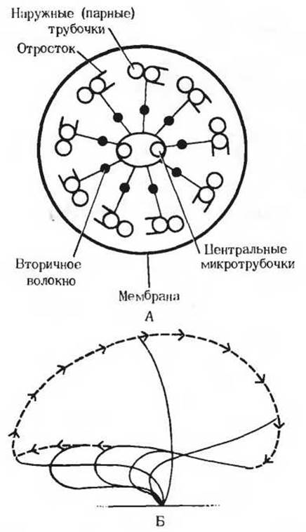
Fig. 14-12. In eukaryotic Cilia and flagella, mechanical force is generated through the utilization of ATP. A. Cross-section of a cilium. These structures consist of nine outer pairs of microtubules forming a ring, and two single central microtubules (the "9 + 2" arrangement; Section 2.16). Cilia are enclosed by a membrane that is an extension of Cell/30.html">The Plasma Membrane. The energy for characteristic ciliary movements—whether wavelike, sliding, or rotational—is supplied by ATP hydrolysis. Cilia perform these movements through the sliding or twisting of paired microtubules, which closely resembles the ATP-dependent sliding of thick and thin filaments past one another observed in Skeletal Muscle. Equally spaced projections or arms, resembling the Myosin heads in muscle thick filaments, extend from the outer (paired) microtubules. These projections consist of dynein molecules, a rather large protein possessing ATPase activity. Dynein-catalyzed ATP hydrolysis provides the energy for mechanical motion, namely the sliding or twisting of microtubules. It has been suggested that the central microtubules regulate the beat frequency of cilia. B. Successive phases of a ciliary beat in the gills of a marine worm, where the cilia are approximately 30 µm long. These characteristic movements are imparted to the cilia by the ATP-dependent sliding of tubular filaments relative to one another.
Thanks to creatine phosphate, the concentration of ATP in muscle Cells is maintained at a constant and fairly high level. This is especially vital for skeletal Muscles, which function intermittently but sometimes undergo intense, high-speed exertion. Whenever a portion of a muscle cell's ATP is consumed during contraction, ADP is formed As a result of ATP hydrolysis. With the participation of creatine kinase, creatine phosphate rapidly transfers its phosphate group to ADP molecules, restoring the normal ATP level. The creatine phosphate content in muscle is 3 to 4 times greater than that of ATP (Table 14-4); therefore, a sufficient quantity of phosphate groups can be stored in the form of creatine phosphate to fully maintain a constant ATP level during brief periods of strenuous muscular activity. Due to the reversibility of the creatine kinase reaction, the accumulated creatine is rephosphorylated back to creatine phosphate by ATP during the recovery period. Because no other metabolic pathway exists for the Synthesis and Breakdown of creatine phosphate, this compound is ideally suited to fulfill its function as a phosphate group reservoir.

Fig. 14-13. Creatine phosphate plays The Role of a storage donor of high-energy phosphate groups in muscles. It acts as a specialized buffer that maintains a constant ATP concentration.
In the muscles of many invertebrates, the role of the reserve energy carrier is performed not by creatine phosphate, but by Arginine phosphate. Compounds that, like creatine phosphate and arginine phosphate, serve as reserve Energy Sources are known as phosphagens.
Last update: 06/08/2026
Editorial and Educational Adaptation: This material has been compiled based on the primary/original source text. The project team performed an editorial review, corrected technical inaccuracies, structured sections, and adapted the content for an educational format.
What was processed:
- elimination of formatting defects (OCR errors, structural breaks, corrupted characters);
- editorial organization of content;
- standardization of terminology in accordance with academic sources;
- verification of factual statements against the original source text.
All mentions of the author, publication year, and origin of the primary text have been preserved in accordance with the source.