Special Histology and Embryology: Practical Course - V. K. Napkhanyuk 2001
Skin and its appendages
Nails
The nail (ungus) is an epidermal appendage. It is a keratinized plate resting upon the nail bed.
The nail bed consists of epithelium and Connective Tissue. The epithelium of the nail bed—the hyponychium—serves as the germinative layer of the epidermis. The nail plate lying directly upon it represents its cornified layer.
The nail (cornified) plate is formed by tightly packed cornified scales containing hard keratin. The nail plate consists of a ROOT, body, and free edge.
The nail bed is bounded laterally and proximally by Skin folds known as the nail folds (posterior and lateral).
Slide 2. Human scalp (Fig. 64).
Low magnification. Examine and sketch the slide.
Locate the epidermis, the thickness of which is significantly smaller than that of hairless skin. Only the germinative and thin cornified layers are distinguishable in the epidermis. The papillary layer of the dermis is less pronounced. The reticular layer of the dermis is much more developed than in hairless skin areas; it is formed by dense irregular connective tissue and contains Hair roots and Skin glands. The hair roots are positioned at a slight angle to the skin surface. The following structures are clearly visible in the hair root: the hair shaft, epithelial root sheath, and connective tissue hair follicle (dermal sheath). Deep within the skin, the hair root terminates in an expansion—the hair bulb—into which the connective tissue dermal papilla projects. Sebaceous Glands are located near the hair root, with their short excretory ducts opening into the root sheath at approximately the level of the upper quarter. These are simple branched alveolar glands with a holocrine secretion type. The secretory units of the sebaceous glands lack a lumen and are filled with pale polygonal Cells containing a small Nucleus. In standard preparations, their Cytoplasm has a porous appearance. The excretory ducts of Sweat Glands, appearing as tortuous dark tubes, can be found at various levels. The secretory units of sweat glands, which belong to the simple tubular type, form dense coils within the dermis and appear in multiple cross-sections on the slide.
Class="center">
Fig. 64. Human scalp. Hematoxylin-eosin staining. x 100:
1 — epidermis; 2 — dermis; 3 — subcutaneous adipose tissue (hypodermis); 4 — secretory unit of sweat gland with excretory duct; 5 — hair root; 6 — internal root sheath; 7 — external root sheath; 8 — hair follicle (dermal sheath); 9 — hair follicle; 10 — hair papilla; 11 — sebaceous gland; 12 — arrector pili Muscle
The dermis of the scalp contains arrector pili Muscles formed by bundles of smooth myocytes. The dense region of the dermis is underpinned by a layer of hypodermis (subcutaneous tissue), formed by an interwoven network of loose connective tissue bundles and adipose tissue.
Label the following on the diagram: 1) epidermis; 2) dermis; 3) hypodermis (subcutaneous tissue); 4) secretory unit of sweat gland with excretory duct; 5) hair root; 6) internal root sheath; 7) external root sheath; 8) hair follicle (dermal sheath); 9) hair follicle; 10) hair papilla; 11) sebaceous gland; 12) arrector pili muscle.
Slide 3. Cross-section of a nail (Fig. 65).
Low magnification. Examine and sketch the slide.
The preparation demonstrates that the cornified plate is formed by densely apposed cornified scales containing hard keratin. The nail plate rests upon the nail bed, which is composed of the germinative layer of the epidermis (with the overlying nail plate serving as its cornified layer) and the connective tissue of the dermis. The cornified layer of the epidermal nail folds transitions into the epithelium of the nail bed, and the stratum corneum extends over the nail from above, forming the so-called hyponychium (or cuticle of the nail fold). Clefts (posterior and lateral) are present between the nail bed and the nail folds.
Label the following on the diagram: 1) nail plate; 2) eponychium (cuticle) of the nail fold; 3) germinative layer; 4) connective tissue of the nail bed with Blood Vessels; 5) finger phalanx; 6) papillary layer of the skin.

Fig. 65. Cross-section of a nail. Hematoxylin-eosin staining. x 40:
1 — nail plate; 2 — eponychium (cuticle) of the nail fold; 3 — germinative layer; 4 — connective tissue of the nail bed with blood vessels; 5 — phalanx; 6 — papillary layer of the skin
Slide 4. Section of the mammary gland during Lactation (Fig. 66).
Low magnification. Examine and sketch the slide.
At this magnification, the distinct lobular architecture of the organ is clearly visible. The lobules vary greatly in shape and size. They are separated by broad interlobular connective tissue septa containing blood vessels and interlobular excretory ducts. The glandular lobules are formed by secretory units (alveoli). The alveoli are partitioned by thin intralobular connective tissue septa containing small vessels and capillaries, as well as small intralobular excretory ducts. Myoepithelial (basket) cells can be identified within the terminal portions of the mammary gland; the preparation reveals only their dark, elongated nuclei pressed against the alveolar epithelium. Small intralobular ducts are characterized by a small caliber and are lined with cuboidal epithelium. Larger interlobular ducts and lactiferous ducts are lined with a double-layered epithelium.
Label the following on the diagram: 1) glandular lobules; 2) secretory unit; 3) interlobular lactiferous duct; 4) connective tissue septa with blood vessels.

Fig. 66. Section of the mammary gland during lactation. Hematoxylin-eosin staining. x 140:
1 — gland lobules; 2 — secretory end piece; 3 — interlobular lactiferous duct; 4 — connective tissue septa with blood vessels
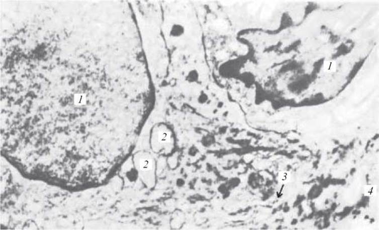
Electron micrograph 39. Cells of the basal layer of human skin epithelium. x 12,500:
1 — nuclei of epithelial cells; 2 — Cell/35.html">Mitochondria; 3 — basement membrane; 4 — Collagen fibers

Electron micrograph 40. Desmosomes in the spinous layer of human skin. x 87,500:
1 — desmosomes; 2 — tonofibrils; 3 — intercellular spaces

Electron micrograph 41. Fragment of an epithelial cell containing pigment granules from human skin. x 37,500:
1 — Nucleus of the epithelial cell; 2 — bundles of tonofibrils consisting of tonofilaments; 3 — Ribosomes
1. Sources of development of epidermocytes (keratinocytes), melanocytes, and Langerhans cells. Structural features and Functions.
2. Development of the dermis.
3. Structural Features of the papillary and reticular layers of the dermis.
4. Structure OF THE epidermis.
5. Skin glands: localization and structure.
6. Structural Features of the mammary gland.
7. Cytology/cytology/67.html">Development and Structure of the hair.
8. The nail. Structural features.
9. Situational Problems
1. In an experimental Setting, the dorsal mesoderm—specifically the dermatome—was damaged during the Embryonic period. How will this affect skin development?
2. On the extensor surfaces of the body (back, shoulders), the reticular layer of the dermis is significantly more developed than on the flexor surfaces (abdomen). Which skin function is this related to?
3. The skin is injured. Through which cellular layers is its epidermis restored?
4. An electron micrograph shows an epidermal skin cell lacking mitochondria and Endoplasmic reticulum. To which layer of the epidermis do these cells belong?
5. The figure shows the fingerprints of two individuals. What determines the individual pattern of fingerprints?
6. Due to illness, the function of the sebaceous glands has decreased. What changes will occur in the epidermis and hair?
7. A nursing mother experienced a decrease in milk secretion. The secretory process in lactocytes is not impaired. A decrease in which hormone is this associated with?
Sample Exam Questions
1. Skin, its structural components, and functional significance. Age-related features.
2. Skin Appendages (hair, glands, nails). Sources of development. Structure and function.
3. Mammary gland. Structural features of the lactating and non-lactating gland. Regulation of lactation.
Last update: 10/08/2026
Editorial and Educational Adaptation: This material has been compiled based on the primary/original source text. The project team performed an editorial review, corrected technical inaccuracies, structured sections, and adapted the content for an educational format.
What was processed:
- elimination of formatting defects (OCR errors, structural breaks, corrupted characters);
- editorial organization of content;
- standardization of terminology in accordance with academic sources;
- verification of factual statements against the original source text.
All mentions of the author, publication year, and origin of the primary text have been preserved in accordance with the source.