Review of Medical Physiology - William F. Ganong 2002
Functions of the Digestive System
Mechanisms of Regulation of Gastrointestinal Functions
Oral Cavity and Esophagus
In the Oral Cavity, food is mixed with saliva to form a bolus, which is then swallowed into the Esophagus. Peristaltic waves of the esophagus propel the food bolus toward The Stomach.
Mastication
During mastication, large food particles are broken down and mixed with saliva, the secretion of the Salivary Glands. Moistened and homogeneous food is swallowed and subsequently subjected to Digestion. Large food particles are sometimes swallowed without proper chewing, which induces strong and often painful contractions of the esophageal Muscles. Smaller, more dispersive particles can also be swallowed without saliva, though with difficulty, as they fail to form a proper food bolus. The optimal number of chewing cycles required to adequately break down food generally ranges from 20 to 25.
Edentulous patients, who typically restrict their diet to soft foods, may experience difficulties when consuming dry products.
Salivary Glands and Saliva
In the salivary glands, secretory (zymogenic) granules containing salivary Enzymes are released from acinar Cells into the ducts (Fig. 26-6). The characteristics of each of the three pairs of salivary glands are summarized in Table 26-4.
Approximately 1500 mL of saliva is secreted daily. At rest, the glands secrete saliva with a pH slightly below 7.0; during active secretion, the pH rises to up to 8.0. Saliva contains two digestive enzymes: lingual lipase, secreted by the Glands of the Tongue, and salivary a-amylase, secreted by the salivary glands. The Functions of these enzymes are described in Chapter 25. Saliva also contains mucins, which are Glycoproteins that lubricate food and protect the oral mucosa. In addition to these components, it contains IgA (see Chapter 27), which provides the first line of immunological defense against Bacteria and Viruses; Lysozyme, which destroys bacterial and viral Cell walls; lactoferrin, an iron-binding protein with bacteriostatic activity; and Proline-rich Proteins, which protect tooth enamel and bind toxic Tannins.
Saliva performs several vital functions. It facilitates swallowing, moistens the oral cavity, dissolves chemical molecules that stimulate taste buds, eases the Movements of the Lips and tongue during speech, and cleanses the ORAL CAVITY AND Teeth. Saliva also exhibits certain antibacterial properties; patients with impaired salivation (xerostomia) are more prone to dental caries than healthy individuals. The buffering properties of saliva maintain the pH in the oral cavity around 7.0. They also neutralize the acidic environment of the stomach, alleviating heartburn in cases of gastric juice regurgitation into the esophagus.
Ionic Composition of Saliva
There are distinct and significant differences in the ionic composition of saliva among individual species, as well as depending on the specific gland producing the secretion. In general, the saliva secreted by the acini is nearly isotonic, with concentrations of Na+, Cl-, and HCO3- close to those in plasma. The excretory ducts, and likely the intercalated ducts connected to them, modify the composition of saliva by reabsorbing Na+ and Cl- while secreting K+ and HCO3-. Because the ducts are relatively impermeable to Water, the saliva becomes hypotonic as it passes through the duct system. Consequently, during slow salivation rates, the saliva entering the oral cavity is hypotonic, slightly acidic, rich in K+, but relatively depleted of Na+ and Cl-. When salivation is intense, there is less time for ionic modification to occur. As a result, the still-hypotonic human saliva becomes more isotonic due to increased concentrations of Na+ and Cl-. The Effect of aldosterone is analogous to its action in the Kidneys—it increases K+ concentration and decreases Na+ concentration in saliva (see Chapters 20 and 38). Addison's disease (aldosterone deficiency) is characterized by a high Na+/K+ ratio in saliva.
Regulation of Salivary Secretion
Salivary secretion is regulated by The Nervous system. Stimulation of the parasympathetic innervation induces profuse secretion of watery saliva with a relatively low organic content. The vasodilation in the gland associated with this secretion results from the local release of VIP. This polypeptide acts as a cotransmitter with acetylcholine in certain postganglionic parasympathetic Neurons. Atropine and other cholinergic blocking agents reduce salivary secretion. Stimulation of the sympathetic innervation causes vasoconstriction; in humans, this results in the secretion of a small volume of saliva rich in organic components from the submandibular glands. The presence of food in the oral cavity triggers reflex salivation, as does stimulation of afferent vagal fibers from the abdominal portion of the esophagus. Salivary secretion also occurs via conditioned reflex mechanisms, as demonstrated by Pavlov's classic experiments (see Chapter 16). In humans, the sight, smell, or even the thought of food can induce salivation ("making one's Mouth water").
Class="center">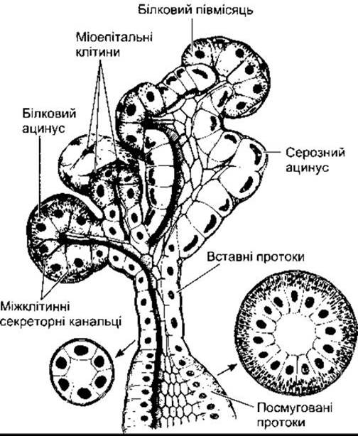
Fig. 26-6. Structure OF THE submandibular glands. Note the cells in the mucous acini, whose basal nuclei have become flattened, whereas the cells in the serous acini possess rounded nuclei and contain apical zymogenic secretory granules. Intercalated ducts transition into striated ducts, where cells are specialized for ion transport.
Swallowing
Swallowing is a reflex response initiated by afferent impulses from the trigeminal, glossopharyngeal, and vagus nerves. These impulses are integrated in The Nucleus tractus solitarius and the nucleus ambiguus. Efferent fibers innervating the musculature of the Pharynx and tongue are conveyed by the trigeminal, facial, and hypoglossal nerves. Swallowing is initiated voluntarily after a food bolus is formed on the tongue and propelled into the pharynx. This action triggers a wave of involuntary contractions in the pharyngeal muscles that propel the bolus into the esophagus. Respiratory arrest and closure of the glottis are integral PARTS OF THE reflex response. Swallowing becomes difficult or even impossible if the mouth is open, as anyone who has spent time in a dentist's chair and felt saliva accumulate in the throat knows well. In healthy adults, swallowing occurs frequently during meals as well as during the interdigestive period. The total number of swallowing acts per day is about 1200. Of these, The ratio of swallowing during eating to drinking is 600:200, 350 swallowing acts occur between meals, and 50 occur during Sleep.
Table 26-4. Characteristics of Human Salivary Glands
Gland |
Histological Type |
Secretion1 |
Share of Total Saliva2 (1.5 L/day), % |
Parotid |
Serous |
Watery |
20 |
Submandibular |
Mixed |
Moderately viscous |
70 |
Sublingual |
Mucous |
Viscous |
5 |
1 Serous cells secrete ptyalin, and mucous cells secrete mucin.
2 The remaining 5% of saliva volume is produced by lingual and other minor oral cavity glands.
At the pharyngoesophageal junction, There is a three-centimeter segment of the esophagus characterized by high resting wall pressure. This region relaxes reflexively during swallowing, allowing the food bolus to enter the body of the esophagus. Once the bolus passes, peristaltic circular Muscle contractions of the esophagus are initiated, and the bolus is rapidly propelled down the esophagus at a speed of about 4 cm/s. When a person is in an upright position, liquid and semi-liquid foods quickly reach the distal esophagus under METABOLISM/18.html">The Influence of gravity, ahead of the peristaltic wave.
Lower Esophageal Sphincter
Unlike the rest of the esophageal musculature, the lower esophageal sphincter (LES) at the gastroesophageal junction is tonically active, yet relaxes during swallowing movements. The tonic activity of the LES prevents the reflux of gastric contents into the esophagus between meals. The LES consists of three components (Fig. 26-7). The smooth Muscles of the esophagus are more prominent at the junction with the stomach (the intrinsic sphincter). The fibers of the crural Diaphragm (the right crus) are skeletal muscles that surround the esophagus in this region (the extrinsic sphincter), exerting a pinch-cock effect on the esophagus. Additionally, the oblique or sling fibers of the gastric wall form a flap-like mechanism that helps close the gastroesophageal junction and prevents regurgitation during increases in intragastric pressure.
The regulation of LES tone is mediated by the nervous system. The release of acetylcholine by vagal fibers causes contraction of the intrinsic sphincter, whereas the release of NO and VIP by interneurons innervated by other vagal fibers induces its relaxation. The contraction of the diaphragmatic crura, innervated by the phrenic nerves, is coordinated with Respiration and the contractions of the chest and Abdominal muscles. Thus, the extrinsic and internal sphincters work in concert to ensure the orderly entry of food into the stomach while preventing the reflux of gastric contents into the esophagus.
Large doses of gastrin increase LES tone, but circulating Blood levels of gastrin achieved after a meal do not reach concentrations sufficient to produce this effect.
Motor Disorders of the esophagus
Achalasia is a condition in which food accumulates in the esophagus, causing it to dilate massively. This leads to an increased resting tone of the LES and incomplete relaxation during swallowing. The loss of the esophageal myenteric plexus contributes to LES dysfunction and impairs the release of NO and VIP. Treatment involves pneumatic dilation of the sphincter or surgical division of the esophageal muscle (myotomy). Inhibiting acetylcholine release by injecting botulinum toxin into the LES is also effective, providing relief for several months.
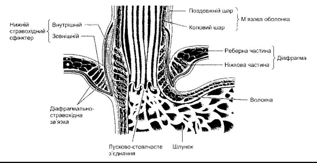
Fig. 26-7. The esophagogastric junction. Note that the lower esophageal sphincter (internal sphincter) is supplemented by the crura of the diaphragm (external sphincter), and they are linked to each other by the phrenoesophageal ligament (reproduced with permission from Mittal RK, Balaban DH. The esophagogastric junction. N Engl J Med 1997;336:924).
The opposite condition is functional insufficiency of the LES, characterized by the reflux of acidic gastric contents into the esophagus (gastroesophageal reflux). This common disorder causes heartburn and esophagitis and can lead to ulceration and esophageal strictures resulting from scarring. In severe cases of gastroesophageal reflux, weakness of the internal, external, or both sphincters develops, whereas in milder cases, intermittent periods of poorly understood impaired neural coordination of both sphincters are observed. Such conditions can be treated by inhibiting Hydrochloric acid secretion with H2-receptor blockers or omeprazole (see below). Surgical treatment is also effective, creating conditions in which the gastric fundus wraps around the distal esophagus so that the LES is enclosed within a short gastric tunnel (fundoplication).
Aerophagia and intestinal gas
Neurasthenic individuals prone to hyperventilation swallow large amounts of air while eating or drinking (aerophagia). Part of this air is regurgitated (belching), some of its constituent gases are absorbed, but the majority passes into the colon. Here, some of the oxygen is absorbed. The action of intestinal bacteria on CARBOHYDRATES and other substances present in the gut produces hydrogen, hydrogen sulfide, carbon dioxide, and methane. These are subsequently expelled as flatus. The odor is largely due to sulfides. The normal volume of gas in the human gastrointestinal tract is about 200 mL, and the daily production ranges from 500 to 1500 mL. In some individuals, intestinal gas causes symptoms of bloating, borborygmi, and abdominal discomfort.
Last update: 10/08/2026
Editorial and Educational Adaptation: This material has been compiled based on the primary/original source text. The project team performed an editorial review, corrected technical inaccuracies, structured sections, and adapted the content for an educational format.
What was processed:
- elimination of formatting defects (OCR errors, structural breaks, corrupted characters);
- editorial organization of content;
- standardization of terminology in accordance with academic sources;
- verification of factual statements against the original source text.
All mentions of the author, publication year, and origin of the primary text have been preserved in accordance with the source.