Review of Medical Physiology - William F. Ganong 2002
Functions of the Digestive System
Mechanisms of Gastrointestinal Regulation
Gastrointestinal Hormones
Biologically active Polypeptides secreted by Neurons and glandular Cells of the mucous membrane exert their effects both via paracrine pathways and humorally, as they simultaneously enter the bloodstream. Experimental studies of their effects and the measurement of their Blood concentrations using radioimmunoassay identify The Role of these gastrointestinal Hormones in regulating the secretion and motility of the gastrointestinal tract. When administered in large doses, the effects of these hormones overlap. Furthermore, their physiological effects appear quite discreetly. Based on structural similarities (Table 26-2) and depending on the degree of functional identity, some of these hormones belong to one of two families: the gastrins, whose main representatives are gastrin and cholecystokinin (CCK), and the secretins, whose main representatives are secretin, Glucagon, glicentin (GLl), VIP, and gastric inhibitory polypeptide (GIP). In addition, there are Other Hormones that do not belong to either of these two families.
Enteroendocrine cells
The mucosa of The Stomach, Small Intestine, and Large Intestine contains more than 15 types of hormone-producing enteroendocrine cells. Many of these secrete only a single hormone and are identified by letters (G cells, S cells, etc.). Cells that produce serotonin (though not all of them) are known as enterochromaffin cells. Cells that produce polypeptides and amines are sometimes called APUD cells (amine precursor uptake and decarboxylation) or neuroendocrine cells, which, apart from the gastrointestinal tract, are found in the Lungs and other Organs. They are also the cells from which carcinoid tumors originate.
Class="center">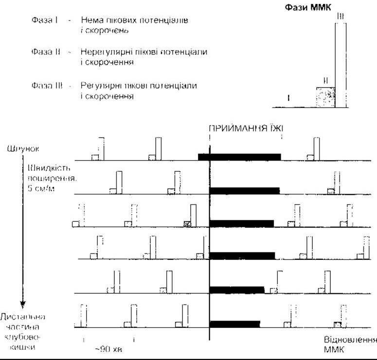
Fig. 26-3. Migrating motor complexes (MMCs). Note that these complexes migrate down the gastrointestinal tract at a normal rate during the interdigestive period, are completely inhibited during food intake, and resume 90-120 minutes after a meal (reproduced with permission from Chang EB, Sitrin MD, Black DD. Gastrointestinal, Hepatobiliary, and Nutritional Physiology. Lippincott-Raven, 1996)
Gastrin
Gastrin is produced by G cells located in the lateral walls of mucosal glands in the antrum of the stomach (Fig. 26-4). These G cells are flask-shaped, with a wide base containing numerous gastrin granules and a narrow apex opening onto the mucosal surface. Microvilli project from the apical end into the lumen. These microvilli bear gastrin receptors that respond to changes in gastrin content. Other hormone-secreting cells of the gastrointestinal tract share similar morphological features. Another type of gastrin-producing Cell is the TG cell, found in both the stomach and the small intestine. They possess G34 and the carboxyl terminal tetrapeptide of gastrin, but have lost G17 (see below).
During embryonic life, gastrin is also present in the islets of the Pancreas. Gastrin-secreting tumors, known as gastrinomas, also occur in the pancreas; however, it remains unclear whether any gastrin is present in the pancreas under normal conditions in adults. In addition, gastrin is produced by the anterior and intermediate lobes of the Pituitary Gland, the Hypothalamus, the Medulla Oblongata, and the vagus and sciatic nerves. Gastrin is a typical representative of polypeptide hormones exhibiting both macroheterogeneity and microheterogeneity. Macroheterogeneity refers to the occurrence of peptide chains of varying lengths in Tissues and Body Fluids, whereas microheterogeneity refers to differences in molecular Structure depending on THE ORIGIN OF individual amino acid residues. Processing of preprogastrin yields fragments of various sizes. The three main fragments contain 34, 17, and 14 amino acid residues, respectively. All of them share the same carboxy-terminal configuration (see Table 26-2). These forms are frequently referred to as G34, G17, and G14 gastrins, respectively. Another form is a carboxy-terminal tetrapeptide that, in the larger form, is extended at the amino terminus by more than 45 amino acid residues. Yet another derivative form involves the sulfation of Tyrosine, which is the sixth amino acid residue from the carboxy terminus. Blood and tissues contain equal amounts of nonsulfated and sulfated forms. Other derivatives are formed by the amidation of the carboxy-terminal phenylalanine.
Table 26-2. Structure of some hormonally active polypeptides secreted by gastrointestinal cells in humans1
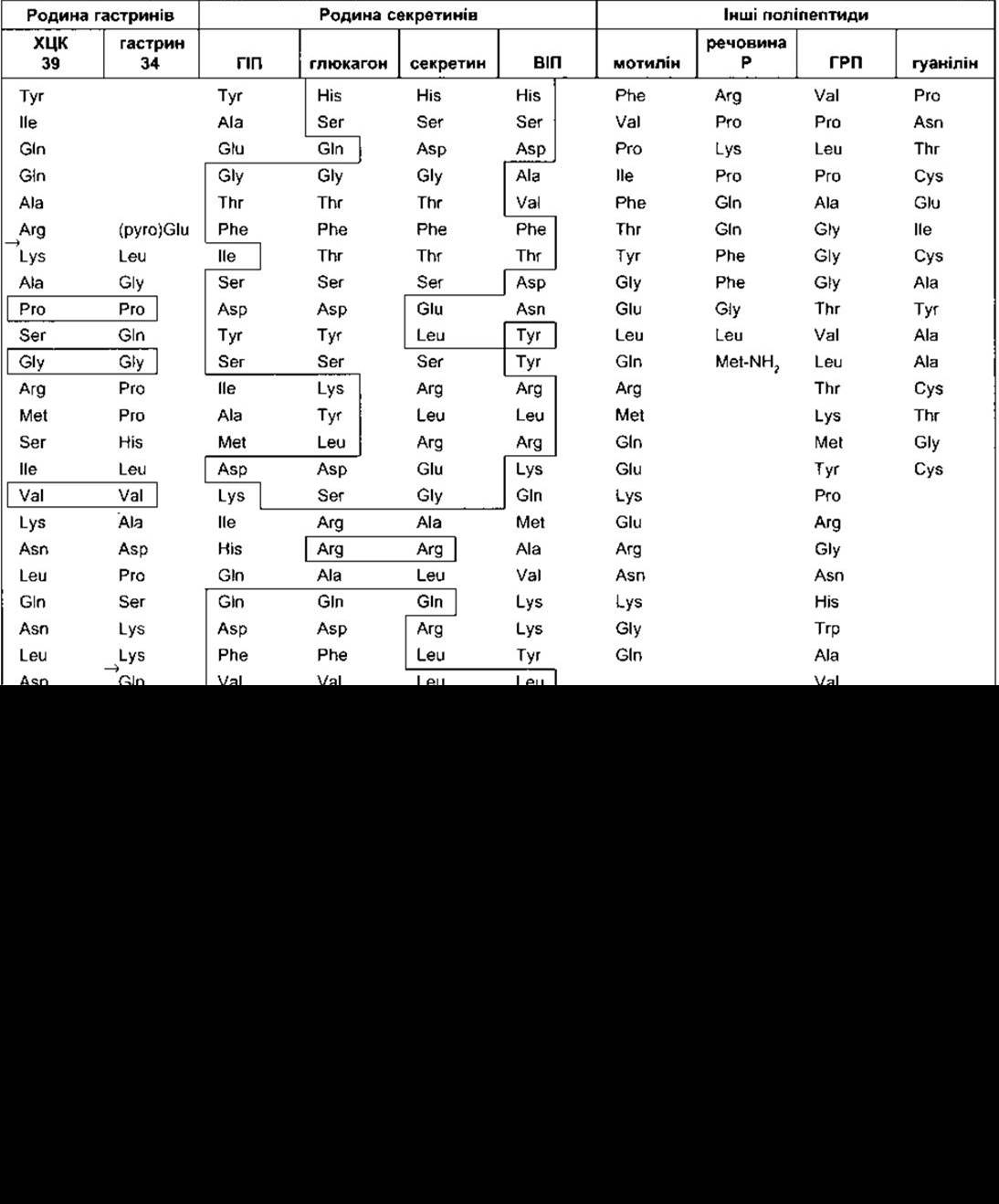
1 Homologous amino acid residues are enclosed within lines extending from one polypeptide to another. Arrows indicate Cleavage sites for the generation of smaller variants;
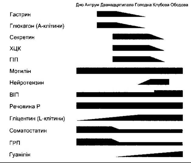
Fig. 26-4. Distribution of Gastrointestinal Peptides along the gastrointestinal tract. The thickness of each marker is proportional to the peptide concentration in the mucosa. Preproglucagon is processed primarily into glucagon by A cells in the proximal region, and into glicentin, GLP-1, GLP-2, and other derivatives by L cells in the distal gastrointestinal tract.
The physiological Significance of the observed heterogeneity is as follows. There is a slight difference in activity among the various components, as well as varying proportions of these components across different tissues where gastrin is present. This is supported by the fact that The formation of different gastrin variants is driven by modified functional demands.
Therefore, it can be stated that G17 is the primary form responsible for gastric Hydrochloric acid secretion. The carboxy-terminal tetrapeptide exhibits all the activities of gastrin, yet possesses only 10% of the potency of G17. The half-life of G14 and G17 in the blood is 2-3 minutes, whereas that of G34 is 15 minutes. Inactivation of gastrins occurs predominantly in the Kidneys and small intestine.
In large doses, gastrin exerts various effects, but its primary physiological action is to stimulate Gastric Acidity and Pepsin secretion, as well as to induce mucosal growth in the stomach, small intestine, and large intestine (trophic effect). Stimulation of gastric motility is another consequence of gastrin action, causing contraction of the gastroesophageal sphincters (see below), although The Significance of this physiological effect remains unclear. Gastrin also stimulates Insulin secretion, but only following the ingestion of a protein meal, not a carbohydrate meal. As the level of circulating endogenous gastrin increases, insulin secretion rises. The Functions of gastrin in the pituitary gland, other Regions of the Brain, and peripheral nerves are not yet fully understood.
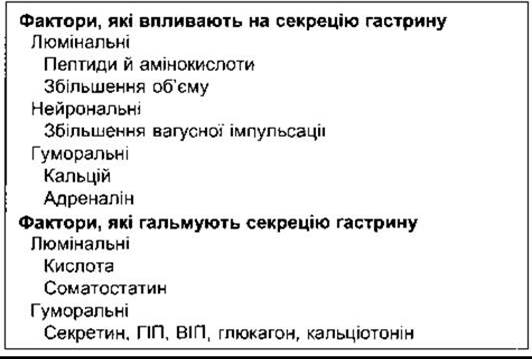
Gastrin secretion depends on ingested food, vagal nerve activity, and various blood-borne factors (Table 26-3). Atropine does not inhibit gastrin release in humans following a test meal because the neurotransmitter secreted by postganglionic vagal fibers innervating G cells is gastrin-releasing peptide (GRP; see below)* rather than acetylcholine. Gastrin secretion is also increased by the presence of digested protein in the stomach, particularly Amino Acids, which act directly on G cells. Phenylalanine and Tryptophan are especially effective.
The presence of acid in the gastric antrum inhibits gastrin secretion, partly through a direct effect on G cells and partly via the release of Somatostatin, a relatively potent inhibitor of gastrin secretion. The Effect of acid on gastrin secretion forms The basis of feedback regulatory mechanisms. Increased hormone secretion leads to an elevation in hydrochloric acid secretion by parietal cells.
An increase in gastric acidity results in the feedback inhibition of gastrin secretion.
The role of gastrin in the pathogenetic mechanisms of duodenal ulcers is described below. In pernicious anemia, when the acid-producing cells of the stomach are damaged, gastrin secretion is chronically elevated.
* Gastrin-releasing peptide is synonymous with bombesin.
Table 26-3. Factors influencing gastrin secretion
Cholecystokinin-Pancreozymin
It was previously believed that the hormone cholecystokinin caused Gallbladder contraction, whereas a completely different hormone, pancreozymin, increased the secretion of enzyme-rich pancreatic juice. It is now known that these represent a single hormone secreted by mucosal cells of the proximal small intestine that performs both functions; hence, its updated name is cholecystokinin-pancreozymin (CCK-PZ, now more commonly referred to as CCK).
Like gastrin, CCK is a compound that exhibits both macro- and microheterogeneity at THE MOLECULAR LEVEL. During processing, pre-CCK yields several fragments. Big CCK contains 58 amino acid residues (CCK-58). In addition, there are several CCK forms with varying numbers of amino acid residues: CCK-39 with 39 residues, CCK-33 with 33 residues, as well as several forms containing 12 (CCK-12) or slightly fewer residues (CCK-8). All these forms, much like gastrin, share Five amino acids at the carboxy-terminal end (see Table 26-2). The carboxy-terminal tetrapeptide (CCK-4) is also found in tissues. The carboxy-terminal ends are amidated, and tyrosine—the seventh amino acid residue from the carboxy terminus—is sulfated. Unlike gastrin, the nonsulfated form of CCK is absent in tissues. Furthermore, other derivatives originating from the remaining amino acid residues of CCK are known to be formed. The half-life of CCK in the Circulation is 5 min, although little is known about its METABOLISM.
CCK is secreted by endocrine cells—specifically, I cells of the proximal small intestine, as well as nerve terminals in the distal ileum and colon. CCK is found in brain neurons, particularly in the Cerebral Cortex, and in nerve fibers throughout The Human Body (see Chapter 4). In the brain, this hormone is involved in The regulation of food intake (see Chapter 14), and its appearance is associated with feelings of fear, anxiety, and analgesia. CCK secreted in the duodenum and jejunum consists predominantly of CCK-8 and CCK-12. In addition to these forms, CCK-58 is present in the gut and circulating blood across many species. Enteric and pancreatic nerve fibers contain primarily CCK-4. The brain contains both CCK-58 and CCK-8.
In addition to the aforementioned effects, CCK potentiates the action of secretin (which stimulates alkaline pancreatic juice secretion), delays gastric emptying, influences pancreatic trophic processes, increases enterokinase secretion, and may enhance the motility of the small and large intestine. In some cases, secretin enhances the contraction of the pyloric sphincter, thereby preventing the reflux of duodenal contents into the stomach. Gastrin and CCK stimulate glucagon secretion during the Digestion of protein meals. Acting individually or together, these hormones serve as intestinal factors that stimulate glucagon secretion (see Chapter 19). As noted in Chapters 4 and 14, two CCK receptors have been identified to date. CCK-A receptors are located predominantly in the periphery, whereas both CCK-A and CCK-B receptors are found in the brain. Both are activated via phospholipase C and produce IP3 and DAG (see Chapter 1). The gastric gastrin receptor has recently been cloned; although very similar, it is not identical to the CCK-B receptor. Despite this, the differences between the peripheral effects of gastrin and CCK and the concentration of CCK-B receptors in the brain remain poorly understood.
CCK secretion increases when digested products, particularly peptides and amino acids, come into contact with the intestinal mucosa, as well as in the presence of Fatty acids containing more than ten carbon atoms in the duodenum. From the moment CCK-stimulated Bile AND PANCREATIC juice enter the duodenum, new portions of protein and fat digestion products are formed, which in turn further stimulate CCK secretion: this is positive feedback regulation. It concludes once the digested products reach the distal regions of the gastrointestinal tract.
Secretin
Secretin holds a unique place in the History of Physiology. In 1902, Bayliss and Starling were the first to describe the excitatory effect of duodenal stimulation on pancreatic secretion by a blood-borne factor. Their research led to the discovery of secretin. The scientists also predicted that many other chemical agents must be secreted by body cells and subsequently transported via the circulation from effector organs located at a considerable distance. Starling coined the term hormone, defining it as a chemical messenger. Modern endocrinology has fully confirmed the validity and correctness of this hypothesis.
Secretin is secreted by S cells located deep within the mucosal Glands of the proximal small intestine. The secretion of secretin (see Table 26-2) differs from that of CCK and gastrin, but is similar to glucagon, GLI, VIP, and GIP. Only a single form of secretin has been isolated, and molecular fragments studied in experiments have been found to be inactive. The half-life of secretin is about 5 min, and little is known about its metabolism.
Secretin increases bicarbonate secretion by the ductal cells of the pancreas and biliary tract, and also stimulates the secretion of a watery, alkaline pancreatic juice. Its effect on ductal cells is mediated via cAMP. Secretin is known to interact with CCK, which is responsible for the secretion of digestive Enzymes by the pancreas.
At the same time, it decreases gastric hydrochloric acid secretion, which leads to contraction of the pyloric sphincter.
Secretin secretion increases with the formation of Protein Digestion products and the irritation of the proximal small intestinal mucosa by acidic contents. The release of secretin in response to acid is another example of feedback regulation. Secretin stimulates the secretion of alkaline pancreatic juice into the duodenum, thereby neutralizing gastric acidity and halting further hormone secretion.
Gastric Inhibitory Polypeptide
Gastric inhibitory polypeptide (GIP) consists of 43 amino acid residues (see Table 26-2) and is produced by K cells in the mucosa of the duodenum and jejunum. GIP secretion is stimulated by glucose and fats present in the duodenum. The production of large amounts of this peptide results in the inhibition of gastric secretory and motor functions—an action that gave rise to its name, gastric inhibitory peptide. However, contemporary data indicate that GIP does not exert this gastric-inhibitory effect when released into the bloodstream in small doses following a meal. At the same time, GIP is known to stimulate insulin secretion. While gastrin, CCK, secretin, and glucagon also produce this effect, GIP alone stimulates insulin secretion to blood levels comparable to those seen after oral glucose ingestion. In this light, GIP is often referred to as glucose-dependent insulinotropic polypeptide. Glucagon derivatives such as GLP-1 (7-36) (see Chapter 19) also stimulate insulin secretion and are more potent in this regard than GIP. Consequently, GIP may also function as a physiological gastrointestinal beta-cell-stimulating hormone.
The Integration of the actions of gastrin, CCK, secretin, and GIP serves to enhance digestion and the utilization of absorbed nutrients (Fig. 26-5).
Vasoactive Intestinal Polypeptide
Vasoactive intestinal polypeptide (VIP) contains 28 amino acid residues (see Table 26-2). It is found in fibers innervating the gastrointestinal tract. Prepro-VIP contains both VIP and a closely related polypeptide (PHI-27 in humans, PHM-27 in other species). The half-life of VIP is 2 min. In the gut, it stimulates intestinal electrolyte and Water secretion. Other effects include relaxation of intestinal smooth Muscle, including sphincters, dilation of peripheral Blood Vessels, and inhibition of gastric hydrochloric acid secretion. VIP is also present in the brain and many autonomic nerve fibers (see Chapter 4), where it frequently colocalizes with acetylcholine in neurons. It potentiates the action of acetylcholine on the Salivary Glands. However, VIP and acetylcholine do not coexist in neurons innervating other PARTS OF THE gastrointestinal tract. VIP-secreting tumors (VIPomas) have been described in patients with severe diarrhea. The Relationship of GIP and VIP to enterogastrone—a hypothetical hormone that inhibits gastric acid secretion and motility—remains not fully elucidated. In addition, peptide YY (see Chapter 19) may also be considered an enterogastrone. Fats trigger its release into the jejunum, and this peptide acts as a potent inhibitor of gastrin-stimulated hydrochloric acid secretion.
Motilin
Motilin is a 22-amino-acid polypeptide secreted by enterochromaffin cells and Mo cells of the stomach, small intestine, and colon. It acts on G protein-coupled receptors of the enteric Nervous system in the duodenum and colon. Administration of motilin induces smooth muscle contractions in the stomach and intestine. Blood levels of motilin rise periodically at 100-minute intervals during the interdigestive period. Motilin is a primary regulator of the migrating motor complex (MMC), which governs gastrointestinal motility between meals. The antibiotic erythromycin binds to motilin receptors, and derivatives of these compounds are clinically significant for patients exhibiting slowed gastrointestinal motility during Treatment with this drug.
Other Gastrointestinal Hormones
Neurotensin is a 13-amino-acid polypeptide produced by neurons and cells located in the ileal mucosa. It is released in response to stimulation by fatty acids.
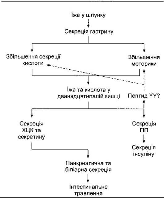
Fig. 26-5. Integrative action of gastrointestinal hormones in regulating digestion and the assimilation of absorbed nutrients. The dashed arrow indicates inhibition. The precise identity of the hormonal or intestinal factors that inhibit gastric acid secretion and motility remains unknown, though peptide YY is a candidate.
The primary effects of neurotensin are the inhibition of gastrointestinal motility and an increase in ileal blood flow. Substance P (see Table 26-1) is found in endocrine cells and neurons of the gastrointestinal tract and may enter the circulation. It enhances small intestinal motility. GRP (gastrin-releasing peptide) contains 27 amino acid residues, with 10 residues at the carboxy terminus being identical to those of amphibian bombesin. It is present in vagal nerve fibers terminating on G cells. Upon their stimulation, bombesin is released, which enhances gastrin secretion. When secretion is markedly elevated, a fraction of the gastrin enters the circulation. Somatostatin, a Growth Hormone-inhibiting hormone, is synthesized not only in the hypothalamus but is also secreted into the circulation by D cells of the pancreatic islets (see Chapter 19) and D-like cells of the gastrointestinal mucosa. As described in Chapter 14, it exists in tissues in two forms—somatostatin-14 (see Fig. 4-27) and somatostatin-28—which are released simultaneously. Somatostatin inhibits the secretion of gastrin, VIP, GIP, secretin, and motilin. Like other gastrointestinal hormones, somatostatin is secreted in large amounts into the gastric lumen and subsequently into the bloodstream. Its secretion increases with rising gastric acidity, and its paracrine action via gastric juice likely mediates the acid-induced inhibition of gastrin secretion. Somatostatin also inhibits exocrine pancreatic secretion, hydrochloric acid secretion, gastric motility, gallbladder contraction, and the absorption of glucose, amino acids, and triglycerides. Gastrointestinal glucagon is responsible (in part) for post-pancreatectomy hyperglycemia. Products generated from preproglucagon in the proximal and distal gut are described in Chapter 19 and shown in Fig. 19-16.
Guanylin is a gastrointestinal polypeptide that binds to guanylyl cyclase C. It consists of 15 amino acid residues (see Table 26-2) and is secreted by intestinal mucosal cells. In humans, guanylin may also be produced by Paneth cells and endocrine cells located near the crypts of Lieberkühn in the small intestine. Stimulation of guanylyl cyclase increases intracellular cGMP concentrations, which likely enhances The activity of CFTR (cystic fibrosis transmembrane conductance regulator) chloride channels, thereby increasing chloride secretion into the intestinal lumen. Guanylin acts primarily in a paracrine manner and is produced by cells spanning from the pylorus to the rectum. An interesting example of molecular mimicry is found in the heat-stable enterotoxin from strains of E. coli that cause diarrhea: it shares structural Homology with guanylin and activates guanylin receptors in the gut.
Guanylin receptors are also found in the kidneys, Liver, and FEMALE Reproductive System. Furthermore, guanylin exerts humoral effects on the regulation of fluid movement in tissues.
TRH and ACTH are found in the gastrointestinal tract; however, these hormones do not appear to enter the Circulatory system from this site, as targeted hypothalamic lesions or hypophysectomy still affect the thyroid and Adrenal Glands provided the gastrointestinal tract remains intact. In addition, locally produced TRH or related peptides are known to be involved in the regulation of secretory Immunity in the gut (see Chapter 25). TRH is also detected in pancreatic islets and likely in beta cells. Urogastrone (named because it was first isolated from urine) belongs to mucosal defense Transmitters and participates in ulcer healing; it is now known as epidermal growth factor (see Chapter 22).
Cells that secrete gastrointestinal polypeptides can form tumors. Gastrinomas account for 50%, glucagonomas for 25% of these tumors, and VIPomas, neurotensinomas, and others are also described in the literature.
Last update: 10/08/2026
Editorial and Educational Adaptation: This material has been compiled based on the primary/original source text. The project team performed an editorial review, corrected technical inaccuracies, structured sections, and adapted the content for an educational format.
What was processed:
- elimination of formatting defects (OCR errors, structural breaks, corrupted characters);
- editorial organization of content;
- standardization of terminology in accordance with academic sources;
- verification of factual statements against the original source text.
All mentions of the author, publication year, and origin of the primary text have been preserved in accordance with the source.