Review of Medical Physiology - William F. Ganong 2002
Functions of the Digestive System
Regulatory Mechanisms of Gastrointestinal Functions
General Principles
The organizational principle of the structures forming the wall of the gastrointestinal tract from the posterior Pharynx to the anus is illustrated in Fig. 26-1. While minor variations exist, the wall generally comprises four layers (from the lumen outward: mucosa, submucosa, muscularis, and serosa). Smooth Muscle fibers are located within the submucosa, whereas the muscularis externa consists of two smooth muscle layers: an outer longitudinal and an inner circular layer. The entire digestive tract is lined with a mucous membrane, except for the Esophagus and the distal rectum, which are covered by a serous coat. This coat is continuous with the mesentery, which houses nerve fibers, Lymphatic vessels, and Blood Vessels surrounding the alimentary canal.
Gastrointestinal Circulation
Blood flow to The Stomach, intestines, Pancreas, and Liver is supplied by a series of parallel vascular beds, wherein blood draining from the intestines and pancreas enters the liver via the portal vein. The physiology of this critical circulatory region is detailed in Chapter 32.
Enteric Nervous system
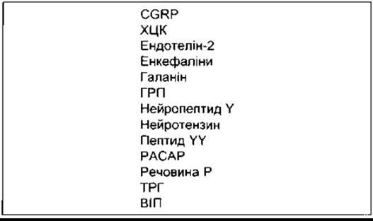
The wall of the gastrointestinal tract contains two intricate networks of nerve fibers: the myenteric plexus (Auerbach's plexus), situated between the outer longitudinal and middle circular muscle layers, and the submucosal plexus (Meissner's plexus), located between the middle circular muscle layer and the mucosa (see Fig. 26-1). Together, these Neurons constitute the enteric nervous system. Comprising over 100 million sensory neurons, interneurons, and motor neurons in humans—surpassing the total number of neurons in the Spinal Cord—this system is perhaps best conceptualized as a "relocated" division of the Central Nervous System dedicated to regulating gastrointestinal Functions. Although linked to the CNS via parasympathetic and sympathetic fibers, the enteric nervous system can operate autonomously in the absence of these connections (see below). The myenteric plexus innervates the longitudinal and circular smooth muscle layers and plays a primary role in controlling gut motility, whereas the submucosal plexus regulates local intestinal secretion. Neurotransmitters of the enteric nervous system include acetylcholine, amines such as noradrenaline and serotonin, Amino Acids like GABA, the purine ATP, the gases NO and CO, as well as numerous Peptides and Polypeptides (Table 26-1). Some of these peptides also act in a paracrine manner, while others enter the bloodstream to function as Hormones. Notably, many of these substances are also found in the Brain.
Class="center">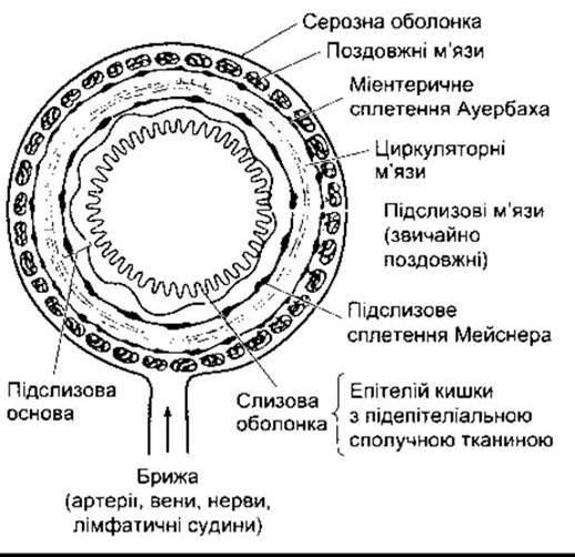
Fig. 26-1. Schematic representation of the wall layers of the stomach, Small Intestine, and colon. The Structure of the esophagus and the distal rectum is similar, except that they lack a serosa and mesentery. Additionally, the musculature in the upper quarter of the esophagus is striated, in the transitional zone it is mixed (containing both smooth and striated muscle), and in the distal section it is exclusively smooth (reproduced by permission from Bell GH, Emslie-Smith D, Paterson CR: Textbook of Physiology and Biochemistry, 9th ed. Churchill Livingstone, 1976).
Extrinsic Innervation
The intestine receives a dual extrinsic innervation: parasympathetic, cholinergic fibers (which generally enhance gastrointestinal smooth muscle activity) and sympathetic, noradrenergic fibers (which produce opposite effects and mediate sphincter contraction). Preganglionic parasympathetic fibers comprise over 2,000 efferent fibers within the Vagus nerve along with contributions from the sacral nerves. These generally terminate on cholinergic neurons within the myenteric and submucosal plexuses. Sympathetic fibers are predominantly postganglionic; however, many terminate on postganglionic cholinergic neurons, where their released noradrenaline inhibits acetylcholine secretion via the activation of α2-presynaptic receptors. Certain sympathetic fibers terminate directly on intestinal smooth muscle Cells. The bioelectrical properties of intestinal smooth muscle are discussed in Chapter 3.
Table 26-1. Main Peptides of the enteric nervous system
Peristalsis
Peristalsis is a reflex response triggered by the distension of the intestinal wall by its contents, occurring throughout the gastrointestinal tract from the esophagus to the rectum. Distension initiates a circular contraction of the smooth muscle aboral to the stimulus and relaxation oral to it. The wave of contraction, moving in a oral-to-caudal direction, propels luminal contents forward at speeds ranging from 2 to 25 cm/s. Intestinal peristaltic activity is modulated automatically by intrinsic neural inputs, but it is also dependent on extrinsic innervation. Furthermore, the forward propulsion of contents is not abolished by transecting and re-anastomosing an intestinal segment in its original orientation, but is blocked only if the segment is reversed. Peristalsis is a prime example of the integrative activity of the enteric nervous system. It involves the release of serotonin in response to local distension, which activates sensory neurons that, in turn, excite the myenteric plexus. Cholinergic neurons run in a retrograde direction within these plexuses and activate neurons that release substance P and acetylcholine, causing smooth Muscle contraction. Concurrently, anterogradely coursing cholinergic neurons activate neurons that secrete NO, VIP, and ATP, leading to the relaxation of regions oral to the stimulus.
Basal Electrical Activity and Motility Regulation
Smooth muscle of the gastrointestinal tract, excluding the esophagus and the proximal stomach, exhibits spontaneous bioelectrical activity. Membrane Potential oscillations range from -65 to -46 mV. This underlying basal electrical rhythm (BER) is generated by interstitial cells of Cajal, stellate mesenchymal pacemaker cells with smooth muscle characteristics that extend long, multiply branched processes to the intestinal smooth muscle. In the stomach and small intestine, these cells are located in the outer circular muscle layer near the myenteric plexus; in the Large Intestine, they are found near the submucosal border of the circular muscle layer. A descending gradient of pacemaker frequency is observed in the stomach and small intestine, whereas in The Heart, the primary pacemaker typically dominates at a higher frequency. The BER itself rarely elicits muscle contractions; spike potentials are superimposed upon the depolarizing phases of most BER waves, thereby increasing muscle tension (Fig. 26-2). The depolarization phase of each spike potential corresponds to Ca2+ influx, and the repolarization phase to K+ efflux. The BER is influenced by numerous polypeptides and neurotransmitters. For instance, acetylcholine increases the frequency of spike potentials and smooth muscle tension, whereas epinephrine decreases both spike frequency and smooth muscle tension (see Fig. 26-2). The BER frequency is 4/min in the stomach, 12/min in the duodenum, and decreases to 8/min in the distal ileum. In the colon, the BER increases from 9/min in the cecum to 16/min in the sigmoid colon. The BER coordinates peristalsis and other motor activities, with contractions occurring exclusively during the depolarizing phase of the waves. For example, following vagotomy or gastric transection, gastric peristalsis becomes irregular and chaotic.

Fig. 26-2. Basal electrical rhythm (BER) of gastrointestinal smooth muscle. Top: Morphology and relationship to muscle contraction. Bottom: stimulatory effect of acetylcholine and inhibitory effect of epinephrine (modified and reproduced with permission from Chang EB, Sitrin MD, Black DD: Gastrointestinal, Hepatobiliary, and Nutritional Physiology. Lippincott-Raven, 1996).
Migrating Motor Complex
During the interdigestive period, the electrical and motor activity of gastrointestinal smooth muscle undergoes cyclical modification. This results in motor activity cycles that migrate from the stomach to the distal ileum. Each cycle, or migrating motor complex (MMC), begins with a period of relative quiescence (Phase I), transitions into a period of irregular mechanical activity (Phase II), and culminates in a burst of regular, high-amplitude activity (Phase III) (Fig. 26-3). The aboral migration of the MMC occurs at a rate of approximately 5 cm/min and repeats roughly every 90 minutes. Although their exact physiological function remains incompletely understood, gastric, biliary, and pancreatic secretions are known to surge during each MMC cycle. They likely serve to sweep residual contents out of the stomach and small intestine, preparing them for the next meal. Upon food ingestion, these complexes cease immediately, giving way to standard peristalsis and other patterns of BER and spike potentials.
Other Aspects of intestinal smooth muscle contractions are highly region-specific and are detailed in the sections dedicated to those specific anatomical segments.
Last update: 10/08/2026
Editorial and Educational Adaptation: This material has been compiled based on the primary/original source text. The project team performed an editorial review, corrected technical inaccuracies, structured sections, and adapted the content for an educational format.
What was processed:
- elimination of formatting defects (OCR errors, structural breaks, corrupted characters);
- editorial organization of content;
- standardization of terminology in accordance with academic sources;
- verification of factual statements against the original source text.
All mentions of the author, publication year, and origin of the primary text have been preserved in accordance with the source.