Molecular Biology: Protein Structure and Functions - Stepanov V.M. 2005
Enzymes
Lysozyme
Lysozyme belongs to the Class of Hydrolases and is a relatively small enzyme that selectively hydrolyzes glycosidic bonds in murein, a complex biopolymer that forms The Cell walls of Bacteria. The structural backbone of murein (Fig. 10.6) consists of polysaccharide chains of alternating N-acetylglucosamine and N-acetylmuramic acid residues linked by 1,4-β-glycosidic bonds. Uniquely structured peptide chains are attached to the carboxyl groups of the lactyl residues of N-acetylmuramic acid, cross-linking the polysaccharide chains into an extensive two-dimensional Structure. Animal lysozymes are known, which are found predominantly in mucous membrane secretions protecting them from microorganisms, as well as bacteriophage lysozymes, which are used to penetrate the bacterial cell and release the mature phage. The most thoroughly studied is hen egg white lysozyme, which will be discussed below. It consists of a single polypeptide chain containing 129 amino acid residues.
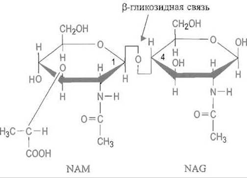
Fig. 10.6. The repeating disaccharide unit in the polysaccharide chain of murein.
Polypeptide bridges connecting the polysaccharide chains are attached to the carboxyl group of the lactyl moiety of N-acetylmuramic acid (NAM). Lysozyme attacks the ß-glycosidic bond between the NAM and N-acetylglucosamine (NAG) residues such that the oxygen atom of this bond remains with N-acetylglucosamine.
Current knowledge regarding the MECHANISM OF ACTION of lysozyme is based primarily on X-ray crystallography data of the enzyme and its complexes with substrate analogues—Oligosaccharides composed of N-acetylglucosamine.
The formation of the complex between the extended polysaccharide and the enzyme results from multiple non-covalent interactions between the enzyme and six sequentially arranged monosaccharide units A–B–C–D–E–F, which fit into the substrate-binding site (Fig. 10.7).
Evidently, substrate unit C, located near the cleavable glycosidic bond, binds most strongly. The enzyme forms four Hydrogen Bonds with it: two via the substrate's acetamide group, which forms a sort of ß-Structure with the carbonyl group of residue 107 and the NH group of residue 59, and two more via the NH groups of the indole rings of Tryptophan residues Trp-62 and Trp-63. Units A and B, which precede unit C, each form one Hydrogen bond with the enzyme.
Van der Waals interactions also play a definite role in the Formation of the enzyme-substrate complex, particularly between ring B and the Trp-62 residue. However, lysozyme mutants in which this residue is replaced by Tyrosine, phenylalanine, or Histidine exhibit even slightly higher activity. Notably, the conformation of the first three polysaccharide chain units—A, B, and C—requires local conformational adjustments in the adjacent Regions of the enzyme. For instance, the indole ring of Trp-62 shifts by 0.7 Å, making room for the tight packing of the substrate.

Fig. 10.7. Substrate binding in the Active Site of lysozyme.
The system of hydrogen bonds (indicated by arrows) is shown, which ensures the binding of a portion of the substrate—rings A, B, and C. These bonds are supplemented by van der Waals contacts, specifically with the indole ring of tryptophan.
There is reason to believe that the formation of the enzyme-substrate complex begins with the binding of monosaccharide units A, B, and C, followed by the accommodation of three additional units—D, E, and F—into the cleft on the enzyme's surface. Direct observation of the binding of this substrate portion is not feasible because the hexasaccharide is rapidly cleaved by lysozyme. However, docking a model of the hexasaccharide substrate into the 3D model of the enzyme demonstrated that units E and F not only easily fit into place but also form a series of non-covalent interactions within the binding site.
The situation is more complex with residue D, specifically because its ß-glycosidic bond with residue E is attacked by the enzyme within the hexasaccharide. This unit cannot be accommodated in the enzyme's binding site in the standard "chair" conformation typical of Monosaccharides—it does not fit into the substrate cleft due to steric clashes caused by overly close contacts between the C-6 and O-6 atoms of this unit and the lysozyme molecule. These Steric hindrances, however, are completely relieved if unit D alters its conformation and transitions into a strained "half-chair" conformation (Fig. 10.8). Thus, substrate binding must promote distortion of the conformation near the C-1 carbon atom of unit D, which directly participates in the reaction. Characteristically, substrate analogues containing a terminal gluconolactone, which also adopts a half-chair conformation, bind very tightly to the enzyme and act as its inhibitors.

Fig. 10.8. The "half-chair" conformation characteristic of six-membered rings (including monosaccharides) with a double bond in the ring.
It is hypothesized that the subsequent action of lysozyme (Fig. 10.9) is driven by the bringing together of the glycosidic bond connecting units D and E and the carboxyl group of the Glu-35 residue, which is part of the enzyme's active site. This carboxyl group resides in a hydrophobic microenvironment; therefore, its dissociation would lead to the energetically unfavorable formation of a negatively charged carboxylate anion in a non-polar medium where its Hydration is hindered. Consequently, the carboxyl group of Glu-35 is a weaker acid compared to other carboxyl groups in lysozyme (its $ ext{pK}_ ext{a}$ is close to 6), and in the free enzyme, it typically retains its proton.
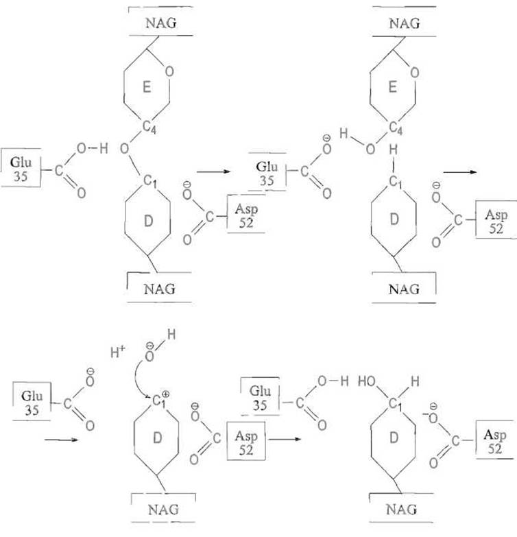
Fig. 10.9. Mechanism of action of lysozyme.
The proton from the carboxyl group of Glu-35 is transferred to the adjacent oxygen atom of the glycosidic bond, forming an oxonium ion. Next, the electron pair that formerly maintained the bond between the C-1 atom of NAG and this oxygen atom shifts to the latter, resulting in the Cleavage of the glycosidic bond. As a result, the C-1 atom becomes positively charged, acquiring The properties of a planar carbocation, which is favored by the half-chair conformation. The charge is additionally stabilized by interaction with the negatively charged carboxylate group of the Asp-52 residue. In the final phase of the reaction, a Water molecule takes THE PLACE OF the departed glycosidic oxygen atom, becomes polarized through interaction with the anionic form of Glu-35, and the resulting hydroxyl ion attacks the positively charged C-1 atom (for further details, see text).
Upon approaching the substrate's glycosidic bond within the enzyme-substrate complex, this group acts as a proton donor, adding to the glycosidic oxygen atom to form an unstable, positively charged oxonium ion:
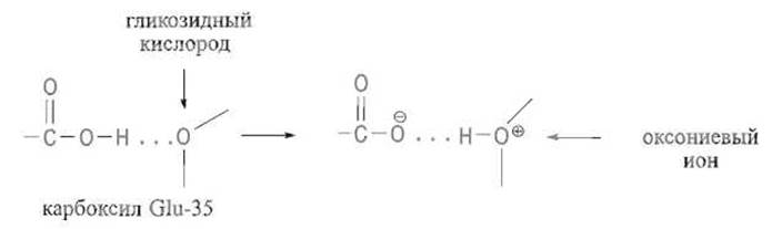
Subsequently, the oxonium ion is stabilized by the Displacement of the electron pair—which previously maintained its bond with the C-1 atom of unit D—toward the oxygen atom. This causes the bond between these atoms, and consequently between units D and E, to break, while the positive charge is transferred to the C-1 atom, forming a carbonium ion. The disaccharide E–F or an analogous fragment of a longer substrate then leaves the binding site, as it is held by only a few non-covalent interactions once the connection to the A–B–C–D fragment is lost.
The Glu-35 residue is critically important for the catalytic mechanism—its replacement with glutamine via Site-Directed Mutagenesis abolishes the enzyme's activity.
The positive charge at C-1 is stabilized by two factors:
1. An electrostatic interaction is established between it and the negatively charged carboxylate anion of Asp-52, which is highly effective in the absence of water (water molecules are expelled from the enzyme's surface upon substrate binding). It is noteworthy that the acidic properties of this residue's carboxyl group, unlike the carboxyl of Glu-35, are enhanced by its hydrophilic environment. This is because the anionic form of the Asp-52 carboxyl is stabilized by incorporation into a network of hydrogen bonds with neighboring residues.
It is worth noting The change in the properties of functional groups—so typical of Proteins in general—caused by differences in their microenvironment. The Role of this group cannot be overstated, yet it is apparently not strictly essential: replacing Asp-52 with asparagine drops the specific activity of the enzyme to roughly 5% of its initial value, without causing its complete inactivation.
2. The positive charge on the C-1 atom is partially offset by the shift of an electron pair from the adjacent ring oxygen atom. Consequently, the O—C+ bond acquires a partial double-bond character, leading to a planar conformation of this entire region of ring D and the transition of the C-1 atom from a standard tetrahedral to a trigonal structure:
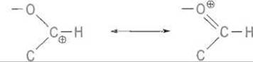
Yet precisely this conformation, which is required to stabilize the carbocation, corresponds to the half-chair structure and is achieved within the enzyme-substrate complex due to the aforementioned distortion of ring D stereochemistry upon oligosaccharide binding. Apparently, the energy needed for this process is "borrowed" from the binding energy of the extended substrate.
Thus, electrostatic and stereochemical effects within the lysozyme catalytic center work in concert, stabilizing the planar carbocation structure characteristic of the Transition State in the enzyme-substrate complex.
Next, the carbocation undergoes an attack by a water molecule, which is bound by the Glu-35 residue anion, occupying roughly the same position previously held by the glycosidic bond oxygen prior to its cleavage.
The Glu-35 anion polarizes the water molecule by abstracting a proton, which aligns with the properties of this residue's carboxyl group as a weak acid. The resulting strong nucleophile—the OH- ion—attacks the positively charged C-1 carbon atom while simultaneously neutralizing the charges. Because the water molecule approaches the planar carbocation from the same side where the glycosidic oxygen was located, the Stereochemistry of the C-1 atom is fully restored, and the hydroxyl group at this atom assumes the ß-position, much like the original glycosidic oxygen:
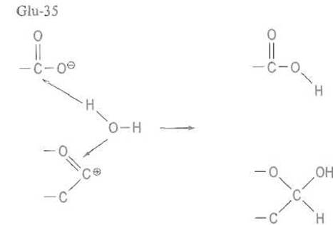
This brings the enzymatic reaction cycle to a close, resulting in the Hydrolysis of the ß-glycosidic bond in murein and leaving lysozyme ready for subsequent cycles of its catalytic mechanism.
Characteristically, T4 phage lysozyme acts on Introduction/37.html">Bacterial Cell wall Polysaccharides in a similar manner, likewise driving the monosaccharide unit adjacent to the attacked bond into a half-chair conformation; however, different functional groups take part in this enzyme's catalytic mechanism. Substrate conformation distortion as a vital element of the catalytic mechanism is also postulated for several Other Enzymes targeting glycosidic bonds, phosphorylase being a notable example.
In Conclusion, let us highlight the core Features of the lysozyme catalytic mechanism:
- the establishment of a network of non-covalent interactions that anchor the extended substrate;
- steric constraints on the binding of the specific substrate unit whose glycosidic bond is cleaved, forcing it into an unusual conformation favorable for stabilizing the carbocation in the transition state;
- the coordinated action of two carboxyl groups, one functioning as a proton donor and the other, in its anionic form, assisting in the stabilization of the carbocation in the transition complex.
Last update: 13/08/2026
Editorial and Educational Adaptation: This material has been compiled based on the primary/original source text. The project team performed an editorial review, corrected technical inaccuracies, structured sections, and adapted the content for an educational format.
What was processed:
- elimination of formatting defects (OCR errors, structural breaks, corrupted characters);
- editorial organization of content;
- standardization of terminology in accordance with academic sources;
- verification of factual statements against the original source text.
All mentions of the author, publication year, and origin of the primary text have been preserved in accordance with the source.