BIOCHEMISTRY - L. Stryer - 1984
VOLUME 1
PART I. CONFORMATION AND DYNAMICS
CHAPTER 7. MECHANISM OF ENZYME ACTION: LYSOZYME AND CARBOXYPEPTIDASE
7.8. Carboxypeptidase A: A Zinc-Containing Proteolytic Enzyme
Let us now turn to carboxypeptidase A, a digestive enzyme that hydrolyzes the C-terminal peptide bond in Polypeptides. Peptides in which the C-terminal residue has an aromatic or large aliphatic side chain are particularly easily hydrolyzed. This enzyme is interesting because its catalytic mechanism is fundamentally different from that of Lysozyme. Before discussing the MECHANISM OF ACTION of carboxypeptidase A in detail, let us note two main aspects.
1. Induced fit. Substrate binding is accompanied by significant Changes in the Enzyme Structure.
2. Electron shift. The Active Site of the enzyme contains a zinc atom and other groups that induce a redistribution of electrons in the substrate, thereby facilitating the Hydrolysis process.
The three-dimensional structure of carboxypeptidase A (Fig. 7.23) at 2 Å resolution was solved in 1967 by William Lipscomb. The enzyme contains a single polypeptide chain of 307 Amino Acids and has a compact shape that can be approximately described as an ellipsoid with dimensions of 50 x 42 x 38 Å. 38 Å. The enzyme contains regions of α-helices (38%) and β-pleated sheets (17%). A zinc ion is tightly bound to the protein, and its presence is essential for enzymatic activity. The zinc ion is located in a cleft near The surface of the molecule, forming tetrahedral coordination bonds with the side chains of two histidines, the side chain of a glutamate, and a Water molecule (Fig. 7.24). Adjacent to the zinc ion, There is a large pocket in the enzyme that accommodates the side chain of the terminal residue of the peptide substrate.
Class="center">Fig. 7.23. Three-dimensional structure of carboxypeptidase A. Only α-carbon atoms and the zinc ion (shaded circle in the center) are shown

Fig. 7.24. The zinc ion located in the active site of carboxypeptidase A forms coordination bonds with the side chains of two histidines and a glutamate. The water molecule occupying the fourth coordination position is not shown here

7.9. Substrate Binding Induces Large Structural Changes in the Active Site of Carboxypeptidase A
Our understanding of how substrates bind to carboxypeptidase A is based on data obtained from structural studies of the complex of this enzyme with glycyltyrosine. Glycyltyrosine is a slowly hydrolyzed substrate. Its binding process (Figs. 7.25 and 7.26) can be represented as five consecutive steps.
Fig. 7.25. Schematic representation of glycyltyrosine binding in the active site of carboxypeptidase A. The postulated catalytically active complex is shown

Fig. 7.26. Spatial arrangement of glycyltyrosine in the active site of carboxypeptidase A. Glycyltyrosine (substrate) is shown in red

1. The negatively charged terminal carboxyl group of glycyltyrosine participates in an electrostatic interaction with the positively charged side chain of Arginine-145.
2. The substrate binds via the side chain of its Tyrosine residue in the nonpolar pocket of the enzyme.
3. The hydrogen of the NH group of the peptide
bond to be cleaved is hydrogen-bonded to the OH group of the aromatic side chain of tyrosine-248.
4. The carbonyl oxygen of the same peptide bond forms a coordination bond with the zinc ion.
5. The terminal amino group of the substrate forms a Hydrogen bond, via an intervening water molecule, with the side chain of glutamate-270. This interaction probably does not occur in the reactive ES complex, and it may be the reason for the extremely slow hydrolysis of glycyltyrosine.
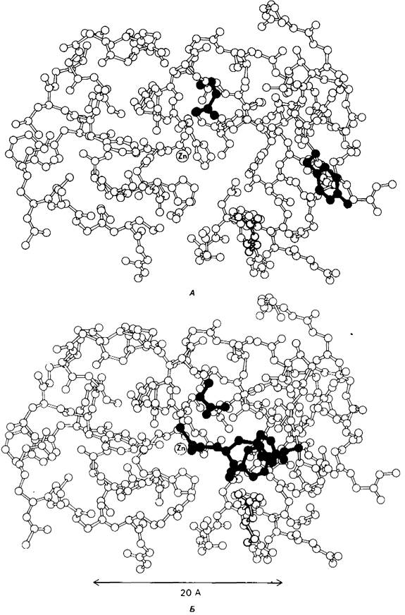
Binding of glycyltyrosine is accompanied by a structural rearrangement of the active site (Fig. 7.27). In essence, only upon binding the substrate do the catalytic groups of the enzyme assume the correct orientation—a concept first postulated by Koshland in his induced-fit model. The guanidinium group of arginine-145, as well as the carboxyl group of glutamate-270, shift by 2 Å. The binding of the carbonyl group of the substrate to the zinc ion displaces a water molecule from its coordination with zinc. At least four more water molecules are displaced from the nonpolar pocket of the enzyme upon binding of the tyrosine side chain of the substrate molecule. The largest conformational change is the movement of the phenolic hydroxyl of tyrosine-248 by 12 Å, which is about 1/4 of the molecular diameter. This movement occurs primarily through free rotation about the single —C—C— bond, bringing the hydroxyl group of tyrosine-248, which was previously On the surface of the molecule, close to the peptide bond of the substrate. As a result, the active site cavity closes, thereby completing its transformation from a water-filled region into a hydrophobic one. All these structural changes are apparently initiated by the binding of arginine-145 to the terminal carboxyl group of the substrate.
Fig. 7.27. The structure of carboxypeptidase A changes upon substrate binding; A - enzyme without substrate (Arg 145 is shown in yellow, Glu 270 in green, Tyr 248 in blue); B - enzyme-substrate complex (glycyltyrosine, the substrate, is shown in red). Only a portion of the enzyme molecule is shown in the figure
7.10. The Rate of Catalysis by Carboxypeptidase A Is Enhanced by Electron Displacement
Based on X-ray crystallographic data, Lipscomb proposed a mechanism for the catalytic action of carboxypeptidase A. The postulated STRUCTURE OF THE reactive ES complex is shown in Fig. 7.28. According to the proposed mechanism, the OH group of tyrosine-248 donates a proton to the NH group of the peptide bond being cleaved. The carbonyl carbon atom of this peptide bond is attacked by the carboxyl group of glutamate-270, which acts in this case as a nucleophilic group. The resulting anhydride of glutamate-270 and the acid component of the substrate is subsequently hydrolyzed.
Fig. 7.28. Postulated mechanism of the catalytic action of carboxypeptidase A: Glu-270 directly attacks the carbonyl carbon atom of the peptide bond being hydrolyzed, while Tyr-248 donates a proton to the NH group of this peptide. The resulting anhydride is subsequently hydrolyzed

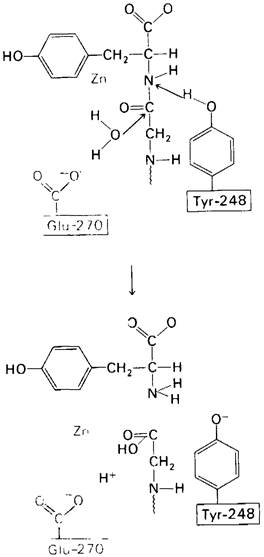
Another mechanism of catalysis is also possible, which is consistent with the X-ray crystallographic data; it is shown in Fig. 7.29. In this scheme, glutamate-270 activates a water molecule. The resulting OH- directly attacks the carbonyl carbon atom of the peptide bond being cleaved. Simultaneously, tyrosine-248 donates a proton to its NH group, and as a result, the peptide bond is hydrolyzed. This catalytic mechanism differs from the one shown in Fig. 7.28 in that it involves direct hydrolysis of the substrate's peptide bond by water, rather than through an intermediate anhydride. Recent chemical and spectroscopic studies indicate that the hydrolysis of peptide substrates proceeds via a direct mechanism, whereas the hydrolysis of esters proceeds through The intermediate formation of an anhydride with glutamate-270.
Fig. 7.29. A second possible mechanism for the catalytic action of carboxypeptidase A.
Tyr 248 performs the same function as in Fig. 7.28. Otherwise, the process proceeds differently: Glu 270 activates a water molecule, which attacks the carbonyl carbon atom of the peptide bond being cleaved. Hydrolysis occurs directly, without the intermediate formation of an anhydride
What is The Role of zinc in these catalytic schemes? The carbonyl group of the peptide bond being cleaved is oriented toward the zinc ion in such a way that the C=O bond becomes more polarized than usual; this makes the carbonyl carbon atom more susceptible to nucleophilic attack. The nonpolar environment of the zinc ion increases its effective charge and thus its ability to induce a dipole. The strong polarization of the carbonyl group is also facilitated by the proximity of the negative charge of glutamate-270. Consequently, carboxypeptidase A induces an electron displacement in the substrate that enhances The rate of catalysis.
We can now appreciate The Significance of the substrate-induced structural changes in the active site of carboxypeptidase A. As a result, the substrate bound to the enzyme becomes completely surrounded by catalytic groups. This enables catalysis for the reasons discussed above. It is quite obvious that only The flexibility of the enzyme structure allows the substrate to enter the sphere of action of the catalytic group system (and the reaction products to exit this system). Overall, a flexible enzyme structure has an advantage over a rigid one in that it possesses a much wider Selection of possible Conformations suitable for catalysis and preserved during selection. Furthermore, induced fit contributes to enhancing the enzyme's Specificity. Indeed, in the case of carboxypeptidase A, the substrate must have a terminal carboxylate ion; the enzyme "checks" for its presence in this way: if the terminal carboxylate ion is present, it forms a salt bridge with arginine-145, which triggers the movement of tyrosine-248 into the catalytically active position; if there is no terminal carboxylate ion, tyrosine-248 remains in place and the enzyme shows no activity. In other words, induced fit can function as a dynamic recognition process.
Last update: 06/08/2026
Editorial and Educational Adaptation: This material has been compiled based on the primary/original source text. The project team performed an editorial review, corrected technical inaccuracies, structured sections, and adapted the content for an educational format.
What was processed:
- elimination of formatting defects (OCR errors, structural breaks, corrupted characters);
- editorial organization of content;
- standardization of terminology in accordance with academic sources;
- verification of factual statements against the original source text.
All mentions of the author, publication year, and origin of the primary text have been preserved in accordance with the source.