BIOCHEMISTRY - L. Stryer - 1984
VOLUME 1
PART I. CONFORMATION AND DYNAMICS
CHAPTER 7. MECHANISM OF ENZYME ACTION: LYSOZYME AND CARBOXYPEPTIDASE
7.6. Carbonium ion intermediate formation is a critical step in catalysis
Based on these structural data, Phillips and coworkers worked out in detail a plausible mechanism for the catalytic action of Lysozyme. They identified the following key steps in the catalytic cycle.
1. The —COOH group of residue 35 transfers an H+ to the bond between C-1 of ring D and the glycosidic oxygen atom; As a result, this bond is cleaved (Fig. 7.14).
Class="center">Fig. 7.14. The first step in the postulated mechanism of lysozyme action: transfer of H+ from Glu 35 to the oxygen atom of the glycosidic bond. This leads to the Cleavage of the glycosidic bond and The formation of a carbonium ion

2. This generates a positive charge on C-1 of ring D. The resulting short-lived intermediate is called a carbonium ion because it contains a positively charged carbon atom.
3. The NAG dimer, consisting of residues E and F, diffuses away from the enzyme.
4. The intermediate carbonium ion reacts with an OH- group from the solvent (Fig. 7.15). The tetra-NAG, consisting of residues A, B, C, and D, diffuses away from the enzyme.
Fig. 7.15. The Hydrolysis reaction is completed by The addition of OH to the intermediate carbonium ion and H+ to the side chain of Glu 35
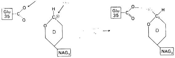
5. Glutamate-35 is reprotonated, and the enzyme is ready to enter a new catalytic cycle.
The essential features of this catalytic pathway are as follows.
1. General acid catalysis. The proton donor is glutamate-35, which is in its un-ionized form and is located at an optimal distance of 3 Å from the glycosidic oxygen atom.
2. Carbonium ion intermediate formation. Two distinct stabilizing factors act on the carbonium ion, greatly facilitating the enzymatic reaction. These are:
a. An electrostatic factor, namely the presence of a negatively charged group close (within 3 Å) to the intermediate carbonium ion. Aspartate-52, in the form of a negatively charged carboxylate ion, electrostatically interacts with the positive charge on C-1 of ring D.
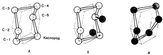
б. A geometric factor, specifically the distortion of ring D (Fig. 7.16). Hexa-NAG fits best into the cleft of the Active Site of the enzyme if the geometry of ring D is distorted from a chair conformation to a half-chair conformation. This distortion accelerates catalysis because the half-chair conformation greatly facilitates the Formation of the carbonium ion. The coplanarity of carbon atoms 1, 2, and 5 and the ring oxygen atom in the half-chair conformation allows the positive charge to be shared between C-1 and the ring oxygen. Thus, by binding the substrate, the enzyme forces it into the transition-state conformation, namely that of a carbonium ion.
Fig. 7.16. Conformational change of ring D of the lysozyme substrate to a half-chair conformation. A—sugar residue in the normal chair conformation; B—upon binding to lysozyme, the ring oxygen and C-5 of sugar residue D shift so that C-1, C-2, C-5, and O lie in the same plane, as shown in C
7.7. Experimental Evidence for the Proposed Mechanism of Enzymatic Catalysis
The hypothesis concerning substrate binding and the catalytic mechanism, based on X-ray crystallographic data, has been tested by numerous chemical experiments. All the results obtained support the proposed hypothesis. Here is some of The most significant experimental evidence.
1. Cleavage pattern of the substrate. In agreement with the assumption that cleavage of the hexamer occurs between the 4th and 5th residues, hexa-NAG is indeed cleaved into tetra-NAG and di-NAG (Fig. 7.17).
Fig. 7.17. Hexa-NAG is hydrolyzed to form tetra-NAG and di-NAG

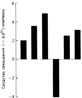
2. Binding affinity. By measuring the binding equilibrium of each of the six sugars with the enzyme, their individual contributions to the total Free energy of binding for the hexamer were determined (Fig. 7.18). A striking phenomenon was revealed: THE CONTRIBUTION OF sugar residue D was negative. Binding of residue D required about 4 kcal/mol. This result Supports the hypothesis that residue D is distorted upon binding to the enzyme: the transition from a chair conformation to a half-chair conformation requires energy. It is also interesting that residue C makes the largest positive contribution to the binding affinity. Indeed, X-ray crystallographic data show that residue C forms A large number of Hydrogen Bonds and Structure/103.html">Van der Waals interactions.
Fig. 7.18. Contribution of each of the six sugar residues of hexa-NAG to the Standard Free Energy of binding of this substrate. Binding of residue D requires energy. Energy is expended to alter the conformation of the residue so that it fits into the active site of the enzyme
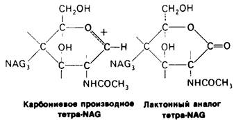
3. Transition-state analogs. METABOLISM/2.html">THE CONCEPT OF distortion of sugar residue D into a half-chair conformation is an important aspect of the postulated mechanism of catalysis, since the half-chair conformation is characteristic of the Transition State. As noted above, this concept is supported by data on the energy cost of binding residue D—energy that is expended on distortion. Another confirmation came from studies of a transition-state analog of the substrate, i.e., a compound that, both before and after binding to the enzyme, has the same geometry as the substrate in the transition state. The D ring of the lactone analog of tetra-NAG (Fig. 7.19) has a half-chair conformation in the tetrasaccharide crystal. Upon binding to lysozyme, the C-1, C-2, C-4, C-6 atoms and the ring oxygen atom of the D ring of this analog lie in a single plane. This 'sofa' conformation is similar to the half-chair conformation postulated for the transition state; this means that the lactone analog, unlike tetra-NAG, undergoes very little distortion upon binding to lysozyme. It turned out that the lactone analog binds to lysozyme (at binding sites A through D) 3,600 times more tightly than tetra-NAG. This suggests that the distortion of the D ring of the normal substrate can increase The rate of cleavage by approximately 3,600-fold.
Fig. 7.19. The D ring of the lactone analog of tetra-NAG has a conformation resembling a half-chair, and in this respect is similar to the transition state intermediate in the reaction catalyzed by lysozyme
The Role of this factor in catalysis was clearly foreseen by Pauling, as evidenced by his lecture delivered in 1948:
“I think that Enzymes are molecules that are complementary in structure to the activated complexes of the reactions that they catalyze, that is, to the molecular structure that is intermediate between the reactants and products of reaction in the catalytic process. The attraction of the enzyme molecule to the activated complex leads to a decrease in the energy of the latter, and hence to a decrease in the activation energy of the reaction and an increase in the rate of reaction.”
4. pH dependence of the catalytic reaction rate. The rate of Chitin hydrolysis reaches its highest values at pH 5 (Fig. 7.20). On either side of this optimum, enzymatic activity drops sharply. The decrease in activity upon shifting the pH to the alkaline side is due to The ionization of glutamate-35, whereas shifting the pH to the acidic side is due to the protonation of aspartate-52. Lysozyme exhibits enzymatic activity only when glutamate-35 is in its un-ionized form and aspartate-52 is in its ionized form.
Fig. 7.20. Rate of chitin (poly-NAG) hydrolysis by lysozyme as a function of pH

5. Selective chemical modification. Lysozyme retains its enzymatic activity if all of its carboxyl groups, except for those of glutamate-35 and aspartate-52, are esterified. Residues 35 and 52 remain unmodified if Esterification is carried out in the presence of the substrate. Upon removal of the substrate, aspartate-52 is esterified (while glutamate-35 remains unchanged). Modification of aspartate-52 leads to complete inactivation of the enzyme. This supports the assumption that a precise orientation of the aspartate-52 carboxylate ion is required to stabilize the carbonium ion intermediate.
6. Transglycosylation. When tetra-NAG is added to lysozyme, slow formation of hexa-NAG and di-NAG occurs (Fig. 7.21). The existence of this reaction, called transglycosylation, confirms one of the key elements of the proposed enzymatic reaction mechanism, namely, the formation of a glycosyl-enzyme intermediate.
Fig. 7.21. The existence of a glycosyl-enzyme intermediate is supported by the ability of lysozyme to catalyze, albeit slowly, a transglycosylation reaction, where NAG4 (shown in red) combines with the glycosyl-enzyme intermediate (shown in blue in Fig. B) to yield NAG6

In normal hydrolytic reactions, this intermediate reacts with OH-. In transglycosylation, a second carbohydrate molecule, ROH, is used in the reaction. The transglycosylation reaction is specific because the acceptor binds to sites E and F in the active site cleft. Furthermore, the resulting glycosidic bond has a β-configuration, just as in the substrate. Thus, these data support the proposed STRUCTURE OF THE catalytic intermediate.
Last update: 06/08/2026
Editorial and Educational Adaptation: This material has been compiled based on the primary/original source text. The project team performed an editorial review, corrected technical inaccuracies, structured sections, and adapted the content for an educational format.
What was processed:
- elimination of formatting defects (OCR errors, structural breaks, corrupted characters);
- editorial organization of content;
- standardization of terminology in accordance with academic sources;
- verification of factual statements against the original source text.
All mentions of the author, publication year, and origin of the primary text have been preserved in accordance with the source.