BIOCHEMISTRY - L. Stryer - 1984
VOLUME 1
PART I. CONFORMATION AND DYNAMICS
CHAPTER 7. MECHANISM OF ENZYME ACTION: LYSOZYME AND CARBOXYPEPTIDASE
7.4. Binding Mode of a Competitive Inhibitor
X-ray crystallographic studies of the Lysozyme-tri-NAG complex established the Location OF THE Active Site and revealed the interactions responsible for specific substrate binding; this provided the basis for a hypothesis predicting the detailed mechanism of lysozyme action. Tri-NAG was found to bind in a cleft On the surface of lysozyme, filling about half of it. Binding is mediated by Hydrogen Bonds and Structure/103.html">Van der Waals interactions. Electrostatic Interactions are absent because tri-NAG contains no ionic groups.
Hydrogen bonds between tri-NAG and lysozyme are shown in Fig. 7.9. The carboxyl group of aspartate-101 forms hydrogen bonds with residues A and B. The most specific and strong Hydrogen bonds are formed between the enzyme and residue C of the inhibitor, where four hydrogen bonds are established. The NH group of the indole ring of Tryptophan-62 is hydrogen-bonded to the O-6 oxygen. The adjacent amino acid residue, tryptophan-63, is similarly bonded to the O-3 oxygen. Upon tri-NAG binding, the ring of tryptophan-62 shifts by 0.75 Å. Strong hydrogen bonds are formed between the CO and NH groups of the acetamido side chain of sugar residue C and the NH and CO groups of the protein backbone belonging to residues 59 and 107, respectively.
Class="center">Fig. 7.9. Hydrogen bonds between tri-NAG and lysozyme. Chemical groups of the substrate involved in hydrogen bonding are shown in blue, and the corresponding groups of the enzyme in red

A large number of van der Waals contacts are formed between tri-NAG and the enzyme. Sugar residue B makes few polar contacts with the enzyme but is closely associated with the indole ring of tryptophan-62. Residue A makes relatively weak contacts with the enzyme.
7.5. From Enzyme Structure to the Mechanism of Enzymatic Action
1. How does substrate binding occur? As previously mentioned, X-ray crystallography cannot directly determine how an active substrate binds to the enzyme. However, data obtained from the X-ray Analysis of the enzyme-competitive inhibitor complex can play a key role in solving this problem. Tri-NAG fills only half of the cleft in the lysozyme molecule. This is a highly promising starting point. It was assumed that the observed binding of tri-NAG as an inhibitor involves the same interactions that occur during substrate binding. It is highly likely that forming a reactive ES complex requires additional sugar residues to fill the other half of the cleft. Indeed, after tri-NAG binds, there is room in the cleft for three more sugar residues. This was encouraging, as the hexamer of N-acetylglucosamine (hexa-NAG) was known to be rapidly hydrolyzed by the enzyme.
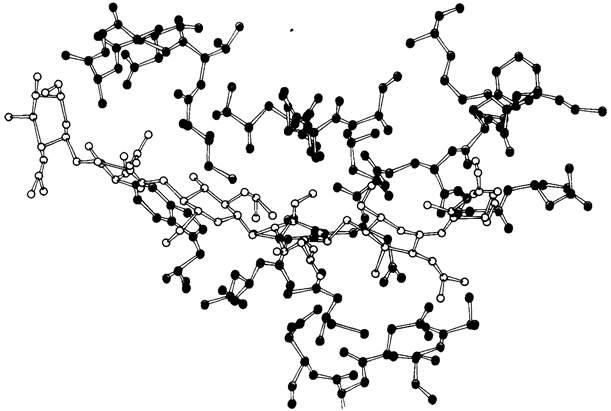
Careful Model Building showed that three additional sugar residues, designated D, E, and F, could fit into the cleft of the enzyme (Fig. 7.10). Residues E and F fit perfectly, forming several strong hydrogen bonds and van der Waals contacts. However, residue D could only fit into the cleft if it was somewhat distorted. In its normal chair conformation, its C-6 and O-6 atoms came too close to certain groups on the enzyme.
Fig. 7.10. Binding mode of hexa-NAG (shown in yellow) to lysozyme. The positions of sugar residues A, B, and C (left) correspond to the location of tri-NAG in the complex with lysozyme; the positions of residues D, E, and F (right) were predicted by model building. Shown in green are the two amino acid residues directly involved in catalysis
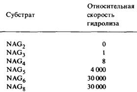
2. Which bond is cleaved by the enzyme? The rate of Hydrolysis of N-acetylglucosamine oligomers increases dramatically as the number of sugar residues increases from 4 to 5, i.e., from NAG4 to NAG5 (Table 7.1). Extending the substrate by one more residue (NAG6) yields an additional increase in Cleavage rate; however, increasing the number of sugar residues in the substrate up to 8 has no further effect. These data are consistent with X-ray crystallographic results showing that six sugar residues are sufficient to fill the cleft containing the active site.
Table 7.1. Efficiency of N-acetylglucosamine oligomers as substrates
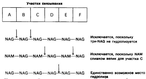
Which bond in hexa-NAG is cleaved by the enzyme? Since tri-NAG is not cleaved, it can be assumed that the A—B bond (i.e., the glycosidic linkage between residues A and B) is not the one targeted by the enzyme. Similarly, the enzyme cannot cleave the B—C bond. A second and decisive piece of evidence that the B—C bond is not cleaved is that NAM cannot fit into site C. While NAG fits perfectly into site C, NAM cannot fit there due to its lactyl side chain. However, in the Introduction/37.html">Bacterial Cell wall polysaccharide, lysozyme cleaves the NAM—NAG linkage. Consequently, the C—D bond also cannot be cleaved, assuming that the bacterial cell wall polysaccharide binds to lysozyme in the same manner as hexa-NAG. The inability of NAM to occupy site C rules out another potential cleavage site, namely the E—F bond. Recall that The Cell wall polysaccharide is an alternating polymer of NAM and NAG; therefore, if NAM cannot occupy site C, it cannot occupy site E either.
From these considerations, it follows that during Enzymatic cleavage of the hexameric substrate, the A—B, B—C, C—D, and E—F bonds cannot be broken. Consequently, the only possible cleavage site of the substrate is the D—E bond (Fig. 7.11).
Fig. 7.11. Line of reasoning demonstrating that the site of enzyme action is the glycosidic bond between sugar residues D and E
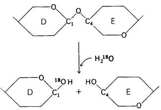
3. Which group on the enzyme directly participates in catalysis? The Conclusion that substrate hydrolysis occurs at the D—E bond allowed researchers to identify the specific groups on the enzyme that directly mediate the hydrolytic reaction. To do this, however, the cleavage site of the substrate had to be localized even more precisely—specifically, by determining on which side of the glycosidic oxygen atom the bond is broken. The answer was obtained from Enzymatic hydrolysis experiments in a medium containing Water labeled with the stable heavy oxygen isotope 18O (Fig. 7.12). In the sugars isolated after hydrolysis, 18O was found attached to C-1 of residue D, whereas the hydroxyl group at C-4 of residue E contained the normal oxygen isotope. It follows that during hydrolysis, the bond is cleaved between C-1 of residue D and the glycosidic oxygen atom adjacent to residue E. This work serves as an excellent example of using isotopes to study the Mechanism of Enzymatic catalysis. Without isotopes, it would have been extremely difficult, if not impossible, to establish the exact site of enzyme action in this case.
Fig. 7.12. Hydrolysis in 18O-labeled water showed that lysozyme cleaves the bond between C-1 and O, but not the bond between C-4 and O (only the backbone of residues D and E is shown)
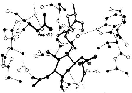
Attention then turned to identifying potential catalytic groups that must be located near the glycosidic bond being cleaved. As mentioned in the previous chapter, catalytic groups refer to those enzyme groups directly involved in the formation or cleavage of covalent bonds. The most suitable candidates for this role are groups capable of forming hydrogen bonds as hydrogen Donors or acceptors. The loss or gain of a proton is a critical step in most enzymatic reactions. In lysozyme, the only residues capable of acting catalytically and located near the glycosidic bond being cleaved are aspartate-52 and glutamate-55. The aspartic acid residue lies on one side of the glycosidic bond, while the glutamic acid residue lies on the other. The environments of these two acidic side chains are completely different. Aspartate-52 is located in a polar environment, where it serves as a hydrogen acceptor in a complex network of hydrogen bonds. Glutamate-35, in contrast, is located in a nonpolar region. It follows that at pH 5, the optimal pH for Chitin hydrolysis by lysozyme, aspartic acid at position 52 must be in the ionized COO- form, whereas glutamic acid at position 35 must be in the un-ionized COOH form. The distance between the glycosidic bond and the nearest oxygen atom of both acidic groups is approximately 3 Å (Fig. 7.13).
Fig. 7.13. Structure of a portion of the lysozyme active site. The D and E rings of the hexa-NAG substrate are shown in yellow. The side chains of aspartate-52 (red) and glutamate-35 (green) are located near the substrate.
Last update: 06/08/2026
Editorial and Educational Adaptation: This material has been compiled based on the primary/original source text. The project team performed an editorial review, corrected technical inaccuracies, structured sections, and adapted the content for an educational format.
What was processed:
- elimination of formatting defects (OCR errors, structural breaks, corrupted characters);
- editorial organization of content;
- standardization of terminology in accordance with academic sources;
- verification of factual statements against the original source text.
All mentions of the author, publication year, and origin of the primary text have been preserved in accordance with the source.