BIOCHEMISTRY - L. Stryer - 1984
VOLUME 1
PART I. CONFORMATION AND DYNAMICS
CHAPTER 7. ENZYME MECHANISM: LYSOZYME AND CARBOXYPEPTIDASE
7.2. Three-Dimensional Structure of Lysozyme
Lysozyme is a relatively small enzyme. Isolated in large quantities from chicken egg white, it consists of a single polypeptide chain of 129 amino acid residues with a molecular mass of 14.6 kDa. The lysozyme molecule contains four cross-linking disulfide bridges that confer stability upon the enzyme. The Amino Acid Sequence of lysozyme is shown in Fig. 7.5.
Class="center">Fig. 7.5. Amino acid sequence of lysozyme from chicken egg white. Residues comprising the Active Site are shown in red

In 1965, David Phillips and co-workers determined the three-dimensional Structure of lysozyme. This was the first high-resolution map obtained for a protein with enzymatic activity. The lysozyme molecule was found to be compact, with an approximately ellipsoidal shape measuring 45 x 30 x 30 Å. As shown in Fig. 7.7, the folding of the polypeptide chain is highly complex. It contains considerably fewer α-helices than Myoglobin and Hemoglobin. In certain regions, the polypeptide chain adopts an extended conformation. In one of these regions, the chain makes a turn and runs back parallel to itself; the two parallel strands are held together by Hydrogen Bonds formed between peptide units. Such "hairpin" loops resemble the aforementioned antiparallel β-pleated sheets—regularly repeating Secondary structure motifs found in Silk Fibroin. The interior of the lysozyme molecule, much like those of myoglobin and hemoglobin, is almost entirely nonpolar. Clearly, as with most other Proteins, hydrophobic interactions play a crucial role in stabilizing the Tertiary Structure of lysozyme.
7.3. Locating the Active Site of Lysozyme
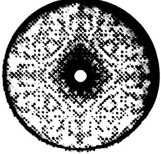
Elucidating the detailed three-dimensional structure of lysozyme did not immediately reveal its catalytic mechanism. Furthermore, examining the electron density map made it difficult to pinpoint even the Location OF THE active site. Unlike myoglobin and hemoglobin, lysozyme lacks a prosthetic group, meaning there is no built-in marker for the active site. Consequently, the essential information required to identify the active site, determine the mode of substrate binding, and elucidate the catalytic mechanism was obtained only through The Use of inhibitors—specifically, via X-ray crystallographic analysis of lysozyme-inhibitor complexes. Once the three-dimensional structure of a protein is known, determining how small molecules bind to it via X-ray crystallography is usually straightforward. These experiments are feasible because protein crystals are highly porous. Even relatively large inhibitor molecules can diffuse through channels between protein molecules to reach specific binding sites. The changes in electron density resulting from the incorporation of an additional molecule can be calculated directly from diffraction intensities (using data previously obtained for the native protein), provided the crystal structure does not undergo significant alterations. This approach is known as the difference Fourier method.
Fig. 7.6. X-Ray Diffraction pattern of a lysozyme crystal
Ideally, one would apply the difference Fourier method to determine The structure of the enzyme-substrate (ES) complex during catalysis. However, under normal conditions, The conversion of the bound substrate into reaction products occurs much faster than the diffusion of new substrate molecules into the crystal. In some cases, this complication can be overcome by slowing down the catalytic process through deep cooling of the crystal (e.g., to —50 °C). This experimental approach is known as cryoenzymology. Another approach involves studying the complex of the enzyme with a substrate analogue that either undergoes no transformations at all or reacts extremely slowly. For lysozyme, tri-N-acetylglucosamine (tri-NAG, or NAG3) proved to be such a substrate analogue, and its structure is shown in Fig. 7.8. Oligomers of N-acetylglucosamine containing fewer than 5 residues are hydrolyzed extremely slowly or not at all. Nevertheless, they bind to the Active Site of the enzyme, which is why tri-NAG acts as a potent competitive inhibitor of lysozyme.
Fig. 7.7. Three-dimensional structure of lysozyme. Only α-carbon atoms are shown
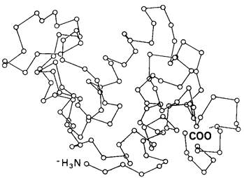
Fig. 7.8. Formula of tri-N-acetylglucosamine (tri-NAG, or NAG3), a competitive inhibitor of lysozyme

Last update: 06/08/2026
Editorial and Educational Adaptation: This material has been compiled based on the primary/original source text. The project team performed an editorial review, corrected technical inaccuracies, structured sections, and adapted the content for an educational format.
What was processed:
- elimination of formatting defects (OCR errors, structural breaks, corrupted characters);
- editorial organization of content;
- standardization of terminology in accordance with academic sources;
- verification of factual statements against the original source text.
All mentions of the author, publication year, and origin of the primary text have been preserved in accordance with the source.