Fundamentals of Biochemistry - Filippovich, Yu. B. 1999
Enzymes
Enzyme Structure
Based on their Structure, Enzymes can be single-component, simple Proteins, or two-component, complex proteins. In the latter case, the enzyme contains an additional non-protein component.
Various terms have emerged over time for the protein portion and the additional group in two-component enzymes. All of them are still found in the literature, for example:
Enzyme as a whole |
Protein part |
Additional group |
Simplex |
Pheron (carrier) |
Agon (active group) |
Holoenzyme |
Apoenzyme |
Coenzyme |
An additional group that is tightly bound and inseparable from the protein moiety is called a prosthetic group; in contrast, an additional group that easily dissociates from the apoenzyme and can exist independently is usually referred to as a coenzyme.
The Chemical Nature of the most important Coenzymes was elucidated in the 1930s thanks to the work of O. Warburg, R. Kuhn, P. Karrer, and others. It turned out that most Vitamins (E, K, Q, B1, B2, B6, B12, C, H, etc.) or compounds synthesized with the participation of vitamins (coenzyme A, NAD+, etc.) function as coenzymes in two-component enzymes. The formulas of the mentioned coenzymes are given in Chapter IV. In addition, compounds such as HS-Glutathione, a numerous group of NUCLEOTIDES and their derivatives, phosphate esters of certain Monosaccharides, and A number of other substances also act as coenzymes.
A characteristic feature of two-component enzymes is that neither the protein part nor the additional group individually possesses noticeable catalytic activity. Only their complex exhibits enzymatic properties. Furthermore, the protein dramatically enhances the catalytic activity of the additional group, which is manifested only to a very slight degree in the free state; in turn, the additional group stabilizes the protein moiety and makes it less vulnerable to Denaturing Agents. Thus, although the prosthetic group forming the catalytic center is the direct executor of the catalytic function, its action is unthinkable without the involvement of the polypeptide fragments of the enzyme's protein part. Moreover, the apoenzyme contains a region characterized by a specific structure that selectively binds the coenzyme. This is the so-called coenzyme-binding domain; its structure is very similar across various apoenzymes that bind to the same coenzyme. Examples include the spatial structures of the nucleotide-binding domains of a number of dehydrogenases (see Fig. 53, p. 119).
The situation is different for single-component enzymes, which lack an additional group capable of coming into direct contact with the compound being transformed. This function is performed by a part of the protein molecule known as the catalytic center. It is assumed that the catalytic center of a single-component enzyme is a unique combination of several amino acid residues located in a specific region of the protein molecule. This is illustrated in Fig. 34, D, which shows the Tertiary Structure of chymotrypsinogen—the precursor of a single-component enzyme: The amino acid radicals of a Serine residue and two Histidine residues, located at different points of the polypeptide chain, are brought close together here within a distance of a few tenths of a nanometer, thus pre-forming the catalytic center of the enzyme. The same figure (B and C) presents the tertiary structures of two other enzyme molecules—Lysozyme and Ribonuclease; they clearly display a binuclear, bilobed molecular architecture with a cleft (groove) at the boundary between the two lobes where the catalytic center is located.
Most frequently, the catalytic centers of single-component enzymes contain residues of ser, his, trp, arg, cys, asp, glu, and tyr. The radicals of these Amino Acids perform the same function here as the coenzyme does in a two-component enzyme.
The amino acid residues that form the catalytic center of a single-component enzyme are located at various points along a single polypeptide chain (see Fig. 34). Therefore, the catalytic center emerges at the moment the protein molecule acquires its characteristic tertiary structure. Consequently, any alteration in the tertiary STRUCTURE OF THE enzyme under METABOLISM/18.html">The Influence of various factors can lead to the deformation of the catalytic center and A change in enzymatic activity.
In addition to the catalytic center, formed by a combination of amino acid radicals or the attachment of a coenzyme, enzymes also feature two other centers: the substrate center and the allosteric center (see below, Fig. 51).
The substrate center refers to the region of the enzyme molecule responsible for binding the substance (substrate) undergoing enzymatic transformation. This region is often called the enzyme's "anchoring pad," where the substrate settles like a ship dropping anchor. In many cases, the attachment of the substrate to the enzyme occurs through interaction with the ε-amino group of a Lysine radical located in the substrate center. The carboxyl group of glutamate, as well as the sulfhydryl group of Cysteine, can also play this role. However, recent studies have shown that hydrophobic interaction forces and Hydrogen Bonds arising between the amino acid radicals of the enzyme's substrate center and the corresponding groups in the substrate molecule are of far greater importance here.
The concepts of the catalytic and substrate centers should not be absolutized. In real enzymes, the substrate center may coincide with (or overlap) the catalytic center. Furthermore, the catalytic center may finally form at the moment of substrate binding. Therefore, reference is frequently made to the active center of the enzyme, representing a combination of the First and Second centers. The active center in enzymes is located at the bottom of the cleft in binuclear structures, such as in lysozyme and ribonuclease, or at the bottom of a deep depression, as in chymotrypsinogen (see Fig. 34).
The allosteric center represents a region of the enzyme molecule upon whose binding to a specific low-molecular-weight (and occasionally high-molecular-weight) substance the tertiary structure of the protein molecule alters. As a consequence, the configuration of the active center changes, accompanied by either an increase or a decrease in the catalytic activity of the enzyme. This phenomenon forms The basis of the so-called allosteric REGULATION OF ENZYMATIC Activity.
Class="center">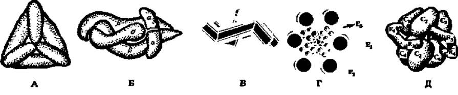
Fig. 46. Structure of certain multimeric enzymes:
A — Glutamate dehydrogenase molecule composed of 6 subunits (M = 56000) arranged along the edges of a regular tetrahedron; molecular mass of the enzyme is 336000; B — model of the RNA polymerase molecule consisting of five subunits: two of type a (M = 40000 each), two of type ß (ß = 150000 and ß' = 155000), and a σ-factor (M = 90000); C — structural diagram of half of a catalase molecule; each subunit is represented by a doubly bent rod-like particle; D — multienzyme complex accelerating The oxidative decarboxylation reaction (see pp. 101, 160, 352–354); E — aspartate carbamoyltransferase composed of 6 catalytic (M = 33500, designated C1–C6) and 6 regulatory (M = 17000, designated R1–R4; R5 and R6 not visible) subunits
The molecular weights of enzymes vary over a wide range: from several thousand to several million. There are several dozen enzymes in nature with relatively small molecules (up to 50 thousand). The structure of some of them is shown in Fig. 34. However, the majority of enzymes are represented by proteins of higher molecular weight, built from subunits (Fig. 46). For instance, catalase (M = 252000) contains six protomers in its molecule, each with M = 42000. The molecule of the enzyme that accelerates ribonucleic acid synthesis (RNA polymerase, M = 475000) consists of 5 unequal subunits. The complete molecule of glutamate dehydrogenase, which accelerates The oxidation of glutamic acid (M = 336000), is constructed from 6 subunits with M = 56000.
The modes of protomer assembly into multimers are diverse. Some of them are illustrated in Fig. 46. Crucially, an enzyme built from subunits exhibits maximum catalytic activity specifically as a multimer: dissociation into protomers drastically reduces enzyme activity. Not all multimeric enzymes are constructed exclusively from catalytically active protomers. Alongside catalytic subunits, Regulatory Subunits are also present in their composition, as, for example, in aspartate carbamoyltransferase (Fig. 46).
Among multimeric enzymes, dimers and tetramers definitely predominate (numbering in the hundreds), hexamers and octamers are less widespread (numbering in the dozens), while trimers and pentamers are exceptionally rare.
In a number of cases, molecules of multimeric enzymes are composed of subunits of two types, conventionally designated as type A and B subunits. They are similar to each other but differ in certain details of their primary and tertiary structures. Depending on The ratio of type A and B protomers within the multimer, the latter can exist in the form of several isomers known as isozymes. Thus, with four subunits, 5 isozymes are possible:

This phenomenon has been thoroughly studied in the enzyme that accelerates The conversion of lactic acid to pyruvic acid and vice versa in Muscles:
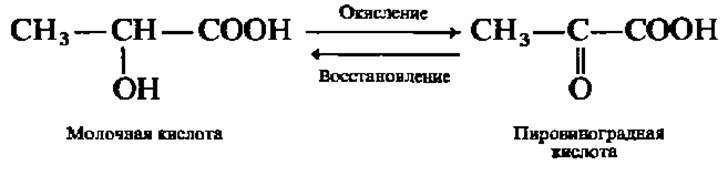
Since the oxidation of lactic acid (acidum lacticum) is accompanied by the removal of H atoms, this enzyme is called Lactate dehydrogenase. The lactate dehydrogenase molecule (M = 140000) is composed of four subunits (M = 35000), which are conventionally designated as H and M (from Heart and Muscle), since types I and V of lactate dehydrogenase have been isolated from The Heart and skeletal muscles, respectively. Consequently, the isozymes of lactate dehydrogenase are as follows:

They differ from one another in their level of activity, Certain physical properties (such as molecular weight and electrophoretic mobility), localization in Organs and Tissues, and so forth. Depending on age, physiological state, and other factors, the Organism establishes a specific ratio of isozymes, which corresponds to a defined overall level of enzyme activity. Altering the isozyme ratio throughout the whole organism or within specific tissues and organs thus represents one of the mechanisms for regulating enzyme action.
Interest in isozymes has surged dramatically in recent years. It turns out that in addition to genetically determined isozymes, There is a large group of enzymes exhibiting multiple forms that arise from post-translational modification (see Chapter VII). Multiple enzyme forms and isozymes in particular are now widely used in medical Diagnostics, for predicting animal productivity, and for selecting parental pairs in breeding to maximize progeny heterosis, among other Applications.
The Significance of the Spatial Organization of enzymes becomes especially apparent when studying the structure of so-called multienzymes—that is, enzymes capable of simultaneously accelerating several Chemical Reactions and driving complex substrate transformations. A prime example is the multienzyme that catalyzes the Oxidative Decarboxylation of pyruvic acid (see Chapter VIII). This multienzyme complex, with an M = 4,500,000, consists of Three types of enzymes. The first of these (E1) accelerates the decarboxylation of pyruvic acid. The complex incorporates 12 dimeric molecules of this enzyme (M = 192,000), shown in Fig. 46, D as large white spheres arranged in pairs around the periphery of the figure. The second and third enzymes, which catalyze the redox processes during pyruvic acid oxidation, are localized inside the multienzyme complex. One of them (E3) is represented by six dimeric molecules (M = 112,000), and the other (E2) by 24 protomers (M = 70,000) (see Fig. 46, shaded large and small spheres, respectively).
When a multienzyme complex drives a single, multi-step process of biochemical transformations, it is referred to as a metabolon (from metabolism). Examples include the metabolons of Glycolysis (see Chapter VIII), The Biosynthesis of a number of amino acids (see Chapter VII), the dicarboxylic and Tricarboxylic Acid Cycle (see Chapter VIII), and others.
As a result of the temporally and spatially coordinated action of all three types of constituent enzymes, the multienzyme transforms pyruvic acid at an immense rate. This cooperative Nature of the catalytic process lies at the heart of what distinguishes biocatalysts from inorganic catalysts, and it is precisely why The rate of biocatalysis exceeds the potency of inorganic catalysts by tens, hundreds, and thousands of times.
Relatively recently, another distinct structural feature of enzymes has been uncovered: some are multifunctional, meaning they possess multiple enzymatic activities yet consist of a single polypeptide chain. Specifically, during The formation of its tertiary structure, this single chain folds into several functionally and sterically distinct globular regions, or domains, each characterized by its own catalytic activity. Relevant examples will be discussed in subsequent chapters.
The Study of multienzyme complexes and multifunctional enzymes has shed light on the most crucial feature of Enzymatic Catalysis—namely, the Relay-like transfer of reaction intermediates from one component of the catalytic system to the next without their release into the medium.
Last update: 06/08/2026
Editorial and Educational Adaptation: This material has been compiled based on the primary/original source text. The project team performed an editorial review, corrected technical inaccuracies, structured sections, and adapted the content for an educational format.
What was processed:
- elimination of formatting defects (OCR errors, structural breaks, corrupted characters);
- editorial organization of content;
- standardization of terminology in accordance with academic sources;
- verification of factual statements against the original source text.
All mentions of the author, publication year, and origin of the primary text have been preserved in accordance with the source.