BIOCHEMISTRY - L. Stryer - 1984
VOLUME 2
PART III. BIOSYNTHESIS OF MACROMOLECULAR PRECURSORS
CHAPTER 21. BIOSYNTHESIS OF AMINO ACIDS AND HEME
21.13. Amino Acids Are Precursors of Many Other Biomolecules
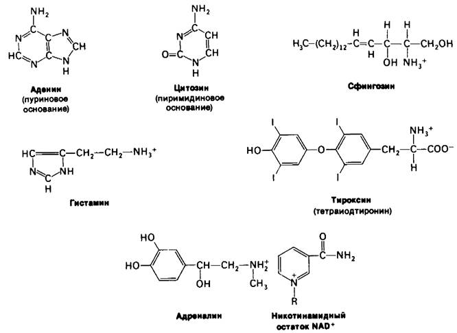
Amino Acids are the Building Blocks of Proteins AND Peptides. They also serve as precursors of many small molecules that play important biological roles. Let us briefly consider some molecules synthesized from amino acids (Fig. 21.19). Purines and Pyrimidines are derived in part from amino acids. The Biosynthesis of these precursors of DNA, RNA, and many Coenzymes will be discussed in detail in the next chapter. Six of the nine atoms of the purine rings and four of the six atoms of the pyrimidine ring originate from amino acids. The reactive terminal moiety of sphingosine, an intermediate in sphingolipid synthesis, is derived from Serine. Histamine, a potent vasodilator, is formed from Histidine by decarboxylation. Tyrosine is the precursor of the Hormones thyroxine (tetraiodothyronine) and epinephrine, as well as melanin, a polymeric pigment. The neurotransmitter 5-hydroxytryptamine (serotonin) and the nicotinamide ring of NAD+ are synthesized from Tryptophan. Glutamine provides the amide group for the nicotinamide moiety.
Class="center">Fig. 21.18. Model of Glutamine Synthetase. Peripheral adenylylation sites are shown

Fig. 21.19. Biomolecules derived from amino acids
21.14. Porphyrins Are Synthesized from Glycine and Succinyl-CoA
The Role of amino acids in the biosynthesis of the porphyrin rings of Hemes and chlorophylls was first discovered in isotopic labeling experiments conducted by David Shemin and his colleagues. In 1945, they showed that after the administration of 15N-Glycine to humans, 15N was detected in the heme molecule, whereas after the administration of 15N-glutamate, label incorporation was negligible. Using carbon-14, which had just become available to researchers at that time, they established that in nucleated duck erythrocytes, 8 carbon atoms of heme originate from the α-carbon of glycine; not a single atom originates from the carboxyl carbons. Further studies showed that the remaining 26 atoms of heme could be derived from acetate. Moreover, 14C atoms from methyl-labeled acetate were found in 24 of the 26 carbon atoms of heme, while 14C atoms from carboxyl-labeled acetate were found in only the other two atoms. Thus, these experiments revealed a clear labeling pattern, based on which Shemin proposed that the heme precursor is formed by the Condensation of glycine with an activated succinate compound. Indeed, the first step in porphyrin biosynthesis is the condensation of glycine and succinyl-CoA to form δ-aminolevulinate.
This reaction is catalyzed by δ-aminolevulinate synthase, a Pyridoxal phosphate-dependent mitochondrial enzyme. As expected, this committed step in porphyrin biosynthesis is regulated. Then, two molecules of δ-aminolevulinate condense to form porphobilinogen. This dehydration reaction is catalyzed by δ-aminolevulinate dehydratase.
Four molecules of porphobilinogen condense in a HEAD-to-tail manner to form a linear tetrapyrrole, which remains bound to the enzyme (Fig. 21.21). One ammonium ion is released for each methylene bridge formed. This linear tetrapyrrole cyclizes with the loss of NH4+. The cyclic product is uroporphyrinogen III, in which the arrangement of side chains is asymmetric. A synthetase and a cosynthetase participate in these reactions. In the presence of the synthetase alone, the symmetric product uroporphyrinogen I is formed. Cosynthetase is required to isomerize one of the pyrrole rings to form the asymmetric uroporphyrinogen III.
Fig. 21.21. Pathway of heme synthesis from porphobilinogen. (Abbreviations: A, acetate; M, methyl; P, propionate; V, vinyl.)

The porphyrin Skeleton is now complete. Subsequent reactions modify its side chains and increase its unsaturation (Fig. 21.21). Coproporphyrinogen III is formed by the decarboxylation of the acetate side chains. After The formation of double bonds in the porphyrin ring and The conversion of two propionic
acid side chains into vinyl groups, protoporphyrin IX is generated. Finally, the chelation of iron yields heme, the prosthetic group of proteins such as Myoglobin, Hemoglobin, catalase, peroxidase, and cytochrome c. The insertion of ferrous iron is catalyzed by ferrochelatase. Iron is transported in Blood Plasma by transferrin, a protein that binds two ferric ions, and is stored in Tissues within ferritin molecules. The large internal cavity of this protein (about 80 A in diameter) can contain up to 4500 ferric ions.
Several factors regulating heme biosynthesis in animals have been identified. δ-Aminolevulinate synthase, The enzyme catalyzing the first committed step of this biosynthetic pathway, is feedback-inhibited by heme, as are δ-aminolevulinate dehydratase and ferrochelatase. In addition, regulatory mechanisms operate at the level of enzyme synthesis.
Heme represses the synthesis of δ-aminolevulinate synthase. Recent studies suggest that the iron atom itself may be an important regulatory factor.
21.15. Porphyrins Accumulate in Some Inherited Disorders of Porphyrin Metabolism
Several Inherited Disorders of porphyrin METABOLISM are known. In congenital erythropoietic porphyria, The activity of uroporphyrinogen III cosynthetase—the isomerase that catalyzes the Formation of the asymmetric isomer during the cyclization of the linear tetrapyrrole—is deficient. To synthesize the required amount of uroporphyrinogen III, the body is forced to accumulate huge amounts of uroporphyrinogen I, a symmetric isomer devoid of any physiological role. In addition, uroporphyrin I, coproporphyrin I, and other symmetric derivatives accumulate. This disease is characterized by premature destruction of erythrocytes. The disease is inherited as an autosomal recessive trait. The urine of patients is colored red due to The excretion of large amounts of uroporphyrin I. The Teeth of patients fluoresce bright red under ultraviolet light due to the deposition of Porphyrins. Furthermore, their Skin is usually extremely sensitive to light.
Acute intermittent porphyria is a disease of a completely different nature. It affects Liver Cells rather than erythrocytes, and the skin of patients is typically not sensitive to light. This disease is caused by a deficiency in uroporphyrinogen synthase activity and is accompanied by a compensatory increase in δ-aminolevulinate synthase activity. Consequently, the concentrations of δ-aminolevulinate and porphobilinogen in the liver are elevated, and large amounts of these compounds are excreted in the urine. Acute intermittent porphyria is inherited as an autosomal dominant trait. Characteristic clinical symptoms include intermittent abdominal pain and neurological dysfunction. As the name of the disease implies, the clinical manifestations are episodic. Acute attacks can sometimes be suppressed by drugs such as barbiturates and estrogens.

Fig. 21.22. Space-filling model of protoporphyrin IX, the immediate precursor of heme

21.16. Biliverdin and Bilirubin Are Intermediates in Heme Degradation
Human red Blood Cells normally have a lifespan of about 120 days. Senescent cells are removed from the Circulation and degraded in the Spleen. Globin is hydrolyzed to its constituent amino acids. The first step in the Conversion of the heme group into bilirubin (Fig. 21.23) is the Cleavage of the α-methene bridge to form biliverdin, a linear tetrapyrrole. This reaction is catalyzed by heme oxygenase. Two features of this reaction are noteworthy. First, the enzyme catalyzing this reaction is a monooxygenase: O2 and NADPH are required for the cleavage. Second, the carbon of the methene bridge is released as carbon monoxide. This pathway of endogenous formation is related to a specific problem concerning the evolution of oxygen carriers (Section 3.7). Subsequently, the central methene bridge of biliverdin is reduced by biliverdin reductase to yield bilirubin. NADPH serves as the reductant in this reaction as well. The color changes in bruises are the most familiar manifestation of these cleavage reactions.
Figure 21.23. Cleavage of heme to bilirubin

Bilirubin, complexed with serum albumin, is transported to the liver, where it is converted into a more soluble state by the attachment of sugar residues to its propionate side chains. These solubilizing sugar residues are glucuronic acid, which differs from glucose in having a COO- group rather than a CH2OH group at the C-6 position. The conjugate of bilirubin and two molecules of glucuronate, called bilirubin diglucuronide, is excreted in the Bile. UDP-glucuronate is formed by The oxidation of UDP-glucose. This is the activated intermediate in the synthesis of bilirubin diglucuronide. Thus, the iron atom of heme is recycled, while the organic portion is converted into a soluble, open-chain form that is excreted from the body.

Last update: 06/08/2026
Editorial and Educational Adaptation: This material has been compiled based on the primary/original source text. The project team performed an editorial review, corrected technical inaccuracies, structured sections, and adapted the content for an educational format.
What was processed:
- elimination of formatting defects (OCR errors, structural breaks, corrupted characters);
- editorial organization of content;
- standardization of terminology in accordance with academic sources;
- verification of factual statements against the original source text.
All mentions of the author, publication year, and origin of the primary text have been preserved in accordance with the source.