Human Biochemistry, Volume 2 - Murray R. 1993
Biochemistry of Intracellular and Intercellular Communication
Mechanism of Hormone Action
Mechanism of Action of Group I Hormones
The General mechanism of action for Hormones of this group is illustrated in Fig. 44.1. Their lipophilic molecules diffuse freely through Cell/30.html">The Plasma Membrane of any cell, but they encounter their specific, high-affinity receptor exclusively within target Cells. This forms a hormone–receptor complex, which subsequently undergoes 'activation'. Driven by Temperature and the presence of salts, this reaction alters the size, conformation, and surface charge of the complex, endowing it with The ability to bind to Chromatin. Whether the formation and activation of this complex take place in the Cytoplasm or The Nucleus remains a matter of debate, though it is not critically important for understanding the overall process. The hormone–receptor complex then binds to a specific region of DNA, thereby activating or inactivating specific genes. Through this selective modulation of Gene METABOLISM/31.html">Transcription and the synthesis of corresponding mRNAs, the levels of specific Proteins change, which ultimately alters various metabolic processes. The Effect of each hormone in this group is strictly specific; as a rule, their influence impacts less than 1% of the target cell's proteins or mRNAs. Here, we focus on the nuclear MECHANISM OF ACTION of steroid and THYROID HORMONES, as this pathway is the most thoroughly characterized. Nevertheless, evidence also points to direct effects of these hormones on cytoplasmic components and various cellular Organelles.
Estrogens and glucocorticoids have been shown to influence mRNA degradation, and it is also known that glucocorticoids affect the post-translational Processing of certain proteins. Even so, the overwhelming majority of data indicates that the primary effect of these hormones is exerted at the level of gene transcription. Although the biochemical mechanism of gene transcription in mammalian cells is not yet fully understood, a general model can nevertheless outline the structural components required for the regulatory influence of steroid and thyroid hormones on this process (Fig. 44.2). Genes undergoing transcription must reside within regions of 'open' or transcriptionally active chromatin (depicted as a puff in Fig. 44.1), as evidenced by their susceptibility to Digestion by DNase I. Current evidence suggests that such genes contain at least two distinct regulatory elements (regulatory sites) located within the DNA sequence adjacent to the 5' end of the Transcription initiation site (Fig. 44.2). The first of these is the premotor element (PE), which is universal in that it appears in some form in virtually all genes. It determines the binding site of RNA polymerase II on the DNA and, consequently, the precision of transcription initiation (THE START OF DNA reading) (see Chapter 41).
Class="center">
Fig. 44.1. A steroid hormone binds to an intracellular receptor, inducing a conformational change. This complex subsequently binds to a specific region on the chromatin, leading to the Activation of a restricted number of genes. Thyroid hormones bind to a receptor that forms an integral part of the chromatin complex. In all other respects, The Mechanism of action of these hormones appears to be identical.
The second element—the hormone-responsive element (HRE)—has been identified in numerous genes regulated by Steroid Hormones. It is located somewhat further from the 5' end than the PE and may consist of several distinct components. The HRE is thought to modulate the frequency of transcription initiation and is less constrained by position and orientation than the PE. In this regard, it bears resemblance to enhancer elements found in other genes (see Chapter 41). Typically, the HRE is located several hundred NUCLEOTIDES upstream of the transcription initiation site, though its exact position varies from gene to gene. In some instances, this element is situated within the transcribed gene itself.
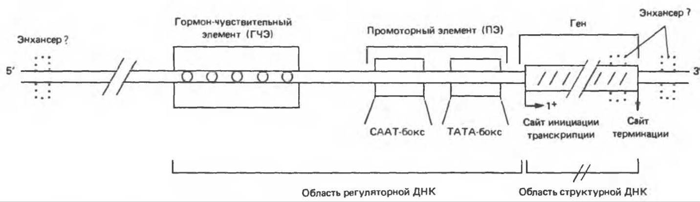
Fig. 44.2. Structural components involved in the steroid Introduction/30.html">Regulation of Gene transcription.
Identifying an HRE requires demonstrating that it binds the hormone–receptor complex with significantly higher affinity than the bulk of nuclear DNA or DNA from an alternative source. Such specific binding has indeed been demonstrated. Furthermore, an HRE must be capable of relaying its hormonal response downstream. To test this, the putative regulatory DNA sequence is fused to a reporter gene. Typically, these chimeric genes incorporate markers that are normally unresponsive to the hormone. Most commonly, the reporter genes used encode globin, thymidine kinase, or bacterial chloramphenicol acetyltransferase. These resulting fusion genes are then introduced into a target cell; if the hormone subsequently drives the Transcription of the reporter, the presence of a functionally active HRE is confirmed. This technique allows researchers to map the precise Location, orientation, and base-substitution sensitivity of the HRE. The exact mechanism by which the interaction between the hormone–receptor complex and the HRE influences transcription is currently an area of intense research. Regulation is presumed to occur primarily at the level of transcription initiation, though effects on elongation and termination processes are also possible. It has been suggested that regulatory sites may reside within the gene itself, or extragenically either upstream of the 5' initiation site or downstream of the 3' end. Finally, trans-active regulatory mechanisms (i.e., influences originating from another chromosome) may also play a role.
Last update: 06/08/2026
Editorial and Educational Adaptation: This material has been compiled based on the primary/original source text. The project team performed an editorial review, corrected technical inaccuracies, structured sections, and adapted the content for an educational format.
What was processed:
- elimination of formatting defects (OCR errors, structural breaks, corrupted characters);
- editorial organization of content;
- standardization of terminology in accordance with academic sources;
- verification of factual statements against the original source text.
All mentions of the author, publication year, and origin of the primary text have been preserved in accordance with the source.