Antibiotics (Properties, Application, Interactions) - M.P. Cherenko 1999
Burns
THERMAL BURNS
A burn (lat. combustio) is an injury to the Skin or mucous membranes, frequently involving underlying Tissues, caused by high temperatures (thermal burn), chemically active substances (chemical burn), or Physical and Chemical factors such as electrical current and radiation (electrical and radiation burns).
Burns account for a significant percentage of open injuries. In the United States, 2 million people sustain burns requiring medical attention annually. In recent years, the incidence of burns in Ukraine has decreased somewhat, amounting to 145,000 cases in 1993 (M.Yu. Povstianyi, 1994).
The most common causes of burns are hot liquids, steam, and flames. Thermal Injury (protein coagulation and Cell death) begins at temperatures of 44 °C, and The rate of tissue damage doubles with each degree increase in Temperature from 44 °C to 51 °C.
Radiation burns include sunburns as well as those caused by X-rays and radioactive rays. Under normal living conditions, radiation burns are very rare; however, in catastrophes (such as atomic bomb or nuclear Reactor explosions), they can assume mass proportions. X-ray burns are occasionally observed during the Treatment of patients with superficial forms of Cancer, predominantly in the postoperative period.
Chemical burns are caused by acids, bases, and other active substances resulting from safety violations during the production, transportation, and storage of such compounds (external skin burns), as well as from accidental or intentional ingestion (burns of the oral mucosa, Esophagus, and Stomach). Electrical burns result from the action of industrial, domestic, or atmospheric (lightning) current. Although local tissue damage can sometimes be profound—even resulting in the detachment of a part of an organ or tissue—the primary threat to the victim's life stems from the systemic effects of the current on the body, particularly the nervous and cardiovascular systems (cardiac arrest, Shock).
The majority of burns occur in domestic settings (approximately one-third of the victims are children) and are associated with violations of safety rules during the operation of gas and electric stoves, other heating appliances, improper storage of flammable items and substances, as well as inadequate supervision of children.
Burns most frequently occur As a result of exposure to hot liquids and open flames. Scalds from hot liquids are especially common among children and women, whereas flame burns are predominantly observed in men. The lower and upper extremities are most frequently affected, while the trunk, face, and neck are involved less often.
In the event of indoor fires or boiler explosions, burns to both the skin and the mucous membranes of the respiratory tract can be caused by hot air or steam.
Since burns primarily affect the skin, understanding burn pathology and successfully treating patients requires a thorough knowledge of its anatomical Structure AND Functions. The surface area of the skin ranges from 1.6 to 1.9 m2, and its mass accounts for approximately 15% of the body weight of a lean individual. Its functions include mediating all types of sensitivity, regulating body temperature, preventing excessive Water loss, and protecting the body against the penetration of infectious agents (microbes) and numerous other aggressive (toxic) factors (barrier function). Skin thickness varies depending on the anatomical site (from 0.5 to 3–4 mm), which is determined by its functional characteristics in different areas. The skin consists of the epidermis and dermis (Fig. 58, a). The epidermis is composed of layers of squamous epithelium with varying degrees of keratinization. The deepest layer is the basal layer of the epidermis—the germinal or Malpighian layer—which produces melanocytes that supply the pigment melanin to keratinocytes for their development. Migrating upward from the base into the upper layers of the epidermis, melanin protects the basal layer from ultraviolet radiation damage. Cells of the superficial layers undergo progressive keratinization leading to complete cornification and desquamation. The developmental cycle of an epidermal cell from its emergence to death lasts 28 days.
Class="center">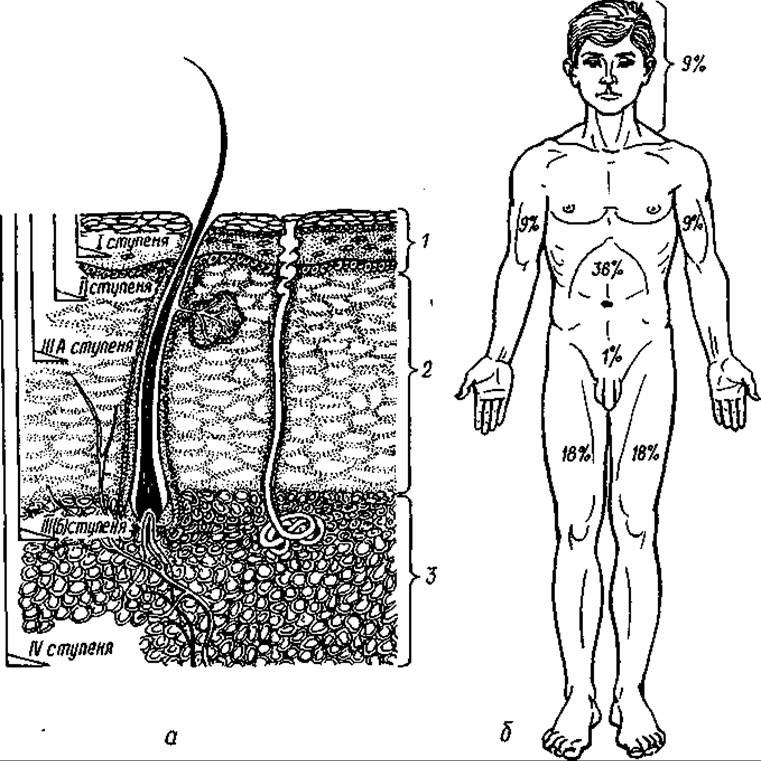
Fig. 58. Depth of tissue damage in a burn (a): 1 — epidermis; 2 — dermis; 3 — subcutaneous tissue. Determination of burn surface area using the rule of nines (b)
Beneath the epidermis lies the dermis (corium), which consists of Collagen and elastic fibers and fat. Its upper layer is dense and papillary, while the lower layer is looser—the reticular layer—which rests upon the subcutaneous tissue. The junction between the dermis and epidermis has a wavy appearance due to papillary projections of the dermis extending into the basal layer of the epidermis, hence the name papillary layer. The loose layer of the dermis houses Blood and Lymphatic vessels, nerves, and epidermal appendages (Hair follicles, sebaceous and Sweat Glands). The epidermis itself is devoid of Blood Vessels and receives nourishment via capillaries and Lymphatic vessels of the dermal papillae.
Due to their deep Location within the skin, the epithelial cells of epidermal appendages are spared in superficial burns and serve as a source for the regeneration of epidermal cells and the restoration of the skin.
Burns cause both local and systemic disorders. Local burns lead to disruption of the skin cover and its tissue functions, as well as an inflammatory response to the trauma of deeper tissues. Systemic disorders are caused by impaired functioning of the body's major systems (nervous, cardiovascular, endocrine, excretory, and metabolic).
The severity of the clinical course of burns and the resulting disorders are determined by the surface area and depth of the skin or mucosal lesion. There are numerous Methods for measuring burn surface area. Methods that provide an estimation of the affected area are simple and applied during the Cytology/cytology/16.html">Early stages of treatment (during antishock therapy under hospital conditions). These include the rule of nines (Berkow) and the palm method. According to the latter method, the area of the palm is assumed to be 1%; according to the rule of nines (Fig. 58, b), the surface area of the upper extremity accounts for 9%, the lower extremity for 18%, the trunk for 36%, and the scalp, face, and neck collectively account for 9% (3% for each). This scheme should not be applied to infants under one year of age, as their HEAD surface area is proportionally much larger than that of an extremity.
The burn surface area can be precisely determined using B. Postnikov’s "transparent imprint" method or by plotting the linear dimensions of the burn in centimeters onto a 17-cm human silhouette (featuring a millimetric grid on its surface). The transparent imprint method involves applying a sterile, transparent cellophane (polyethylene) film onto the burn surface and outlining the burn contours with a dye (such as methylene blue or brilliant green). After cleansing the wound of exudate and remnants of necrotic epidermis, the film is placed over the grid to determine the area of the outlined burn.
According to G.D. Vilyavin’s method (linear dimension plotting), the dimensions of the burn are mapped onto a 17-cm human silhouette covered with a millimetric grid. Each square centimeter of the burn corresponds to one square millimeter on the diagram. This method has the limitation that the human skin surface area and height are assumed to be standard values—17,000 cm2 and 170 cm, respectively.
Depending on the depth of the burn, 3 (Boyer) or 5 (Kreibig) degrees of injury are distinguished. In our country, a four-grade scale is used to assess burn depth. The numerical value of the degree increases with the depth of the burn. First-degree burn (combustio erythematosa) is characterized by damage exclusively to the superficial layers of the epidermis (stratum corneum, stratum lucidum) and clinically manifests as a burning pain, erythema, and mild skin edema.
Second-degree burn involves a deeper lesion of the epidermis (extending to the basal layer) with The formation of blisters filled with a clear fluid on the skin surface (combustio bullosa). Blisters arise as a result of a sharp increase in vascular permeability, accumulation of the liquid portion of the blood within the epidermis, and detachment of its superficial layers. The blister roof is pale, gray, and insensate to irritation, whereas the blister base (the deep basal layer of the epidermis) is red, highly sensitive, and painful.
First- and second-degree burns are predominantly sunburns and scalds caused by high-temperature liquids.
Third-degree burn is a necrotic form (combustio escharetica) subdivided into grades IIIA and IIIB.
In grade IIIA burns, necrosis extends through the entire thickness of the epidermis and the superficial papillary layer of the dermis (predominantly partial involvement of the latter).
Grade IIIB is characterized by Necrosis of the entire dermis, including the embedded epidermal appendages—sebaceous and sweat glands, and hair follicles. Third-degree burns are caused by boiling liquids, flames, and high-temperature metals. These thermal agents can also produce mixed burns ranging from grade I to IV.
In third-degree burns, depending on their depth, blisters and areas of dry skin necrosis—eschars, mostly white-gray or grayish-yellow in color—are also observed.
During the first hours and even days, it is difficult to differentiate between second- and third-degree burns, as The process of cell necrosis in the skin often continues long after the thermal agent has ceased to act, driven by vascular and Metabolic Disorders and The Development of infection.
The most reliable test for determining burn depth is assessing the sensitivity of the burn surface (excluding blister bases) to mechanical or chemical stimuli. In third-degree A (IIIA) burns, surface sensitivity is preserved, much like in second-degree burns, whereas in third-degree B (IIIB) burns, sensitivity is completely absent to both needle pricks and alcohol-swabbed cotton balls. A very pale burn surface—particularly one resembling manufactured artificial leather—indicates full-thickness skin destruction. Blisters indicate partial-thickness skin damage.
In fourth-degree burns, due to the rapid evaporation of tissue water, the affected skin turns a dark gray or brown color and transforms into a cracked eschar. The area of the affected skin decreases sharply, and surface sensitivity is entirely absent. In fourth-degree burns, necrosis extends through the entire thickness of the skin and underlying tissues, resulting in tissue charring. Such burns are typically caused by flames, electrical currents, molten metal, or similar agents.
Burns of the first, second, and IIIA degrees are classified as superficial, while IIIB and fourth-degree burns are classified as deep.
The vast majority of thermal burns are superficial—grades I, II, and IIIA. A smaller proportion consists of IIIB and fourth-degree burns, with fourth-degree burns accounting for about 5%. In adults, superficial first-degree burns make up 30%, while grades II and IIIA account for up to 15%. Deep burns covering up to 10% of the body surface are considered localized, whereas larger burns are termed extensive and classified as severe, typically involving the clinical course of burn disease. In children under 12–15 years of age and elderly individuals (60 and older), superficial burns covering 10–15% and deep burns exceeding 5% are considered severe. All inhalation injuries are also classified as severe.
Deep burns covering 50% or more are generally considered incompatible with life, although exceptions do occur, particularly in recent years.
PATHOPHYSIOLOGICAL CHANGES IN BURNS
Thermal injuries (cell destruction due to Protein Denaturation and coagulation, accompanied by circulatory failure) lead to increased capillary permeability within just 15 minutes. This permeability rises rapidly during the first 12 hours and gradually decreases by 48 hours. This process results in the shift of exudate fluid (water, salts, and Proteins with molecular weights up to 150,000) from the vessels into the perivascular space. In superficial localized burns, the exudate drains outward through the burn wound into the surrounding tissues, whereas in severe burns, it also accumulates in anatomically uninjured Organs (primarily skeletal Muscles), causing edema. Fluid loss through the wound can reach 200 ml/m2 per hour (compared to a normal rate of 15 ml/m2 per hour). Damage to the skin, which acts as a primary antimicrobial barrier, can be complicated by local wound infection and systemic infection, or Sepsis.
Fluid loss through the wound surface and its sequestration in tissues due to impaired drainage and lymphatic outflow (tissue edema) lead to a sharp decrease in circulating blood volume, or hypovolemia. In extensive burns, hypovolemia frequently triggers shock unless the lost fluid (plasma) is promptly replenished. Although red Blood Cells are destroyed within the burn zone, their loss relative to plasma is minor, resulting in hemoconcentration. Furthermore, patients with burns experience a sharp surge in metabolic processes, primarily driven by the hyperproduction of catabolic Hormones (catecholamines, cortisol, somatotropin, Glucagon), and secondarily by heat loss resulting from the destruction of the skin and water evaporation. High Energy Expenditure (up to 7,000 kcal daily) in extensive burns, particularly due to protein loss, leads to progressive weight loss.
CLINICAL MANIFESTATIONS OF BURNS
The clinical course of burns depends on their size (area and depth), anatomical location, individual patient characteristics (age, overall health status), and The Nature of the thermal agent. The burn surface area is generally the determining factor.
Localized burns follow the course of a standard local wound process, accompanied by minor systemic symptoms (headache, slight tachycardia, mild fever, leukocytosis, and a slight increase in hematocrit).
Extensive burns progress as burn disease, which is characterized by distinct phases. Within the first 24–72 hours, many patients (up to 50%) with extensive burns develop hypovolemic shock of varying severity, accompanied by its classic manifestations: hypothermia, lowered blood pressure, decreased urinary output, tachycardia, collapse of superficial Veins, pale gray and clammy skin, and others. Unlike Other forms of shock (such as traumatic or hemorrhagic), burn shock frequently features a prolonged erethitic phase dominated by psycho-emotional and motor agitation, elevated blood pressure, and accelerated Heart rate.
With proper first aid Organization and effective management of hypovolemia and pain, the incidence of shock can be significantly reduced, as evidenced by the experience of specialized burn units. At 48 to 72 hours post-burn, the so-called toxemia phase develops, typically lasting about two weeks. This phase is associated with the absorption of breakdown products from damaged tissues and metabolic disturbances, occurring in approximately 25% of patients with extensive burns. Patients become agitated or lethargic, complaining of nausea, vomiting, headaches, loss of appetite, and fever. Dystrophic and inflammatory complications emerge (Pneumonia, nephrosis, hepatitis, peptic ulcers of The Stomach and duodenum—known as Curling's ulcers, enteritis, etc.). In many cases (especially when measures to close the burn wound and prevent infection are inadequate), the toxemia phase transitions into septicotoxemia. This phase is typical of deep, extensive burns. Intoxication is further exacerbated by the development of wound infection, the resorption of microbes and their toxins, and purulent exudate.
Infection may remain confined to the wound surface with systemic toxic symptoms (so-called purulent-resorptive fever) or, less commonly, lead to sepsis (septicemia or septicopyemia). Infection remains the leading cause of death in burn patients.
Sepsis occurs predominantly in patients with extensive grade IIIB–IV burns, particularly in the presence of comorbid conditions (Diabetes Mellitus, atherosclerosis, anemia, hypoproteinemia, hypovitaminosis, circulatory failure, etc.). Although effective antibacterial therapy and early surgical intervention (excisional debridement of necrotic tissue and early biological closure of burn wounds) have reduced the incidence of sepsis in recent years (to about 7% of patients with deep extensive burns), the efficacy of preventive and therapeutic measures against sepsis remains far lower than that for burn shock. The primary pathogens responsible for burn wound infection and sepsis are staphylococci, *Pseudomonas aeruginosa*, and, less frequently, hemolytic streptococci and *Escherichia coli*.
BURN TREATMENT, FIRST AID, AND ANTI-SHOCK MEASURES
The management of thermal burns comprises pre-hospital first aid and specialized treatment in surgical burn centers.
A patient's prognosis often depends on the quality of first aid administered. This involves halting the action of the thermal agent, providing the victim with fresh air, and preventing wound contamination by microflora.
Burning clothing is best extinguished by smothering the flames with a blanket, coat, or cloak, or by pouring water on the victim. The patient should be laid flat so that rising flames do not spread to the head and face. Running while wearing burning clothing is strictly prohibited, as it only fans the flames. Once extinguished, the clothing must be removed immediately after being doused with cold (but not ice-cold) water. This applies equally to burns caused by boiling water or hot liquids. However, hypothermia must be avoided at all costs, as it contributes to the development of shock. Dousing the burn with cold water also serves a therapeutic purpose: it relieves pain and prevents the deeper spread of thermal tissue destruction—specifically the progression of IIIA burns to IIIB—as well as reducing edema and suppuration. For this reason, cold water irrigation is particularly recommended during first aid administration (no later than 30 minutes post-burn) for localized burns (especially of the limbs). Cooling the burn surface with water should continue for about 2 hours.
To ensure a supply of fresh air, the victim is moved from the fire scene (or a smoke- and toxic-gas-filled room) to an open area or a room with clean air and then warmed. In cases of asphyxia, artificial pulmonary ventilation (Mouth-to-mouth or mouth-to-Nose) is performed.
The final step of first aid—preventing infection of the burn surface—is achieved by wrapping the victim in a clean sheet, gauze, or plastic film (for large burns) or applying an aseptic bandage for localized burns. Analgesics are administered intravenously, and the patient is transported to the hospital immediately.
For patients with extensive deep burns who are at risk of shock or are already in shock, the infusion of anti-shock solutions (saline solutions, rheopolyglucukin, lactate or Ringer-Locke solutions, etc.) is initiated in the ambulance.
During first aid, all jewelry (rings, wedding bands, bracelets, etc.) should be removed from the patient's hands to prevent a tourniquet effect.
Upon arrival at the medical facility (typically a specialized burn unit), the patient should ideally be placed on a specialized air-fluidized bed. The airway status is assessed first; if airway patency is compromised (indicated by shortness of breath, cyanosis, coughing, or hoarseness), tracheal intubation is performed.
Patients with limited superficial and deep burns without the risk of developing shock undergo wound cleansing under analgesia (primarily via intravenous administration of morphine hydrochloride, non-opioid analgesics, or narcotics).
Hospitalization is required for: patients with extensive burns, airway burns, and limited II–IV degree burns covering more than 3–5% of the body surface area, as well as extensive I degree burns covering more than 30% in children; patients with II–IV degree burns of the head, face, Perineum, hands, and feet, regardless of the affected area.
Patients with limited superficial burns outside of the aforementioned critical areas are referred to an outpatient clinic for follow-up care (wound dressing and treatment) after initial medical assistance is provided.
In cases of extensive burns that have caused or threaten to cause shock, anti-shock therapy is initiated or continued alongside hemodynamic monitoring (blood pressure, pulse, central venous pressure), blood tests (hemogram, hematocrit, electrolyte and urea levels, etc.), and urinalysis (hourly diuresis via an indwelling urinary catheter; urine composition). Body temperature is monitored, and infection Prevention measures are implemented. Wound cleansing and dressing in such patients are withheld until shock and the threat thereof are fully resolved.
Treatment for shock focuses on correcting hypovolemia (plasma volume deficit) and fluid-electrolyte imbalances through the administration of balanced electrolyte solutions (especially Ringer's lactate), specialized solutions (such as rheopolyglucukin), and protein supplements (plasma, albumin, protein, and blood after 24 hours). Infusion solutions must contain sodium, as burn patients frequently experience hyponatremia due to increased secretion of antidiuretic hormone (vasopressin) and sodium retention in the interstitial space.
Hyperkalemia is typically not observed in these patients despite significant cellular destruction, owing to enhanced renal potassium excretion induced by hypercorticism.
The volume of fluid replacement is calculated based on multiple criteria (hemodynamics, urine output, biochemical test results, etc.). In clinical practice, simplified formulas are predominantly used. For instance, according to the Parkland formula, a patient is administered 4 mL of Ringer's lactate per 1 kg of body weight during the first 24 hours, multiplied by the percentage of the total body surface area burned. Half of this volume is infused within the first 8 hours, and the remaining half over the subsequent 16 hours. To determine the volume of plasma and other colloidal solutions—particularly in the UK—the Muir-Barkley formula is used, where the volume of infused plasma equals the burn area multiplied by body weight (in kg) and divided by 2, i.e., 0.5 mL/kg per 1 percent of the burn.
To replenish fluid deficits, a 5% glucose solution is administered intravenously. If the patient has no gastrointestinal dysfunction, drinking water, tea, and juices are recommended.
For analgesia, intravenous analgesics are administered (opiates such as morphine, omnopon, fentanyl, etc.), along with high doses (100–500 mL per day) of cortisol (hydrocortisone), energy substrates such as hypertonic glucose solutions (10–20%) with Insulin, ascorbic acid, vitamin B1, cardiovascular medications (Glycosides such as strophanthin, corglycon), beta-blockers, etc.
Adequate energy support is of paramount importance in the management of burn patients. METABOLISM in these patients is characterized by hypermetabolism, predominantly driven by accelerated catabolic processes. Hypermetabolism is primarily linked to the stimulation and activation of the sympathoadrenal system, as well as significant heat loss resulting from the destruction of a large skin surface area. It manifests as increased oxygen consumption, elevated heart rate and minute ventilation, higher core body temperature, weight loss,
and urinary nitrogen wasting. Therefore, nutritional support must be high in calories.
One of the formulas used to calculate the caloric requirements of a patient is the Curreri formula, according to which the daily energy intake should be 25 kcal per 1 kg of body weight plus 40 kcal multiplied by the percentage of the total body surface area burned.
Hyperalimentation is achieved via enteral or intravenous routes, or more commonly a combination of both, provided the patient has no gastrointestinal complications. For parenteral Nutrition, the nutritional regimen relies primarily on hypertonic glucose solutions (not exceeding 3 g of glucose per 1 kg of body weight) along with lipid emulsions. Patients are administered 2 g of protein per 1 kg of body weight. For the prevention of Curling's ulcers, patients are prescribed H2-receptor blockers (cimetidine, ranitidine, famotidine, etc.).
LOCAL TREATMENT
Local treatment typically begins immediately after the patient is admitted to the healthcare facility, or once patients in a state of shock or at risk of this complication have been successfully resuscitated.
For first-degree burns, the affected surface is treated with ethyl alcohol (56–76%) and left uncovered by a dressing. After 3–5 days, the necrotic superficial epithelium desquamates and is replaced by new tissue, which rapidly acquires a pink coloration.
Second-degree burns are treated after administering analgesia (intravenous analgesics). The skin surrounding the burned area is disinfected with a 0.5% chlorhexidine solution, 1% iodonate, or 76% ethyl alcohol. The burn surface is gently washed with cotton-gauze Sponges soaked in an antiseptic solution or warm saline, and then dried with sterile swabs. Blisters are evacuated of exudate and completely excised. A moist-drying dressing is usually applied to the burn surface using a furacilin solution (1:5000), 0.5% chlorhexidine (preferably its alcoholic solution, hibitane), a hydrophilic ointment (levosyn, levomekol, etc.), or flamazine (1% silver sulfadiazine). If there is no strike-through (monitored daily), the dressing is left undisturbed for 7–10 days. If the wound remains uninfected, it heals during this timeframe. Typically, upon removal of the dressing, the burn surface is completely covered by newly formed pink epithelium. If exudate strikes through, the dressing is replaced with a new one.
Second-degree facial burns are treated using the open method. After washing and drying the burn surface, it is treated with astringent or tanning agents (3% potassium permanganate solution, 5% tannin solution, etc.), or less frequently with film-forming compositions (plastubol, etc.).
Second-degree burns of the hands, after treatment of their surface with an antiseptic (preferably in ointment form), are either dressed or placed in sterile plastic bags tied at the wrists. These bags must be changed daily. This method does not restrict finger mobility or impair Blood Circulation in the hand, as can occur with circumferential bandaging. Second-degree burns of the extremities or trunk can also be treated openly—without dressings—within specialized aero-therapeutic chambers, such as isolation bags for limbs and air-treatment units (ATUs) supplied with dry, sterile air.
Third-A degree burns are treated in the same manner as second-degree burns. In uncomplicated cases, they heal within 2–3 weeks.
Local treatment of third-B and fourth-degree burns is administered depending on their surface area.
Very limited burns in appropriate anatomical locations may be excised with primary closure using mobilized local tissue. Moderately sized burns of uniform depth can be tangentially excised during primary wound care (primary necrectomy) and closed with a split-thickness skin graft harvested using a dermatome. However, this primary necrectomy is accompanied by significant blood loss, and the resulting wound bed is not always sufficiently smooth and dry for the skin graft to adhere properly without the risk of hematoma or exudate accumulation beneath it. Although perforating the graft minimizes this complication, primary necrectomy with immediate autologous skin grafting is rarely performed and is largely restricted to specialized burn units.
In most cases, during the initial treatment of deep burns—particularly those of mixed depth—the surface eschar is either covered with an aseptic dressing until a clear demarcation line of necrosis and subsequent Separation appears (which can be accelerated by applying Proteolytic Enzymes or 40% salicylic acid to the eschar surface), or necrectomy followed by autografting is performed a few days post-burn once demarcation is established. In circumferential deep burns of the chest or extremities (particularly the lower legs or thighs), the eschar must be longitudinally incised in multiple places during the initial local wound care to relieve compression, thereby restoring normal Respiration and circulation.
For extensive deep burns of uniform depth, the entire necrotic area (eschar) within healthy skin borders is tangentially excised during the first week (post-burn days 3 to 7) and temporarily covered with a "biological" dressing, such as preserved porcine skin, synthetic skin, or less commonly, preserved allograft skin (the latter is used less frequently due to the risk of HIV and other infections). Such temporary closure should be performed sooner when the necrotic area is larger and the risk of mortality is higher. Temporary coverage of the burn wound prevents infection and plasma loss, and accelerates the preparation of the wound bed for definitive autografting with split-thickness skin. This Procedure can be performed either in a single stage or in multiple steps. Concurrently, other measures are employed to prevent infection and stimulate tissue regeneration (ultrasound, laser irradiation, etc.).
Prior to skin grafting for deep burns, anemia, hypoproteinemia, and other metabolic disturbances must be corrected, and the microbial flora on the wound surface—particularly streptococci—must be neutralized and verified via microbiological testing. Early closure of deep burn wound surfaces, combined with The Use of Antibiotics, constitutes the most critical measure in preventing burn sepsis.
The surgical closure of deep, extensive burns is primarily performed using split-thickness skin grafts harvested with a dermatome (Fig. 59). Compared to full-thickness skin grafts, their advantages include better engraftment due to enhanced nutrient uptake and closer adherence to the wound bed. Split-thickness grafts are also advantageous because they can be re-harvested from the same donor site on the patient's body within 2–3 weeks, as this area rapidly epithelializes from skin appendage epithelium—hair follicles, Sebaceous Glands, and sweat glands. Such repeated skin harvesting is especially crucial for patients with massive burns who experience a shortage of healthy skin.
For free skin grafting during the acute phase, split-thickness grafts are almost invariably used. The disadvantages of these grafts include their low tolerance to physical stress—even "physiological" trauma such as pressure and friction—as well as a tendency toward contraction and deformity.
Due to these characteristics, rather than using grafts for areas of the skin subject to significant mechanical stress (palms, soles) or areas of cosmetic importance, surgeons use full-thickness grafts or composite vascularized skin flaps. The latter are employed to reconstruct burn surfaces with exposed bones, joints, and tendons. The closure of even large-area burns in specialized medical facilities is achieved within 2–3 months.

Fig. 59. Dermatome:
a — manual Paget-type dermatome; b — electrodermatome (1 — attachment; 2 — stop ring; 3 — gear housing; 4 — protective cover; 5 — power cord; 6 — pedal; 7 — blade; 8 — protective cap)
At the same time, patient treatment and the rehabilitation period should last for at least a year, as scar evolution continues for a year or even longer. This period can be complicated by severe physical and cosmetic defects (contractures, ectropion, etc.) as well as psychological trauma. Both the grafted skin areas and the donor sites during this period should be treated with emollient creams and subjected to gentle massage. For areas with contractures, physical therapy Procedures (UWF, paraffin therapy, laser therapy, mechanotherapy, etc.) are advisable. To prevent hypertrophic scarring, elastic garments and physical therapy are widely used, and if the patient is prone to developing such scars, corticosteroid medications (prednisolone, hydrocortisone—both as ointments and intramuscularly) are prescribed.
Individuals with significant cosmetic defects and contractures require psychological and social rehabilitation.
INHALATION INJURIES
Inhalation injuries (burns of the respiratory tract) have become increasingly common recently. Regardless of the degree and extent of mucosal damage, these injuries are classified as severe because they pose a significant threat to the patients' lives. Burns extending to the subglottic region of the Larynx are particularly dangerous. Inhalation injuries occur during fires in residential buildings, ships, steam boiler explosions, burning of fuel oil tanks, pipelines, etc., and are caused by the inhalation of hot air, steam, and smoke. They are frequently combined with poisoning by toxic chemicals released during the combustion of household appliances, which are predominantly made of plastic components.
Since inhalation injuries are almost invariably combined with skin burns, which initially draw the most attention, timely Diagnosis of respiratory tract burns is critical. The Clinical symptoms of these burns in the early period (the first 8 hours) are limited to laryngeal Muscle spasm, and later to edema and inflammation of the respiratory mucosa, leading to airway obstruction and impaired breathing. Diagnosis is based on the following clinical signs and symptoms:
1) presence of Head and Neck burns;
2) singed nasal hairs;
3) hoarseness;
4) dyspnea and cyanosis;
5) "brassy" ( Croup-like) cough;
6) constricting chest pain along the Sternum;
7) inflammation of the oropharynx;
8) expectoration of sputum mixed with soot and carbon.
A sharply increased amount of bronchial secretions (bronchorrhea).
All such patients require mandatory hospitalization. If necessary, the diagnosis can be verified using flexible fiberoptic bronchoscopy.
Treatment for these patients consists of humidified Oxygen therapy, bronchial hygiene (toilet of the Bronchi), sometimes combined with endoscopy, and the inhalation and administration of bronchodilators and mucolytics. Cortisol or prednisolone (50 mg per day) is also prescribed as an anti-edematous and anti-inflammatory agent, along with antibiotics. In cases of escalating asphyxia, tracheal intubation and mechanical ventilation are performed. In some cases, tracheostomy is performed, but it is used under strict indications because it increases the risk of sepsis. Considering this complication, many authors, particularly American ones, do not recommend corticosteroids. However, domestic combustiologists do not share this view and administer such drugs in high doses (R.D. Ryabaya, R.I. Kravchenko, 1980; N.E. Povstyanoy, G.P. Kozinets, 1984).
CHEMICAL BURNS
Chemical burns are caused by chemically active substances—acids (sulfuric, hydrochloric, nitric, acetic, etc.), bases, phosphorus oxides, as well as gasoline, kerosene, and certain oils upon prolonged contact with the skin. These substances can damage both the skin and the mucosa of the digestive tract if ingested. In most cases, chemical burns occur in industrial chemical plants and laboratories and are associated with workers violating safety regulations during the pouring, handling, and transportation of reactive chemicals. Chemical burns in domestic settings are rare and usually result from accidental or intentional ingestion of chemicals.
Chemical skin burns in everyday life are even rarer than mucosal burns and typically occur in individuals with hypersensitivity (allergies) to such substances.
In terms of the depth of the lesion and the affected area, chemical burns are classified, similarly to thermal burns, into 4 degrees, as well as localized and widespread. Burns of the esophagus are divided into 3 degrees: superficial, involving the full thickness of the mucosa, and involving the full thickness of the wall. Areas of physiological narrowing within the esophagus are the most vulnerable.
Superficial burns of the I–II degree typically occur when the skin and mucous membranes come into contact with low-concentration acids and bases, or from prolonged exposure to kerosene or gasoline. Superficial skin burns are the most frequently observed. Among esophageal burns, deep burns predominate; they result from the action of concentrated acids and, especially, bases, as well as phosphorus. Quantitatively, third-degree burns are the most common. Industrial burns are dominated by acid burns, and somewhat less frequently by bases or phosphorus.
Based on their MECHANISM OF ACTION, chemically active substances are divided into those that cause protein precipitation and coagulation through the rapid extraction of water from tissues (acids, heavy metal salts, etc.), and those that have a liquefying effect (bases). In low concentrations, substances that promote protein coagulation—due to the rapid shift of cellular water beneath the epidermis—cause edema and blistering of the skin (second-degree burn). In high concentrations, they cause dry skin necrosis with eschar formation. Liquefying substances damage the epidermis, break down proteins, and form insoluble soaps with tissue Lipids, leading to liquefactive necrosis. They act more slowly than acids, but penetrate deeper, forming a moist white eschar whose detachment is accompanied by ulceration. The deeper the ulcerations, the higher the concentration of the base. Protein breakdown products are toxic, easily absorbed, and lead to systemic intoxication. The Effect of acids and bases of varying concentrations on the mucosa of the digestive tract causes burns of the same depth and character as those on the skin.
When low-concentration acids or bases are ingested, mucosal burns are superficial yet cover a larger surface area. Conversely, more concentrated substances cause significantly deeper, though somewhat smaller, burns due to a sharp spasm of the esophagus that limits the downward spread of the solution into the distal parts. In general, chemical burns tend to be localized (affecting no more than 10% of the body surface area).
Clinical presentation of chemical burns.
General clinical manifestations of these burns are considerably less pronounced than those of thermal burns. Due to their predominantly local nature, shock and toxemia are rare. In deep acid burns, body temperature also remains almost unchanged owing to the dry nature of necrosis and the absence of formation and absorption of toxic necrotic products. Only in superficial burns with blister formation can body temperature rise transiently. Deep burns caused by bases are accompanied by significant intoxication resulting from the formation of toxic protein breakdown products and their absorption. Burns of the digestive tract mucosa follow a significantly more severe clinical course than skin burns.
Superficial skin burns of the I–II degree are characterized by inflammation with hyperemia, edema, and blister formation (in second-degree burns). The burn surface has clear margins and an irregular shape, often with traces of the chemical solution On the surface. Deep skin burns caused by acids, heavy metal salts, and phosphorus are accompanied by dry necrosis—an eschar of varying color depending on the chemical agent. Nitric acid produces a light yellow eschar; sulfuric acid creates an initial white eschar that later turns brownish-black (due to Hemoglobin degradation). Hydrochloric acid leaves a whitish-gray eschar on the skin surface, while bases produce a soft, whitish-gray eschar. The surface of deep chemical burns is painless. The detachment of eschars is not accompanied by Hemorrhage, and both the epithelium and Connective Tissue regenerate slowly.
The diagnosis of chemical burns is straightforward and based on a thoroughly taken Anamnesis and the Specific characteristics of the burn. In this regard, In addition to the color of the eschars, uniformity of the lesion (its color, blister density, etc.) is significant in superficial burns. This does not occur in thermal burns, which typically present a combination of different degrees of burns (many blisters, both intact and ruptured). Burns of the oropharynx, esophagus, and rarely the stomach primarily manifest as sharp retrosternal pain (along the course of the esophagus), vomiting of mucus mixed with blood (or gastric contents in the case of gastric burns), signs of agitation, cardiovascular dysfunction (tachycardia, less frequently decreased blood pressure and shock), and later, fever. The absorption of the chemical substance, as well as protein breakdown products, causes systemic intoxication, leading to secondary dysfunctions of The Nervous system, Liver, Kidneys, etc. Patients present in a depressed state. Toxic hepatitis (Hyperbilirubinemia and jaundice), nephrosonephritis, and anemia are characteristic.
Deep burns of the esophagus, especially those caused by bases, can be complicated by necrosis and perforation of the esophageal wall, leading to severe purulent (posterior) mediastinitis. Signs of the latter include a sharp rise in BODY TEMPERATURE AND chills, increasing pain in the Mediastinum, worsening cardiovascular symptoms, and respiratory disorders. Such a clinical course requires immediate mediastinal drainage and intensive therapy with antibiotics (both systemic and local) and other modalities.
In most cases, esophageal burns are not accompanied by perforation and are treated conservatively. On the 2nd to 3rd day, the necrotic esophageal mucosa begins to detach, usually completing by the 5th to 6th day (sometimes resulting in the shedding of tubular casts of necrotic mucosa). This is followed by the granulation phase of the burn surface. The reparative process of burn surfaces on the skin and the mucosa of the digestive tract (Pharynx, esophagus, stomach) is characterized by the hyperproduction of fibrous tissue, which leads to the formation of hypertrophic, deforming scars and distortion of the affected organs (ectropion of the eyelids, narrowing or strictures of the nasal, oral, pharyngeal, esophageal, and pyloric orifices). Such deformations and strictures require complex plastic surgeries.
First aid for chemical burns consists of thorough irrigation of the burn surface with water under moderate pressure for 10 minutes until the chemical substance is completely washed away (and the odor disappears). Water not only washes away the chemical agent but also cools the tissues, eliminates the exothermic effect, and prevents the necrosis from spreading deeper. If the causative agent is identified, irrigation is performed using an appropriate neutralizing solution.
For burns of the oropharynx, esophagus, and stomach, after pain management, the patient's stomach is lavaged with water or isotonic sodium chloride solution, or—if the causative agent is known—with a solution that neutralizes its action: sodium bicarbonate solution for acid burns, and a 2–3% citric or acetic acid solution for burns caused by bases. Phosphorus burns are extinguished by submerging the affected area in water and removing the particles in a dark room. After irrigation, the burn surface is covered with a dressing moistened with a neutralizing substance, and in the case of phosphorus burns, with a 3–5% copper sulfate solution. Individualized general therapeutic measures are prescribed (analgesics, anti-shock, sedative, and detoxifying agents, etc.). If the burns are deep, particularly with dry necrosis, early necrotomy (incision of the eschar), partial necrectomy, and the application of necrolytic agents (proteolytic enzymes, 40% salicylic acid) are performed to accelerate the sloughing of necrotic masses. Surfaces cleansed of dead tissue are closed surgically. Limited burn areas may be excised in the first few days and closed with skin flaps. Due to the tendency of scars resulting from chemical burns to undergo hypertrophy, inflammation, and organ deformation, physical therapy is of great importance. In patients with esophageal burns, alongside the aforementioned local and general measures, antibiotic therapy is administered, and enteral nutrition is established via a nasogastric tube (or via a gastrostomy in severe cases, particularly with esophageal perforation, as well as in young children). Patients showing signs of renal failure undergo hemodialysis and other supportive procedures. 5 to 7 days after the injury, esophageal bougienage is initiated to prevent cicatricial strictures. In patients with a gastrostomy, so-called permanent bougienage, or "endless bougienage," is performed, which the patient can carry out themselves. For this purpose, a bougie is tied to the end of a strong thread passed through the oropharynx and esophagus (the thread is introduced into the stomach by swallowing a small bead tied to its end), while another bougie is tied to the gastric end. The treatment outcomes can only be evaluated after 2–3 months or later. If cicatricial stenosis of the esophagus or the gastric outlet develops, surgical interventions are performed (esophageal replacement with a bowel loop or stomach, pyloroplasty, or even gastric antrectomy). In the long-term period following an esophageal or gastric burn, malignant tumors frequently develop. Mortality from skin burns is low, whereas mortality from esophageal burns, especially in children, is high.
The prevention of chemical burns in industry and laboratories is based on the strict adherence of workers to safety regulations. In everyday life, it is primarily ensured by storing chemically active substances in places inaccessible to children, clear labeling and secure closing of bottles containing such substances, and careful handling thereof, among other precautions.
RADIATION BURNS
The widespread use of radiation sources in the national economy, defense (power plants), medical practice, and research has made both radiation sickness and radiation burns (often in combination with other injuries) possible. Alpha, beta, gamma, and X-rays in therapeutic doses cause local tissue damage—burns. Their severity depends on the radiation dose (B.S. Vikhryayev, V.M. Burmistrov, 1986):
I — erythematous dermatitis, developing after local irradiation with a dose of 800–1200 rad. Acute inflammation appears 2–3 weeks post-irradiation, accompanied by pain, hair loss, and pigmentation. It heals with complete epithelial regeneration within 2–3 weeks;
II — bullous dermatitis, beginning one week after irradiation with a dose up to 2000 rad. Necrosis extends to the full thickness of the skin (epithelium and dermis). The clinical course is protracted, with healing occurring within 1.5–2 months, resulting in scarring, epilation, and skin pigmentation. It is frequently accompanied by a systemic reaction (weakness, fever, headache, dyspeptic symptoms);
III — gangrenous dermatitis, occurring at doses exceeding 2000 rad. Local manifestations appear within a few hours. Erythema and skin edema develop and persist for 2–3 days, and after a week, necrosis of the entire skin and underlying tissues ensues. It is frequently accompanied by radiation sickness. The clinical course is very sluggish, leading to chronic ulcers and unstable scars at the burn site. A tendency toward malignant transformation is observed.
In addition to acute radiation burns, chronic burn dermatitis of the hands is sometimes observed, most commonly among radiologists.
During an atomic bomb explosion, alongside general and local radiation injuries, so-called profile burns of exposed skin areas (face, neck, hands) caused by light, thermal, and ultraviolet rays are observed. These burns can account for a massive proportion of combined injuries (up to 80%). Their Clinical Presentation and treatment are fundamentally no different from those of conventional thermal burns.
Treatment of grade I–II radiation burns consists of conservative local measures: applying dressings with antiseptic agents after cleansing the burn surface (washing with sterile solutions—isotonic sodium chloride or 0.25–0.5% ammonia, excising blisters, and drying the surface).
For deep burns, Surgical treatment is employed (necrotomy, necrectomy, and defect plasty after wound cleaning). Alongside local measures, supportive general therapy, detoxification agents, immunostimulants, and symptomatic treatment aimed at resolving radiation sickness and potential complications are prescribed.
Last update: 08/08/2026
Editorial and Educational Adaptation: This material has been compiled based on the primary/original source text. The project team performed an editorial review, corrected technical inaccuracies, structured sections, and adapted the content for an educational format.
What was processed:
- elimination of formatting defects (OCR errors, structural breaks, corrupted characters);
- editorial organization of content;
- standardization of terminology in accordance with academic sources;
- verification of factual statements against the original source text.
All mentions of the author, publication year, and origin of the primary text have been preserved in accordance with the source.