Antibiotics (Properties, Applications, Interactions) - M.P. Cherenko 1999
Open Injuries. Wounds. Insect Stings and Snakebites
OPEN INJURIES
A large group of open injuries, the main characteristic of which is a breach of the integrity of the General Integument—the Skin and external mucous membranes—primarily encompasses wounds, as well as thermal and Electrical injuries, microtraumas (excoriations, abrasions, needle pricks), Snakebites and Insect stings, open fractures, and the like.
The Clinical significance of this type of injury is determined primarily by two circumstances. First, it is associated with the risk to the lives of victims resulting from such complications as massive Hemorrhage and infections. Second, a wound is an inevitable element of all surgical therapeutic interventions, which form The basis of modern surgery with the aforementioned potential complications and threats to the patient. Therefore, Structure/149.html">The problem of wounds is considered one of the principal ones in surgery, and a surgeon's qualification is largely determined by their ability to treat wounds and prevent all kinds of complications.
WOUNDS
Definition and Classification. A wound is a sudden mechanical injury to the skin or the external mucous membrane of the body (mostly accompanied by deeper tissue layers) involving a breach of their integrity.
A wounding (or severe wound) is a deeper open tissue injury, often involving trauma to Organs, or a set of injured Tissues and organs along the wound tract (S.S. Girgolav, 1956).
The Pathogenesis of accidental wounds is determined by the interaction between the energy of the mechanical traumatic agent (with all its characteristics—size, mass, shape, direction of action, etc.) and the Anatomical and physiological properties (elasticity, resistance to stretching and compression) of tissues, primarily the skin and mucous membrane. The skin of a healthy person, as well as tendons, bones, and Muscles, possesses significant resistance to mechanical factors. The mucous membrane exhibits considerably lower resistance, especially the subcutaneous base. Various pathological conditions in humans sharply weaken the elasticity and strength of the skin and other tissues. In particular, skin resistance to tearing is greatly reduced in Vitamin C Deficiency, hypercortisolism (Itsenko-Cushing disease and syndrome; hypercortisolism associated with the THERAPEUTIC USE OF corticosteroids, for example, in the Treatment of Bronchial Asthma, certain collagenoses, etc.), Diabetes Mellitus, and other Metabolic Disorders and diseases.
The sharper the object (tool), the more easily a wound occurs and the more limited the tissue damage it causes. Conversely, blunt objects cause wounds when acting with greater force than sharp ones, but the zone of tissue damage in such cases is larger.
Depending on the circumstances of occurrence, wounds are divided into accidental and intentional. The latter refers to wounds inflicted for therapeutic purposes, i.e., surgical incisions. Based on the presence or absence of microbial contamination, wounds are classified as aseptic, clean, and contaminated with microflora. Only surgical wounds are considered aseptic, whereas all accidental wounds are microbially
contaminated. In shape, wounds can be linear, punctured (fenestrated), flap-like, scalped, etc., and in number—single or multiple (plural). Wounds may occur with or without tissue loss, involving only a breach of integrity. Depending on the traumatic instrument, wounds are divided into two large groups: gunshot wounds, inflicted by firearms, and non-gunshot wounds, which include all other injuries, including those inflicted by edged weapons. Due to the specific features, complexity, and severity of gunshot wounds, their study and treatment constitute a separate branch of surgery—military field surgery.
All non-gunshot wounds are classified According to the specific type of injuring object and The Mechanism of their occurrence. These include incised, chop, stab, contused, lacerated, crushed or mangled, bite, poisoned, and mixed wounds.
Incised wounds (vulnus incisum) are inflicted by sharp objects such as a knife, razor blade, scythe, Glass, sheet metal, etc.
Chop wounds (vulnus caesum) are those inflicted by heavy sharp objects—an axe, sword, spade, etc.
Stab wounds (vulnus punctum) are caused by pointed thin objects (a needle, nail, awl, pen, fish fin spine, fish or bird bone, etc.).
Contused wounds (vulnus contusum) occur As a result of blunt force trauma or a fall of the body onto a hard blunt object.
Lacerated wounds (vulnus lacerated) are inflicted by sharp-toothed or hook-shaped implements, especially if they move rapidly (saws, hooks, etc.), as well as by heavy objects acting at an acute angle to the body surface, during falls of the body at such an angle onto a hard surface, or during explosions. They occur when limbs get caught in machine drums and are accompanied by tears and often avulsion (traumatic amputation) of fingers, feet, hands, or even an entire upper or lower limb. A variant of lacerated wounds is scalped or flap wounds. They are caused by heavy objects falling at an acute angle to the body surface, often possessing a large tangential area. A wide flap of skin and subcutaneous tissue is torn away from the underlying tissues, completely losing its connection to the native bed or remaining attached by a single pedicle. Such wounds are encountered, for example, on the scalp as a result of Hair getting caught in machinery due to safety violations. In the distant past, such wounds (scalping the Skull) were inflicted by American Indians on their captive enemies.
Crushed or mangled wounds (vulnus conquassatum) are formed when tissues, predominantly of the limbs, are compressed between hard, massive objects (such as concrete slabs, blocks, or machinery falling onto the legs or arms). These wounds frequently occur in traffic accidents.
A bite wound (vulnus morsum) is a wound resulting from an animal bite (dogs, cats, foxes, rats, etc.) or a human bite. In shape, it may be lacerated.
A poisoned wound (vulnus) is a wound into which toxic substances enter at the moment of infliction, or which results from the bite of venomous snakes or the prick of venomous fish. Poisoned wounds containing chemical venom are primarily classified as gunshot wounds.
A mixed wound (vulnus mixta) is caused by a combined mechanism. For instance, a stab-incised wound may be inflicted by a knife, while a contused-lacerated wound may result from dropping a heavy metal object onto the FOOT.
Gunshot wounds (vulnus sclopetaria), depending on their origin (type of weapon), are divided into bullet, shrapnel, poisoned, and combined wounds. The latter are caused by the action of various factors (firearms, thermal energy, blast wave, and penetrating radiation). Multiple wounds are most commonly contused, bite, and gunshot wounds.
Every wound is accompanied by tissue damage outside the wound tract or cavity. This damage can be mild and reversible, manifesting as cellular concussion, or severe, involving profound Circulatory Disorders that may subsequently lead to tissue necrosis. Depending on the size and zone of tissue injury, a distinction is made between wounds with a small zone of injury—incised, stab, and some chop wounds—and wounds with a large zone of injury—contused, mangled, bite, lacerated, and, especially, gunshot wounds. Wounds featuring both an entry and an exit opening are called through-and-through wounds. Wounds accompanied by damage to major trunk vessels, nerve trunks, joints, Body Cavities, and Internal Organs, or those that become infected or are accompanied by trauma of another nature (Burns, etc.), are termed complicated. They are significantly more dangerous than uncomplicated ones.
Wounds with a large zone of tissue injury and a complex shape of the wound tract (cavity) are also referred to as complex wounds. Wounds with a small zone of injury are considered simple. Complex wounds are predominantly lacerated, mangled, and gunshot wounds.
Taking into account adjacent body cavities or joints, wounds are divided into penetrating (into a cavity—abdominal, pleural, cranial, or joint) and non-penetrating.
Wounds are also classified according to the time elapsed since their occurrence. Wounds inflicted no more than 12–24 hours ago are considered fresh, while those inflicted more than 24 hours ago are regarded as stale.
The Clinical presentation of a wound, its course and healing type, as well as the patient's prognosis, are determined by the individual CHARACTERISTICS OF THE patient, The Nature of the wound, the degree and type of microbial contamination, the time elapsed since injury, and its anatomical localization.
Wound clinics. A wound causes both general and local disorders in the patient's body, which vary widely in intensity and frequency depending on the characteristics and condition of the patient, The Nature and localization of the wound, and environmental factors.
The main manifestations of a wound are pain, bleeding, gaping of the wound or tissue defect, and impaired organ (or systemic) function. Among systemic disturbances, pain syndrome is the most frequent. It is absent only in individuals with Central Nervous system dysfunction resulting from disease or trauma (such as syringomyelia, tertiary Syphilis, traumatic or neoplastic blockade of pain sensitivity, or interruption of pathways at the Spinal Cord level). Occasionally, people do not feel pain at the moment of injury if they are in a state of severe emotional stress, or alcohol or drug intoxication.
Pain is more pronounced in individuals with an unstable nervous system characterized by a predominance of excitation processes, as well as in children and young people. It is also more severe in persons with large areas of tissue damage and blunt-force trauma (involving massive damage to nerve elements), or when wounds are localized on the hands, face, chest, genitals, or Perineum. Pain is perceived more acutely on the anterior surface of the trunk, and on the inner aspects of the limbs. Other frequent systemic reactions to the wound include tension in the nervous and endocrine systems (with hypersecretion of catecholamines), cardiovascular and metabolic dysfunctions (pallor, tachycardia, thirst, etc.), which in some patients lead to syncope or Shock. Fainting occurs more frequently in individuals with a labile nervous system (neurocirculatory dystonia) and may be observed even with a minor wound. It is caused by acute cerebral hypoperfusion and manifests as nausea, darkening of the Vision, tinnitus, pallor, and brief loss of consciousness. The patient recovers upon being placed in a horizontal position, receiving stimulants, and being provided with a free flow of fresh air.
Shock occurs primarily in cases of severe injuries (contused, lacerated, and crush wounds with traumatic amputation or scalping, which are accompanied not only by severe Brain irritation but also by hemorrhage).
Wound localization also plays a major role in The Development of shock. In particular, shock most frequently complicates wounds with a large zone of damage localized on the extremities (foot, hand), in the articular regions, perineum, and genitals, as well as the abdomen. Many wounds, especially those with a small zone of tissue damage, are not accompanied by clinically noticeable systemic disorders, aside from pain.
The local manifestations of wounds and their clinical signs largely depend on their origin, nature, the orientation of the wound's long axis relative to Langer's lines, and the General condition of the patient.
The main local symptom of a wound is bleeding. However, in injuries to small vessels—provided the Blood clotting system is intact (no hemophilic syndrome)—bleeding is usually not profuse and soon stops spontaneously due to The formation of thrombi that seal the vessel openings. Other things being equal, bleeding is always greater from incised and stab-cut wounds than from blunt injuries. This is due to minor tissue damage in the former and massive damage in the latter. Blunt trauma triggers the release of numerous blood-clotting factors and causes the curling of the inner lining of vessels. Wounds of the face, HEAD, perineum, and genitals are accompanied by heavier bleeding than wounds in other locations due to the rich blood supply of these areas. Incised and stab-cut wounds have even edges, and their surfaces bleed profusely. Contused, lacerated, and crush wounds have uneven, jagged, and crushed edges, with hemorrhages in the wound and surrounding tissues. Blood clots are present within these wounds. If large vessels are undamaged, such wounds may even appear "dry".
Scalped and flap wounds bleed profusely. Puncture wounds are almost unaccompanied by bleeding if the wound opening is blocked by a blood clot. Animal bite wounds also bleed very little. Poisonous wounds are accompanied by minor bleeding if the venom (from a snake or chemical) enhances blood clotting; conversely, if the venom impairs clotting (as happens with rattlesnake or viper bites), the bleeding can be severe.
Wound gaping (opening) depends on the direction of its axis relative to skin tension lines, the orientation of elastic fibers, tissue elasticity, and the depth and volume of lost tissue. Incised and stab-cut wounds that involve no tissue loss and are inflicted parallel to the direction of skin elastic elements (Langer's lines) and Muscle fibers do not gape. Conversely, their gaping is pronounced when the wound axis is perpendicular to these fibers.
If part of the tissue is torn away during the injury, wounds will gape regardless of the direction of their long axis relative to the skin's elastic fibers. However, even these wounds gape to a greater extent when their long axis lies perpendicular to the skin tension lines.
All the aforementioned general and local signs apply only to fresh wounds. The clinical picture of stale (neglected) wounds generally differs somewhat from that of fresh ones. First and foremost, the signs of an inflammatory infectious process must be taken into account. Depending on the stage of its development, particularly the Nature of the exudate, a stale wound may be infected or purulent. The former is characterized by the appearance of a serous inflammatory exudate, while the latter features a purulent one. Furthermore, in a stale wound, foci of tissue necrosis (the boundary between traumatized and healthy tissues) can already be discerned visually.
Impairments of overall bodily Functions and those of a specific organ depend on the size and nature of the wound, as well as its localization. Even a small wound on a finger causes dysfunction of the entire limb. In certain professions (such as musicians or surgeons), such a wound can lead to temporary disability. Some wounds, particularly complex ones, can result in persistent organ dysfunction and disability, and occasionally even the patient's death, especially if severe complications develop.
Wound healing is a highly complex biological process involving a series of pathophysiological, histomorphological, and biochemical reactions, The ultimate outcome of which is the restoration of the anatomical integrity of damaged tissues (elimination of the tissue defect) and their function. It occurs through regeneration—that is, the reproduction and recreation of tissue similar to the original. Only certain so-called simple tissues are capable of this: the epithelium of the skin and mucous membranes, Connective Tissue, nerve fibers, bones, and, among compact organs, the Liver. All other highly specialized tissues cannot regenerate, and therefore defects in them are repaired via a process of reparation, meaning the Filling of the defect with connective (granulation) tissue, which eventually forms a scar during The final stage of healing. Even newly formed skin epithelium does not fully restore the original angio-architectonics; it lacks Sweat Glands and hair follicles. The wound healing process goes through multiple stages. There are numerous classifications dividing the wound healing process into stages. In the vast majority of classifications, the healing process is divided into three stages or phases, and only in a few into two. However, the names of the stages vary somewhat depending on the criterion chosen by a particular author—whether pathophysiological, clinico-anatomical, or clinico-biochemical. The First stage is most commonly called the stage of inflammation, cleansing, or the preparatory phase; according to the two-stage classification by I.G. Rufanov and his followers, it is termed the Hydration phase. The Second Stage is called the filling, proliferation, or fibroplasia phase, or the dehydration phase according to Rufanov; the Third Stage is scarring, fibrotization, or scar remodeling and epithelialization. The most widespread and scientifically grounded approach is to distinguish three stages of wound healing based on clinical-histopathophysiological criteria. The most widely accepted classification is the one proposed by M.I. Kuzin and B.M. Kostyuchenok, who distinguish the inflammation stage, the regeneration stage (proliferation and maturation of granulation tissue), and the stage of scar remodeling and epithelialization.
The first stage—inflammation—is a phylogenetically determined response of living tissue to injury, regardless of its nature (mechanical, physical, chemical, or biological). Therefore, it is not strictly specific to wounds. This stage is primarily cleansing in nature (clearing the wound of breakdown products from damaged tissues and exogenous contamination, particularly microbes, through vascular reactions, phagocytosis, and proteolysis) and prepares the tissues for healing. The processes occurring during this period are aimed at ridding the wound of dead Cells, foreign bodies, and microbes, as well as stimulating the reparative process. The inflammation stage begins with the reflex (trauma-induced) activation of the sympathoadrenal system and the hyperproduction of catecholamines (adrenaline/epinephrine and noradrenaline/norepinephrine), which cause vasoconstriction aimed at arresting hemorrhage. Alongside catecholamines, this short-lived vascular reaction involves A number of chemical mediators or Transmitters (histamine, serotonin, Prostaglandins, Leukotrienes, etc.) released from various connective tissue cells, Blood Cells (platelets, basophils), and other tissues within the wound zone. The action of histamine is brief, lasting about 30 minutes. Serotonin is generally of minor importance as a mediator of the vascular reaction. The action of kinins and prostaglandins is more prolonged. Although the latter are rapidly inactivated (with a biological half-life of 20 seconds), they act longer than histamine and serve as the final mediators of the inflammatory reaction.
When only small vessels are injured and blood clotting reactions are normal (in the absence of coagulopathic syndromes in the patient), this reaction results in the cessation of bleeding. If large vessels are damaged, spontaneous cessation of bleeding either does not occur at all or happens belatedly under conditions of lowered blood pressure that facilitate it.
Vascular constriction and the reduction of blood flow into the tissues surrounding the wound rapidly induce Hypoxia and acidosis in the latter. In interaction with chemical mediators—kinins, prostaglandins, and other substances—these factors shift vasoconstriction to vasodilation (or vasoplegia), accompanied by an influx of blood into the Vessels of the tissues surrounding the wound and impaired venous outflow. The walls of blood-engorged capillaries and venules become overly permeable not only to the liquid constituents of blood (Water, electrolytes, Proteins) but also to cells—primarily leukocytes and, to a lesser extent, erythrocytes (intercellular Pores in the Capillary Wall increase 2-6 fold, from 38 angstroms to 50-250, and the volume of fluid lost by capillaries can increase up to 7-fold). Such high permeability of capillaries and venules is caused by kinins (kallidin, bradykinin), prostaglandins, and The Complement System (the latter, in combination with kinins, triggers the release of prostaglandins synthesized from arachidonic acid by local cells). These inflammatory mediators and leukotrienes are also responsible for leukocyte chemotaxis into the wound. Vascular congestion and the extravasation of the liquid blood component (and cells) into the extravascular space lead to tissue Swelling within the wound, the accumulation of protein-rich exudate in its cavity, skin reddening, and an increase in tissue Temperature around the wound. Lysosomes—"the starting pads of inflammation," in the graphic phrasing of A.I. Strukov—emerge from damaged cells in the wound. They release a large amount of Proteolytic Enzymes (proteases) that activate catabolic processes and protein lysis. As a result of these processes, profound physicochemical changes occur in the wound and adjacent tissues, leading to hypertonicity, depolymerization, hyperosmolarity, the accumulation of K and H cations, and escalating acidosis (the latter increases The activity of many proteases), the transition of proteins from a gel to a sol state, and tissue hydration. This increases tissue edema in the wound and disrupts BLOOD AND Lymph Circulation due to escalating compression of micro-Blood Vessels and lymph vessels (in the latter, pressure rises sharply), leading to cellular anoxia and the death of some cells that were not directly destroyed by trauma but whose function was impaired. Thus, inflammation in the wound is accompanied by an expansion of the necrosis zone. These manifestations of tissue edema and physicochemical changes within the wound lead to the exudation of the liquid blood component and cells into the wound cavity, forming an exudate. Depending on its composition, the exudate may be serous or purulent. When blood serum components (water, electrolytes, protein) predominate with few cells and little fibrin, it is called a serous exudate; if the exudate is also rich in fibrin and leukocytes, it is termed purulent.
The cellular reaction of the inflammatory process, occurring parallel to the vascular reaction described above, consists of the emigration of leukocytes—primarily polymorphonuclear ones, which previously occupied a marginated position in the dilated, congested vessels—out of these vessels through amoeboid movements into the extravascular space (i.e., into tissues adjacent to the wound and into its cavity). The exact mechanism governing this leukocyte migration is not yet fully understood. Microbes, elements of dead cells, fibrin, and foreign bodies act as triggers for the chemotaxis of these and other cells. Polymorphonuclear leukocytes establish antimicrobial defense; neutrophils, known as microphages, engulf (phagocytose) primarily microorganisms, thereby executing antimicrobial defense in the wound. A certain number of other blood cells also exit through the vessel walls, among which monocytes are the most functionally significant. Monocytes penetrate the wound both from the blood and from the connective tissue (its wandering cells). Monocytes are also crucial phagocytic cells and, unlike neutrophils, are called macrophages. They predominantly engulf debris from dead cells, fibrin, and foreign bodies. The exudate cleanses the wound of tissue breakdown products, microbes, and their toxins partly mechanically (by flushing), partly physico-chemically (via oxidation), and through enzymatic and immunological pathways (proteases, Antibodies, phagocytosis).
Class="center">
Fig. 53. Granulating wound of the hand
Phagocytes laden with engulfed Materials and undergoing senescence are themselves subjected to lysis—breakdown by proteolytic enzymes released both from wound cells and from the dying phagocytes themselves.
A positive role in preventing the spread and development of microflora in the wound during this stage is played by fibrin, which forms in the wound as a result of the coagulation of exudate fibrin. It creates a protein "wall-capsule" around the microbes. This fibrinous capsule around microbes in the wound appears as early as 6 hours after injury. However, the fibrinous sheath around microbes also serves as a protective barrier for the microbes themselves, as it hinders both their phagocytosis and the action of Antibiotics, thereby shortening the so-called golden period of antibiotic efficacy, restricting it to the first 6 hours. This does not mean, however, that antibiotics should be abandoned in subsequent hours. Another beneficial role of fibrin lies in gluing together the edges of many wounds, as well as forming a protective wound covering (nearly impermeable to external microbes) and a framework over which epithelium migrates across the wound surface.
Already during the inflammatory stage of wound healing (by 16 hours), the second stage—the regenerative-reparative stage—begins. It consists of the migration and proliferation of epithelial and connective tissue cells, predominantly fibroblasts. The source of the latter is the adventitial cells of blood vessels.
Through the proliferation of vascular endothelial cells and fibroblasts, so-called granulation tissue is formed, which fills the wound cavity (Fig. 53). It consists of loops formed during the proliferation of capillary vascular endothelium, surrounding fibroblasts, and a small number of other cells, such as histiocytes and lymphocytes. Through The production of Collagen by fibroblasts (collagen forms bundles that bind the dermis edges and deeper wound structures, ensuring wound contraction and tensile strength) and the regression of capillaries, granulation tissue transforms into scar tissue.
Granulation tissue has a bright pink color, a finely granular surface (the granularity is formed by loop-like convoluted capillaries), and a soft consistency. It exudes a clear fluid—the so-called wound juice—which possesses bactericidal properties. Furthermore, lacking Lymphatic vessels, it is impermeable to microbes. Thanks to these factors and a rich blood supply, healthy granulations constitute a reliable barrier against the spread of pathogenic microflora. The filling of the wound defect with granulations is observed by the 5th to 7th day post-injury. The total duration of the second stage is 10 to 15 days, and often significantly longer. Due to the formation of collagen in the wound, its polymerization, and reorientation along tension lines, the wound contracts, or closes, by 2 weeks.
For the maturation of granulation tissue and the formation of collagen, an adequate supply of ascorbic acid to the body is of utmost importance, without which The conversion of Proline to hydroxyproline is inhibited. The tensile strength of the scar increases steadily, reaching a near-peak by the 6th week.
In the third stage of wound healing—following scar formation and epithelialization of the wound—alongside fibroblast involution, the ongoing restructuring and remodeling of collagen continue for 3–12 months, including the reorientation and compaction of its fibers. The number of capillaries decreases and they undergo obliteration (regression), leading to atrophy, stretching, and pallor of the scar.
TYPES OF WOUND HEALING
The described phases (stages) of the wound healing process are inherent to the healing of any wound.
However, the course of biochemical and morphological processes in a wound differs quantitatively and qualitatively between clean-cut wounds with closely apposed edges—primarily surgical (sutured) wounds—and gaping wounds, especially contused, lacerated, or tissue-defect (skin) wounds.
While the former (clean-cut with apposed edges) heal rapidly and without suppuration, averaging about a week, the latter (gaping, suppurating wounds) take 2–4 weeks or more. These clinically distinct types of wound healing were recognized by physicians in antiquity, and their characteristics were scientifically formulated in the 13th century. Bruno Longobucco introduced the terms healing by first intention (per primam intentionem) and healing by second intention (per secundam intentionem).
In healing by first intention, the inflammatory response in the wound is mild: tissue edema is negligible, hyperemia is absent, and any exudate is short-lived and serous in nature. A barely noticeable layer of fibrin forms between the wound edges, effectively blocking microflora from entering the wound and transforming into an epithelial covering within just 24 hours. In the deeper layers of the wound, very little granulation tissue forms (Fig. 54), which differs markedly from standard granulation tissue; its capillaries develop via sprouting from the wound edges through fibrin clefts, without forming blind loops. Wound tensile strength develops more rapidly and reaches its maximum sooner than in healing by second intention. The wound heals within 5–10 days. The scar is barely noticeable (especially when its axis coincides with the lines of skin tension), and anatomical and functional tissue restoration is virtually complete, although the architecture of the epithelial scar differs from normal tissue by the absence of sweat glands and hair follicles.
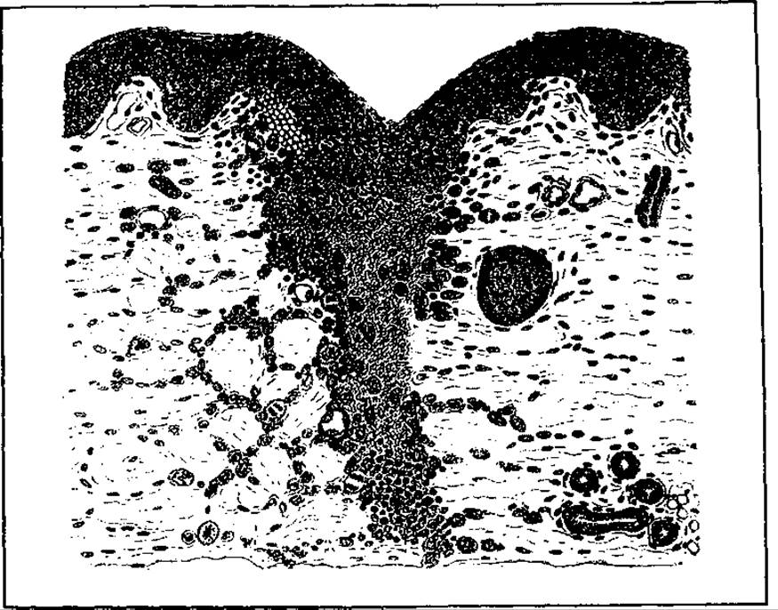
Fig. 54. Microscopic appearance of a wound healing by first intention
Healing by first intention occurs under the condition of tight apposition of even, minimally damaged wound edges, a minimal amount of necrotic tissue in the wound, the absence or negligible presence of microbes, and the absence of foreign bodies or blood clots (hematomas), provided active bleeding has ceased. However, the decisive factor is the tight apposition of the wound edges. Without this—that is, in the presence of wound gaping—it cannot heal by first intention. Surgical and many incised wounds typically heal by first intention. General systemic conditions are also of great significance for achieving healing by first intention. If a patient's general condition is impaired (diabetes mellitus, hypovitaminosis, anemia and hypoproteinemia, immune system disorders, impaired innervation in the wound area, or infectious diseases), or in the presence of negative local factors such as microbial contamination or excessive tension from sutures, healing by first intention may fail. In special cases, wounds may fail to heal altogether, progressing to chronic ulcers.
All gaping wounds, regardless of their origin, heal by second intention. These are primarily wounds heavily contaminated with microflora and necrotic tissue, containing foreign bodies, hematomas, etc. Since these characteristics are typical mainly of lacerated-contused wounds or wounds resulting from significant tissue (skin) loss, Western literature classifies such wounds (termed excision and avulsion wounds) separately from incised wounds.
Table 3. Differences between Primary and secondary wound healing
|
Primary intention |
Secondary intention |
|
Minimal tissue loss, including necrosis-related loss Wounds are typically free of microflora (sterile or minimally contaminated) Healing occurs rapidly Exudate is absent or minimal and serous Concludes with virtually no macroscopically visible granulation tissue Wound contraction is minimal (achieved primarily via sutures) Re-epithelialization is minimal |
Significant amount of necrotic tissue and tissue loss Wounds are heavily contaminated with microflora or infected Healing occurs slowly Exudation is abundant; exudate is purulent Granulation tissue forms a broad scar Wound contraction is substantial, and the wound closes through contraction Re-epithelialization is extensive, and the wound cannot close without contraction |
In secondary wound healing, the processes differ quantitatively and qualitatively (Table 3). Inflammation is pronounced: the wound edges are edematous, the skin is hyperemic, exudation is abundant and purulent, and fibrin covers the entire wound. During the proliferation phase, granulations are clearly visible; sometimes they become excessive and protrude above the skin level—so-called "proud flesh" (commonly seen in the presence of foreign bodies). The epidermis surrounding the wound margin is clearly visible as a whitish-gray ring or disc. Following wound healing and the completion of epithelialization, a deforming scar forms, which may sometimes impair not only the local anatomy but also organ function (contracture).
In addition to primary and secondary intention, healing under a scab (crust) is also distinguished. This is a variant of secondary healing in superficial skin wounds with significant tissue defects, which avoids suppurating inflammation thanks to the formation of a biological dressing on the wound surface—a crust composed of blood clots, necrotic cells, and fibrin that rapidly dries into a solid mass. However, the wound defect is ultimately repaired through the proliferation of connective tissue and epithelium.
Healing by first intention is the most biologically and economically rational type of healing, as it results in the nearly complete restoration of tissue anatomy and normalization of organ function in the shortest possible time. Therefore, the primary goal of medical practice is to achieve primary wound healing whenever possible.
Clean wounds include those resulting from soft-tissue surgery and operations on organs unaffected by infection, which do not serve as a natural environment for microbial proliferation. Examples include the removal of peripheral soft-tissue tumors, hernia repair, strumectomy, splenectomy, and vascular, joint, and bone surgeries.
Clean-contaminated wounds are those slightly contaminated with endogenous microflora due to surgical Procedures performed on the upper gastrointestinal tract—the Esophagus, Stomach, and duodenum—for degenerative or neoplastic diseases.
Contaminated wounds are associated with interventions on the small and, especially, Large Intestine involving the opening of the lumen, as well as operations on thoracic and abdominal organs affected by inflammatory and infectious processes (appendicitis, cholecystitis, chronic lung abscesses and Bronchiectasis, apostematous nephritis, purulent infections of the fallopian tubes and Ovaries, etc.).
There are many reasons for the rising incidence of postoperative wound infections, which reaches 10% or more. They can be divided into objective factors—independent of surgeons—and iatrogenic factors related to deficiencies in the Organization of surgical care and surgical technique. Objective causes include: 1) a sharp increase among surgical patients of advanced and senile age with metabolic and circulatory disorders caused by vascular atherosclerosis, diabetes mellitus, etc.; 2) a dramatic increase in the duration of surgical interventions, reconstructive and cardiovascular surgeries, transplantation procedures, etc., which lead to severe air and wound contamination by exogenous pathogens and changes in Organ and tissue status (circulatory disorders, desiccation, etc.); 3) an increase in virulent microbial strains in surgical wards due to the widespread use of antibiotics; 4) a decline in immune defense caused by environmental degradation (radiological and chemical pollution, etc.); 5) an increase in cases of Polytrauma and acute surgical conditions (cholecystitis, pancreatitis, etc.), among others.
Among iatrogenic causes, gross violations of asepsis protocols in both hospital wards and operating suites take precedence.
The development of wound infection—whether pyogenic (to which humans have developed considerable tolerance), anaerobic, or specific—is facilitated by certain predisposing conditions.
COMPLICATIONS OF WOUNDS AND THE HEALING PROCESS
Wound complications are numerous and occur at various stages of healing. They can be local or systemic. Infectious complications are the most common. Scar- and dystrophy-related complications in the wound area are also frequent—hypertrophic and keloid scars, scar contractures limiting movement, scar hernias and diastasis resulting from muscle atrophy, trophic ulcers, and rarely, tumors. Infections typically manifest as local purulent processes, less frequently putrid (purulent and putrefactive inflammation), and much more rarely anaerobic, spore-forming (gas gangrene and Phlegmon), or specific (tetanus, diphtheria). The causative agents of the first two forms (purulent and putrefactive) are staphylococci, streptococci, Escherichia coli, Proteus, Pseudomonas aeruginosa, and non-spore-forming anaerobic clostridia like Bacteroides, among others.
Systemic wound infections primarily present as acute specific toxicoses (tetanus, rabies) and non-specific pyogenic infections—Sepsis (acute and chronic with wound cachexia).
Both local and generalized infectious wound complications (sepsis) can lead to external hemorrhage due to vascular erosion, internal bleeding (into the gastrointestinal tract and interstitial tissue hemorrhages), and thromboembolic disorders. Hemorrhagic and thromboembolic syndromes primarily arise from impaired Blood Coagulation caused by infection. Infectious complications typically occur in accidental wounds, but they are also frequently observed (in up to 10%) in surgical wounds—both clean (rarely) and contaminated ones.
For a local purulent infection to develop in the tissues of a healthy person, the bacterial load must be at least 106 per 1 g of tissue. In most cases of wound infection, however, microbial contamination is lower, and the development of infection is promoted by various predisposing factors or conditions. The primary local condition is impaired tissue Blood Circulation within the wound area, which is observed in many wounds and particularly in those with a large zone of damage (caused by blunt objects, etc.). Circulation in the wound is compromised due to both vascular injury, thrombosis, and compression from inflammatory edema. Circulatory disorders also occur in clean surgical wounds and may result either from traumatic surgical access (inadequate incision and excessive wound stretching with metal retractors, causing tissue damage and vascular thrombosis), or from tissue crushing with clamps that are applied too roughly over a large mass of tissue, or from very tight wound closure with densely placed sutures close to the wound edges. Animal experiments have demonstrated that in tightly closed wounds, an infection can develop with just 10-2 Bacteria per 1 g of tissue, compared to 10-6 under normal blood circulation. Severe circulatory impairment is also caused by suturing a wound under high tension and the overuse of electrosurgery for hemostasis. Other factors contributing to infection include general anemia, hypoproteinemia (reduced immunological defense of the tissues and the body as a whole, associated with protein compounds), hypovitaminosis (especially vitamin C deficiency), metabolic disorders (diabetes mellitus), immunodeficiency states, certain endocrine disorders (Cushing's syndrome and disease), and iatrogenic hypercorticism (during treatment for bronchial asthma and collagenoses). Alongside impaired wound circulation, a major role in the development of local (both purulent and anaerobic) infection is played by the presence of large amounts of necrotic tissue and closed, poorly ventilated pockets and channels that hinder oxygen access. Under such conditions, microbial associations of aerobes and anaerobes mutually reinforce each other; specifically, aerobes consume oxygen, making the environment even more anaerobic and thus promoting the development of anaerobic infection.
A high susceptibility to wound infection is observed in elderly individuals due to atherosclerosis and reduced mobility (ultimately leading to diminished tissue blood supply), as well as degenerative tissue changes and lowered overall resistance of the Organism.
Other things being equal, infectious wound complications develop more frequently in anatomical areas with poor tissue vascularization and higher microbial skin contamination, and conversely, less frequently in areas with better vascularization and lower contamination. For instance, wounds on the lower extremities suppurate much more often than wounds on the face, neck, head, and hands. Wounds penetrating deeply into the subcutaneous adipose tissue, which exhibits low resistance to infection, become complicated more frequently than superficial, cutaneous wounds. A narrow wound channel that impedes the drainage of blood and exudate also promotes infection. This explains the higher incidence of suppuration and Other types of infection in small puncture wounds compared to incised wounds.
Particularly negative roles are played by non-sterile suture material, which causes implantational infection, and a number of other factors. Along with breaches in asepsis and iatrogenic causes, careless tissue handling during surgery also contributes to the development of wound infection.
The development of local infection in a wound is closely linked to other local complications affecting both the wound and the resulting scar. Highly virulent infection may cause the dissolution of blood clots within vessels or (more rarely) of the unaffected vascular wall at the Base of the wound, leading to secondary erosive hemorrhage.
Deformed and hypertrophic scars are also frequent consequences of wound infection. Hypertrophic scars rise above the skin level; they are tense, red, painful, and itchy. They often form when wound edges are subjected to strong tension, mechanical irritation (numerous ligatures, clothing friction, etc.), and unfavorable wound locations, such as the sternal area (rigid base and tightly bound skin). Hypertrophic scars (Fig. 55) eventually regress, atrophy, and fade. Large scars covered by a single layer of epithelium are unstable, highly fragile, and prone to ulceration. This can potentially lead to Squamous Cell Carcinoma.

Fig. 55. Hypertrophic scar following an abdominal incision
Keloid scars must be distinguished from hypertrophic scars. Unlike the latter, keloids extend beyond the BOUNDARIES OF THE original injury into undamaged skin. The direct cause of both hypertrophic and keloid scars is the hyperproduction of collagen in the wound by fibroblasts.
Keloid scars (Fig. 56) are mainly associated with individual patient characteristics and genetically determined reactivity. Individuals with dark skin are particularly prone to them. Keloids occur more frequently in burn wounds.
Large, wide scars on the anterior abdominal wall, which typically result from wound and peritoneal infection (Peritonitis), become the sites of ventral hernia formation. Beyond infection, other factors significantly influence the healing process. Healing is hindered by tissue hypoxia within the wound, caused primarily by Impaired blood supply resulting either from excessive tension on the wound edges during closure and closely spaced sutures, or from wound edema and tight bandaging (the tourniquet effect of a dressing). Conversely, increasing the partial pressure of oxygen in the local environment promotes healing by enhancing collagen synthesis. In this regard, direct local oxygen delivery works better than Hyperbaric Oxygenation, which induces vasoconstriction. General and local radiation exposure of the wound delays healing, as do high doses of corticosteroid Hormones. Lidocaine, procaine (novocaine), and cyclopropane inhibit wound healing because they interfere with the conversion of proline to hydroxyproline. Vitamin C and zinc deficiencies sharply impair collagen formation and wound tensile strength (scar rupture resistance). In individuals who suffered from scurvy, old wounds have been known to reopen even years after they had healed.
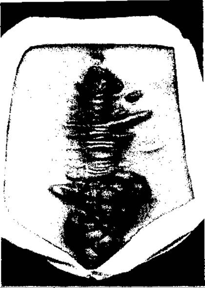
Fig. 56. Keloid scar following an abdominal incision
Animal experiments (guinea pigs, rats) conducted by R. Niedner (1995) established that among the antiseptics and antibiotics used topically to treat purulent wounds, many suppress granulation tissue development to varying degrees (exerting toxic effects on fibroblasts, DNA Synthesis, hydroxyproline production, etc.).
Among antibiotics, tetracycline has the most pronounced negative impact on granulation, followed to a lesser extent by Aminoglycosides (gentamicin) and chloramphenicol. Neomycin and, in particular, bacitracin practically do not inhibit granulation growth.
Among antiseptics, iodopyrone and silver nitrate inhibit granulation to a minor degree—by 18% and 25%, respectively. Chlorhexidine has the most detrimental effect on reparative processes in the wound: even at a concentration of 0.05%, it suppresses granulation growth by 57%, disrupting DNA and hydroxyproline synthesis. Furthermore, it is toxic.
Foreign bodies in the wound, including non-absorbable sutures (silk, linen, synthetic threads such as capron, nylon, dacron, etc.), frequently lead to delayed healing due to the formation of purulent fistulas caused by microbial contamination of the foreign materials (implantational infection). Suture contamination is facilitated by capillarity (or the wick effect), which is particularly characteristic of organic sutures (silk, cotton, and linen threads). Tissue reaction (aseptic infection—edema and granulation tissue proliferation) to these sutures is also quite pronounced. Synthetic threads, especially monofilaments, exhibit low capillarity, and tissue reaction to them is minimal. Animal-derived threads (catgut) are absorbed while eliciting a predominantly exudative tissue reaction. Direct subcutaneous and cutaneous placement of catgut favors the development of hypertrophic scars (collagen hyperproduction). Resorbable synthetic threads (made of propylene glycol) do not affect tissues in the same way as catgut. The properties of suture material must be carefully considered when treating wounds. To approximate subcutaneous tissues in microbially contaminated, particularly infected and purulent wounds, absorbable sutures that provoke minimal tissue reaction should be used.
Wound healing, being largely an enzymatic process, can be disrupted by the stimulation (rarely) or inhibition (mostly) of enzymes that control collagen synthesis, particularly the formation of cross-links between its fibers (D. Metzler, 1980). Such inhibitors include beta-cyanoalanine, found in sweet pea seeds (Lathyrus odoratus). Procollagen peptidase deficiency inhibits the formation of hydroxylysine, which is required for cross-linking collagen fibers, thereby suppressing collagen synthesis and delaying wound healing. The Inhibition of enzymatic collagen synthesis underlies Ehlers-Danlos syndrome and osteolathyrism, which manifest as spinal and joint deformations and aortic rupture. Both inadequate collagen formation and its hyperproduction require careful management during wound treatment. Suppressing this process is critically important, particularly when treating esophageal mucosal burns, in order to prevent luminal narrowing and stricture formation. Penicillamine is among the inhibitors of collagen synthesis (D. Metzler, 1980). The development of excessive granulation tissue in a wound—that is, an overabundance of collagen—leads to the formation of massive, wide, nearly avascular scars covered by a single layer of squamous epithelium. This epithelium is fragile and frequently undergoes necrosis under even minor trauma, forming poorly healing ulcers that sometimes fail to heal entirely or even undergo malignant transformation into a malignant tumor. This necessitates timely and active surgical intervention (placement of secondary early or late sutures with excision of granulation tissue) for wounds healing by secondary intention, preventing the formation of large collagen masses (extensive scars), as conservative measures (ultraviolet therapy, UHF therapy, Applications of cortisol, silver nitrate, and other agents) have low efficacy.
The core tenets of modern surgery are organ preservation and physiological operations. To achieve optimal physiological and aesthetic (cosmetic) surgical outcomes, the correct anatomical placement of surgical incisions and the proper debridement of accidental wounds are of paramount importance. Surgical incisions (and wound debridement incisions wherever possible) should run parallel to skin tension lines—that is, along or at an acute angle to the elastic skin elements and muscle fibers. These are the so-called normal or physiological incisions, which typically have a transverse or oblique-transverse direction relative to the longitudinal axis of the body. Whenever possible, these incisions should be placed within natural skin folds where they remain hidden (camouflage), or along hairline boundaries, etc.
Determining skin tension lines (Langer's lines) is facilitated by observing the direction of skin folds and wrinkles during facial expression, neck flexion, and trunk or limb movement. For this purpose, the skin and subcutaneous tissue are pinched between the fingers (the largest and most easily formed fold corresponds in its axis to the skin tension line of that particular area).
The objective of surgery in wound management is to avert or minimize the threat posed by the wound to the body. Above all, clinicians strive to alleviate pain, stop bleeding, prevent systemic disorders (such as shock) and potential subsequent infectious complications, and eliminate the consequences of the injury—namely, to achieve wound healing. Wound healing occurs due to the living organism's capacity to regenerate certain tissues, primarily connective tissue and epithelium, whereby either the original tissue structure is restored or (more commonly) tissue defects are filled with connective tissue "patches" that organically merge with the surrounding defect edges (repair).
Since we cannot yet artificially induce or direct regeneration, wound treatment consists of sanitation, complication Prevention, and the creation of optimal conditions for the body's intrinsic healing mechanisms to function.
The primary conditions for wound healing are ensuring normal blood circulation throughout the body and specifically within the wound area—meaning an adequate supply of blood and oxygen to the tissues—as well as metabolic normalization. After all, trauma is very frequently accompanied by significant disruptions in both general and local blood circulation and METABOLISM.
WOUND TREATMENT
The history of surgery is, to a certain extent, the history of wound management. Even today, a surgeon's competence is largely evaluated by their ability to treat a wound. Every historical period in surgery has been characterized by specific levels of knowledge, Methods, and approaches to wound care. However, until the 20th century, all these methods were conservative and bloodless, although individual physicians long before had expressed the view that surgical intervention was expedient. For instance, Y.O. Charukovsky, a military physician in the Russian army and a veteran of the Russo-Turkish War of 1828–1829, wrote in his book Voenno-pokhodnaya meditsina (1836–1837) that "every contused wound must be converted into an incised wound and treated for rapid union." What a vivid, content-rich, and concise Definition of the essence of primary wound debridement—an operation that became the cornerstone of fresh wound management almost a century later. It must be noted, however, that this brilliant premise could not be put into practice at the time because it lacked a theoretical foundation due to ignorance of the causes of wound suppuration. For centuries, conservative wound treatment remained entirely empirical. The remedies used for wound care varied widely—ranging from PLANTS AND THEIR components or processed products (plant leaves and juices, PLANT AND ANIMAL ashes), animal tissues and secretions (fat, blood, Bile, urine, etc.) to chemical substances (wine, alcohol, sulfur, iodine, copper sulfate, oils, carbolic acid, etc.) and the application of high heat (cauterizing the wound with a red-hot iron, pouring boiling water or boiling oil into it, etc.).
Only after the causes of wound putrefaction and suppuration were elucidated and the pathogens of wound infection were discovered (L. Pasteur, J. Lister, R. Koch, and others)—that is, from the second half of the 19th century onward—did conservative wound treatment acquire a scientific basis. Yet even then, the conservative method, which long remained the sole approach to wound treatment (effectively until World War I), failed to yield desired outcomes because it could not halt the proliferation of microflora and the development of infection within the wound. A major impetus for surgical intervention came from the experiments of P. Friedrich (1898), who established that after a wound is contaminated with microflora, the bacteria adapt to the wound environment for up to 6 hours without multiplying. This led to the Conclusion that infection could be prevented if the microorganism-colonized wound edges were excised within the first hours (6 hours) of occurrence, that is, before microbial proliferation begins. Advances in antisepsis and asepsis, progress in microbiology, and the empirical experience of isolated successful cases of wound treatment via incision provided the scientific foundation for the Introduction of surgical management of fresh gunshot wounds by French surgeons (Gaudier, Lamaitre, and others) as well as certain Russian army surgeons (M.M. Petrov, V.A. Opel, O.P. Krymov) during World War I. In the French army, this was facilitated by a dense railway network and high concentration of populated areas (cities), which allowed wounded soldiers to be rapidly transported to military hospitals, operated on, and treated without evacuation. This Procedure was named debridement (a term proposed earlier by Desault, literally meaning unbridling—that is, opening or incision of a wound, sometimes with partial removal of crushed tissues). It still bore little resemblance to modern primary surgical wound debridement, but when supplemented by the method of continuous wound irrigation with Carrel-Dakin solution (chlorinated water), it produced significantly better outcomes than all previous methods of wound care. Over two decades, surgical wound management was refined and expanded. By the eve of World War II, its principles and techniques were largely established. It was determined that without removing necrotic, dead tissues from the wound, it is impossible to sanitize it from microflora and prevent the development of infection. Furthermore, achieving the removal of dead tissues through mere incision is impossible; the incision must be supplemented by cutting the edges and base of the wound. If complete removal of damaged tissues is impossible, at least a partial excision must be performed. Although the introduction of surgical wound management sharply increased treatment effectiveness, it did not mean other therapeutic methods could be abandoned. On the contrary, experience showed that complete excision of damaged tissues in a wound is impossible in most cases, meaning that surgical intervention alone cannot always create conditions to prevent infection. All of this necessitates The Use of non-Surgical methods of wound treatment alongside surgery, aimed specifically at suppressing microflora and creating conditions that preclude its development.
Today, wound treatment is comprehensive and differentiated depending on the nature of the wound (operative, aseptic, or accidental; fresh, infected, or purulent), its origin, and the individual characteristics of the patient's body. Comprehensive wound management incorporates both local and general therapeutic measures. Tactically, wound treatment is carried out using local and general measures simultaneously, or local measures against the Background of general therapy.
Management of Fresh Wounds
The main goal in treating fresh wounds is to prevent infection and create conditions for closure and primary healing. The most effective way to achieve this is through timely surgical wound debridement.
This is a complex procedure that evolved from the simpler operation of wound incision (debridement). Ensuring a free outflow of inflammatory exudate and necrotic particles of damaged tissue from the wound, along with access of oxygen to its depths, helps reduce the incidence of anaerobic infections (tetanus, gas gangrene) as well as purulent (banal) infections, or diminishes their scale and threat to the body.
The vast majority of accidental wounds require surgical management. However, even during World War II, 20% of gunshot wounds did not require surgical debridement (S.S. Girgolav). Surgical debridement is not required for: a) small, superficial incised and contused wounds without gaping edges; b) superficial stab wounds of soft tissues that do not penetrate body cavities and involve no damage to vital structures (vascular and neural trunks); c) through-and-through stab and gunshot wounds of soft tissues and even the thoracic cavity without injury to vital structures, hemorrhage, interstitial hematoma, or hemothorax; d) multiple small, superficial shrapnel wounds and certain other wounds with similar characteristics.
Wounds that do not undergo primary surgical debridement are covered with a dressing (primarily an adhesive bandage) containing antiseptic-soaked dressing material after the surrounding skin is cleaned. The dressings are changed daily During the first few days.
The procedure of primary surgical debridement, aimed at preventing infection and achieving primary healing, is based on microbiological research initiated at the end of the 19th century (1898) by P. Friedrich. He established that pathogenic microflora entering a wound remains concentrated on its surface for up to 6 hours without multiplying. Only after this period do microbes generally begin to multiply and penetrate the tissues. Based on these studies, surgeons concluded that microbial proliferation in a wound can be prevented by excising its edges and base within healthy tissue limits during the first 6 hours after injury.
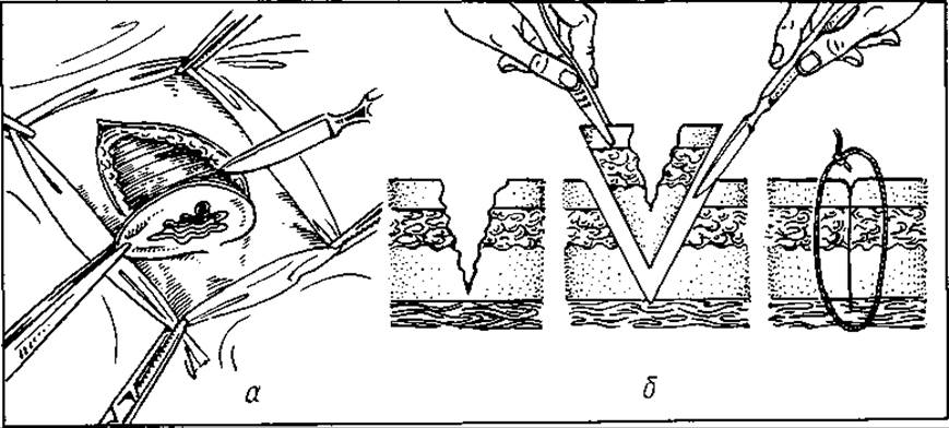
Fig. 57. Surgical wound management (a, b). Excision of the edges and base; wound closure by suture
As soon became apparent, complete excision of the wound edges and base is often impossible; however, removing damaged tissues along with microbes drastically reduces the microbial load in the wound, halts their proliferation, and thus generally creates conditions for primary wound healing. The introduction of highly effective antibiotics into clinical practice for the prophylaxis of wound infection, particularly purulent infection, has allowed this procedure to be performed at later stages—even 24 hours or more later (in the absence of signs of wound infection). Such surgical wound debridement is referred to as delayed.
Modern primary surgical wound debridement consists of the following steps:
1) disinfection of the operative field within a radius of up to 10 cm around the wound;
2) anesthesia (general or local, depending on the wound and the patient's condition);
3) incision of the wound along its long axis down to the base;
4) revision of the wound cavity by inspection (the wound is opened using spiked retractors);
5) removal of foreign bodies from the wound (metal fragments, wood, clothing, stones, soil, etc.);
6) excision of the damaged wound edges and base within healthy tissues using a second scalpel, stepping back 0.5–1.5 cm from the margins (the exact distance depends on the wound localization, i.e., the nature of the tissues—whether there are vital vessels, nerves, or organs in the area; Fig. 57);
7) if complete removal of the wound base (as well as its edges) is impossible, only the most heavily damaged tissues are excised to the anatomically feasible extent;
8) achievement of hemostasis in the wound after the surgeon changes gloves and instruments, by ligating vessels with threads (preferably absorbable ones) or through electrocoagulation;
9) irrigation of the wound with chemical antiseptics (solutions of furacilin, chlorhexidine, iodopyrone, etc.);
10) insertion of a drain into the wound—a rubber strip or a PVC or silicone tube (depending on the nature of the wound and the degree of microbial contamination);
11) closure of the wound with sutures after thorough removal of damaged tissues. Exceptions are frequently made for wounds located in the gluteal and thigh regions. If there are doubts regarding the radicality of damaged tissue removal or in cases of heavy microbial contamination, the wound may be left open for 2–4 days and closed subsequently with delayed primary sutures.
Large lacerated, contused, and crushed wounds cannot, in most cases, be radically cleared of damaged tissue during surgical debridement nor closed with primary sutures. Therefore, they are usually widely incised, the most traumatized edges are excised until bleeding appears from the newly created surface, and they are then left unclosed for 2–4 days under a protective dressing. Dressings and wound revisions are performed every 24 hours. If the wound shows no signs of infection (purulent inflammation), it is closed with sutures. If signs of progressive necrosis and infection appear, the wound undergoes repeat surgical debridement.
Primary delayed sutures are particularly often used when treating wounds on the legs. Due to heavy contamination with pathogenic microflora, including anaerobic bacteria, as well as a poorer blood supply compared to other body areas, these wounds are more frequently complicated by the development of various types of infection.
Primary surgical debridement of fresh wounds complicated by bone fractures, damage to major blood vessels and nerve trunks, and extensive skin loss serves simultaneously as a surgical procedure (primary wound debridement) and a reconstructive intervention: the fracture is reduced and fixed using one of the established methods (primarily osteosynthesis with Ilizarov apparatus, etc.), blood Vessels and nerves are sutured, and skin defects are closed via autoplasty, local transposition of skin-subcutaneous flaps, or split-thickness skin grafts. Such wounds are typically drained with active inflow-outflow irrigation of their cavities using antiseptics. In cases of degloving INJURIES OF THE lower leg with a triangular skin defect or avulsion of the distal Phalanges of the fingers, skin defect closure cannot be successfully achieved by simply stitching back the cyanotic, pedicle-suspended flap—the scalp—as it will undergo necrosis. Therefore, such defects must be closed either with a free split-thickness skin graft or via plastic surgery, such as suturing the injured finger to a surgically prepared skin flap on the torso. Reconstructive interventions involving skin grafting are usually followed by immobilization of the limb using a plaster cast or another rigid dressing.
Chemical antiseptics are used as part of comprehensive wound management both for a single irrigation of the wound following its debridement and for continuous irrigation over several days post-debridement (for example, in wounds complicated by fractures with bone fragment fixation via osteosynthesis). If antiseptics are unavailable, the wound must be rinsed upon completion of debridement with a sodium chloride solution (0.9%) or novocaine.
During surgical wound debridement, the wound may also be irradiated with UV light or a defocused laser, although this is more commonly employed in the treatment of infected or purulent wounds.
Due to the availability of highly effective antimicrobial agents, primarily antibiotics, the indications for primary surgical wound debridement have expanded significantly: it is performed under the cover of prophylactic antibiotic administration to the injured person, even 24 hours or more after injury (in the absence of infection signs or with minimal manifestations, the wound is closed with primary or primary delayed sutures).
In cases of incomplete (non-radical) primary surgical debridement, a repeat debridement of the fresh wound may be performed. Typically, for wounds that are radically debrided within the first 6–12 hours following trauma (early surgical debridement), prophylactic antibiotic administration to prevent infection is unnecessary. In such instances, local chemical and physical antiseptics are employed (wound irrigation after debridement, proper drainage, and application of an aseptic dressing).
Following surgical debridement, a fresh wound is closed—much like an aseptic postoperative wound—by placing sutures and closing it. In recent years, opinions have been expressed regarding the feasibility of closing wounds (bringing their edges into contact) using plastic materials (such as adhesive tape), which, unlike sutures, do not impair local blood circulation. However, due to technical complexities in execution (The Need for careful wound monitoring, lower reliability in immobilizing wound edges, and potential misalignment), this method has not yet gained widespread clinical practice.
Clean (aseptic) postoperative wounds are generally managed by closure with sutures that completely eliminate the dead space between the edges without drainage, and less frequently with short-term (24-hour) drainage using a rubber strip.
Clean-contaminated postoperative wounds, as well as simple, uncomplicated fresh incised accidental wounds (the latter often after extension and exploration), are closed with sutures after rinsing the cavity with antiseptics (furatsilin, iodopiron, etc.) and placing a rubber strip drain for 24–48 hours.
Contaminated surgical wounds are treated either similarly to clean-contaminated ones or left unclosed (subcutaneous tissue and skin layers) for 2–3 days. If no infection develops during this period, they are closed with primary delayed sutures.
Both surgical and accidental fresh wounds, following debridement and suture closure, are managed primarily under gauze dressings (adherent pads), and rarely under rigid polymer films (adhesive compositions). Suture-closed wounds with well-adapted edges become impervious to microbes within just 2 hours due to fibrin sealing.
Among local treatment measures for fresh wounds (both following surgical debridement and without it, when such debridement is not required), physiotherapeutic and chemical methods are widely employed to reduce traumatic inflammatory tissue edema and pain, suppress microflora proliferation, stimulate the wound regeneration process, and accelerate healing. These include UHF therapy, prostaglandin inhibitors (acetylsalicylic acid), defocused laser beams, alcohol compresses (particularly combined with dimexide—dimethyl sulfoxide), and Vitamins, especially B-complex and C, among others.
As noted previously, wound treatment is carried out through a combination of local and systemic measures, since a wound, alongside localized tissue damage, is accompanied by general and systemic disorders whose severity varies widely depending on the wound size, its origin, microbial contamination, patient constitution, and many other factors.
Wound treatment primarily involves analgesics (opiates—omnopon and promedol; non-opioid analgesics—analgin, ibuprofen, tylenol), sedatives (bromides, motherwort and valerian tinctures, tranquilizers—diazepam, meprobamate, etc.), and hypnotics (barbiturates—barbamil, glutethimide, etc.). When necessary, solutions for various purposes are administered: plasma-substituting or hemodynamic solutions (sodium chloride, sodium lactate, rheopolyglucin, etc.); energy-supplying and metabolic-correcting solutions (5% and 10% glucose solutions, Amino Acids, etc.). In cases of significant blood loss (exceeding 20% of circulating blood volume), i.e., complicated by traumatic and hemorrhagic shock, blood transfusions (250 to 500 ml or even more) are administered, along with protein preparations (albumin, protein fractions) and rheological/detoxification solutions (polyvinylpyrrolidone, rheopolyglucin, hemodez, etc.), alongside large doses of hydrocortisone (100–300 mg or more).
In the event of a forced delay in surgical wound debridement or non-radical debridement involving a substantial zone of tissue damage and microbial contamination, systemic antibiotic therapy is initiated (broad-spectrum drugs are prescribed prior to identifying the nature of the microflora and its antibiotic sensitivities). It is particularly crucial to correct protein and Carbohydrate Metabolism disorders—hypoproteinemia and diabetes mellitus-related changes (administration of proteins, amino acids, and Insulin)—as well as vitamin deficiencies, especially vitamins C, B-complex, and A, the lack of which commonly accompanies protein and carbohydrate metabolism disturbances.
For patients with impaired cardiovascular, respiratory, or urinary functions, appropriate medications are administered (cardiac Glycosides; blood pressure regulators; pulmonary antiseptics, Diuretics, bronchodilators, and mucolytic agents; Oxygen therapy, etc.).
Immunological disorders are corrected using immunomodulators, specifically immunostimulants (tactivin, thymalin, thymoptin, vilozen, levamisole, etc.).
Depending on clinical indications, systemic agents are also used (anticoagulants and coagulants, anabolic drugs like retabolil, etc.), along with nonspecific biostimulants (solcoseryl solution, splenine, methyluracil) and desensitizing drugs (taвеgyl, suprastin, diazolin, diphenhydramine, calcium gluconate, etc.).
A separate mention should be made of the management of so-called combined open injuries. Wounds in casualties who have suffered external radioactive irradiation alongside mechanical trauma (combined injury) require a special effort to be timely debrided and healed by primary intention, as radiation sickness may develop over time (manifesting days or even weeks post-irradiation, depending on the dose). This is necessitated by the fact that radiation sickness severely suppresses Immunity, hemopoiesis, and regenerative-repair processes, thereby promoting septic wound complications. Technically, surgical debridement of a wound in victims with external irradiation—or even those with radioactive surface contamination—differs little from standard debridement (except for operating room safety protocols), yet it must be even more radical regarding the removal of traumatized tissue within the wound. Mere radioactive contamination of the wound is insufficient to cause radiation sickness, although it can exert adverse local effects on tissues in the wound area.
Therefore, alongside meticulous surgical debridement, a radioactively contaminated wound must be thoroughly rinsed with antiseptics, particularly agents like hydrogen peroxide, to remove residual radioactive materials. In patients with external irradiation, surgical wound debridement must be combined with appropriate systemic measures aimed at detoxifying the body from metabolic waste and cellular debris, and boosting host defense mechanisms (immunotherapy, transfusions of detoxifying protein and carbohydrate solutions, occasionally blood components; vitamin therapy, sorption therapy, antibiotic therapy, etc.).
Treatment of Wounds Complicated by Purulent and Putrefactive infection
Purulent infection in a wound typically develops within the first 3–5 days following injury.
Among wounds complicated by purulent infection, a distinction is made between infected and purulent wounds. An infected wound exhibits clinical signs of infectious inflammation of a serous character: mild edema and erythema of the wound edges, along with serous exudate discharge.
The principal manifestation of a purulent wound, alongside other pronounced signs of inflammation, is the presence of purulent exudate.
This classification holds practical significance, particularly for treatment tactics and predicting the healing trajectory. Depending on its origin, size, and Location, an infected wound can be managed either actively via surgery, like a neglected accidental wound (i.e., treated 24 hours post-injury), or immediately managed using Conservative methods.
Small, previously untreated infected wounds with a limited area of surrounding tissue damage are actively treated via primary delayed debridement.
Surgical intervention for such a wound involves comprehensive debridement, primary closure with cavity drainage, and the postoperative use of Physical and Chemical antiseptic methods combined with systemic antibiotic therapy. Alternatively, the wound may be left open after debridement, followed by the application of primary delayed sutures.
An infected postoperative wound requires initial conservative therapy: UHF therapy or defocused laser irradiation of the wound; alcohol compresses (50–55%); dressings with sorbents (granular or textile), chemical antiseptics, or dimethyl sulfoxide (DMSO); systemic antibiotic therapy; and occasionally, cavity revision using a thin probe (to facilitate exudate drainage). If such therapy yields no effect within 2 days and inflammation progresses, surgical intervention is indicated—opening the wound (partially or fully, depending on the severity of inflammation and general systemic manifestations) to ensure free and complete outflow of exudate, alongside all local and systemic measures to suppress infection (microflora) and combat intoxication.
Compared to fresh (and simply infected) wounds, purulent wounds are more difficult to treat. This is due to the deep-seated action of microbes and their toxins, systemic wound intoxication, and a decrease in the body's resistance to aggression. Therefore, the treatment of such wounds primarily involves the elimination of purulent intoxication and its source (the wound infection) through systemic and local measures, and only subsequently achieving wound healing, closure, and the restoration of normal organ function.
Treating such wounds combines conservative and surgical measures of both general and local character. All local measures are performed against the backdrop of systemic management. Among the latter, the most crucial are: 1) systemic detoxification by binding and eliminating toxic substances—microbial toxins, as well as products of tissue and microbial breakdown; 2) metabolic correction and systematic antimicrobial (antibiotic) therapy.
Specific measures to achieve these goals primarily include infusion therapy: intravenous administration of isotonic sodium chloride solution (0.9%) or sodium lactate, other electrolyte solutions, and a 5% glucose solution (up to 2 liters per day depending on the degree of intoxication); administration of polyvinylpyrrolidone, rheopolyglucukin, or their analogues (400 ml per day); and occasionally the administration of amino acids, albumin, protein, or plasma, which, alongside other benefits (detoxification and stimulation), compensate for protein losses. Additional measures include the administration of insulin (1 IU per 4 g of glucose) and vitamins alongside the glucose solution, particularly ascorbic acid, vitamin A, and B-complex vitamins; maintaining a caloric and nutritionally balanced diet; and intramuscular, intravenous (sometimes even intra-arterial), endolymphatic, or oral administration of antibiotics tailored to the nature of the microflora and its antibiotic sensitivity. Among the latter, the most effective agents to date include Semisynthetic Penicillins (ampicillin, amoxicillin, ampiox, etc.), second- to sixth-generation Cephalosporins, aminoglycosides (gentamicin), thienam, and—in cases of anaerobic non-clostridial infection—metronidazole, clindamycin, ciprofloxacin, etc.
Local wound treatment is performed primarily using a closed method under a dressing. A wound dressing must meet the following requirements:
1) remove exudate and toxic products without dehydrating the wound itself;
2) protect the wound from secondary infection;
3) maintain adequate moisture within the wound to ensure normal Cell Division and migration, and keep the temperature at body level (temperature reduction inhibits phagocytosis);
4) permit gas permeability, which is important for superficial wounds. In deep wounds, minor acidosis and hypoxia stimulate granulation tissue development;
5) be free of toxic products;
6) be removable without damaging granulations and epithelium.
Naturally, all currently available dressings and materials do not fulfill all of these requirements. However, the most physiological among them are gauze dressings (bandages and adhesive pads).
Local measures for treating purulent wounds during the first (inflammatory) stage are aimed at: a) reducing wound intoxication through surgical intervention (opening the wound, removing purulent exudate and necrotic tissues) and establishing free outflow of exudate and microbes; b) reducing edema in the wound (and surrounding tissues); c) creating a hyperosmotic environment in the wound during The first phase of healing; d) reducing the microbial load in the wound (using bactericidal and bacteriostatic agents); accelerating the clearance of necrotic tissue elements; e) promoting phagocytosis and regeneration.
In the second phase, the goal is the earliest possible wound closure with maximum anatomical and physiological restoration of tissues within the defect area.
These objectives in the first phase of wound healing are achieved by: a) adequate opening of the wound cavity and its pockets, removal of necrotic tissues, rinsing with antiseptic solutions, and drainage; b) applying dressings impregnated with osmotically active and antimicrobial antiseptic solutions (1:5000 furacilin solution, 0.1% furagin, 0.5% iodopiron, etc.) and utilizing antiseptic baths, particularly oxidizing agents (potassium permanganate in a 1:1000–1:500 ratio, 1% hydrogen peroxide solution) for purulent, especially putrid, infections in wounds localized on the limbs; as well as sorbents (granular—dextranomer, and textile). Dressings with ointment compositions are also widely used (B.M. Datsenko et al., 1995): hydrophilic-base antibiotics and antiseptics (levosin, levomekol, dioxizol, etc.). They target microflora in the wound and adjacent tissues (reducing wound edema and pain). Applications of proteolytic enzymes (Chymotrypsin, Papain, etc.) are used to accelerate necrolysis in the wound; metabolic and wound-healing stimulants (solcoseryl gel, etc.) stimulate phagocytosis (when suppressed) via leukocyte mass (neutrophil) administration. If local antiseptics (including topical antibiotics) prove ineffective within 3 days, their continued use is irrational.
To suppress wound microflora and stimulate reparative processes, wound irradiation with UV rays, defocused laser light, and ultrasound (ultrasonic cavitation) is also employed, alongside open-air wound management in a sterile isolation or hyperbaric oxygen chamber—especially in cases complicated by anaerobic infection, whether clostridial or non-spore-forming putrid infections.
In such an environment, wound pain and tissue edema rapidly disappear, the wound is cleared of microorganisms more efficiently, and the processes of tissue cleansing, connective tissue proliferation, and vascular endothelial regeneration are accelerated. Due to the absence of a dressing and its tourniquet effect (vascular compression), Blood Circulation in the wound area is significantly better than under conventional dressings, which inherently accelerates healing. Both dry warm air and elevated oxygen concentrations contribute to this process.
Over the past two decades, the active surgical method has gained widespread acceptance in the treatment of purulent wounds and suppurative inflammatory processes, which are conventionally managed by incision (conversion into an open purulent wound). Its essence lies in the complete excision of the purulent wound or suppurative focus within healthy tissue boundaries, the insertion of drains into the wound cavity, and wound closure followed by continuous through-and-through irrigation of the cavity with antiseptic solutions; or alternatively, surgical debridement involving the removal of necrotic tissues, infiltration of adjacent tissues with antibiotics, placement of tubular drains, wound closure, and subsequent through-and-through irrigation or active drainage (A.A. Fedorovsky, 1962; M.M. Kanshin, 1981, etc.). This method has proven particularly effective in treating various types of localized purulent infections, notably purulent mastitis (M.P. Cherenko et al., 1985).
In the second phase of the wound process—the regenerative-reparative phase—local measures are aimed at accelerating the development of granulation tissue, protecting it from trauma and secondary infection, achieving the earliest possible wound closure, and minimizing the NEGATIVE IMPACT OF the future scar on organ function. For this purpose, dressings with oil-balsamic (hydrophilic-base) preparations are used, containing antiseptics, repair stimulants, and agents that absorb excess wound moisture. Such preparations include streptonateol, methyldioxyline, levosin, levomekol, synthomycin emulsion, solcoseryl ointment, and foam preparations such as hypozol and suliodazosol. They protect granulations from infection and mechanical damage.
Dressings are changed infrequently—every 1–2 days. Once the wound surface (edges and base) is covered with granulations (even in the presence of isolated fibrin deposits), it is closed with early secondary sutures, or the edges are approximated with adhesive strips. Such sutures are typically applied on days 7 to 14. During this timeframe, wound edges are easily approximated, and the granulation tissue layer is thin, requiring no excision and preventing the formation of coarse scars. If the wound edges are fixed to underlying tissues and inflexible (which occurs when early secondary sutures are omitted for various reasons), they must be mobilized surgically by excising the scar-granulation tissue, followed by the placement of late secondary sutures, usually applied after 3–4 weeks.
Today, synthetic dressings capable of creating an optimal microenvironment for wound healing are increasingly recommended. These include polyurethane dressings, xerogels, hydrogels, hydrocolloids, foam generators, etc. They are primarily used for superficial wounds, particularly burns, on anatomically complex body regions, the face, etc.
In the third stage—scar transformation and epithelialization—physical methods are recommended: UHF therapy; ultraviolet irradiation (UVI); micro-resonant therapy; gentle self-massage of tissues surrounding the scar; ointment dressings with low concentrations of corticosteroids—prednisolone or hydrocortisone (especially when the scar tends to hypertrophy); local warm baths with potassium permanganate, and less frequently, paraffin or ozokerite applications, etc.
Deficits in immunological and regenerative processes frequently lead to delayed wound healing, slow and inadequate development of granulation tissue and epithelialization, secondary wound infection, necrosis of granulations, erosive hemorrhages, wound dehiscence, surface ulceration, and even sepsis. Excessive stimulation of granulation development leads to hypertrophic and keloid scars. Therefore, wound treatment requires dynamic monitoring of the healing process. This is accomplished through Traditional Methods—such as microbiological and cytologic examination using the surface impression (granulation) smear method of M.P. Pokrovskaya and M.S. Makarov as modified by M.F. Kamaev—as well as more modern techniques: ultrasound (sonography), epiluminescence Microscopy (capillaroscopy), measurement of interstitial (transcutaneous) oxygen tension, thermography, computed tomography, Magnetic Resonance imaging, etc.
The information obtained from clinical evaluation and the aforementioned auxiliary methods for monitoring wound healing forms the basis for applying corrective treatment measures, immune system stimulants, general tonics, antimicrobial agents, etc.
Hypertrophic and keloid scars, as a manifestation of excessive connective tissue proliferation and collagen production, require correction. Hypertrophic scars (which do not spread or spread very little beyond the borders of the scar, are red and itchy) are treated by applying a tight silicone film-bandage in combination with topical (ointment) or intralesional administration of hydrocortisone. Keloid scars, which extend beyond the boundaries of the scar with branching projections, are treated using cryosurgical methods and other modalities.
INSECT AND SNAKE BITES
Insect and snake bites, as open microtraumas, can be conditionally classified as poisoned wounds, although their harmful effect on the victim's body is primarily due to local and systemic reactions to toxins. Complications directly resulting from the wound itself are possible but rare.
Among the numerous insect bites, the most dangerous are those of bees, wasps, bumblebees, black widow spiders (karakurt), tarantulas, and scorpions. Insect bites are accompanied by varying intensities of both local and systemic disorders.
Humans most frequently suffer from bee and wasp stings. Single stings usually do not lead to severe disorders, although fatal cases have been described even after a single insect sting due to the development of anaphylactic shock. A much greater threat is posed by multiple stings—a swarm attack. The stings of 500 bees are considered lethal to humans. A bee or wasp sting is usually accompanied by acute pain and local tissue edema around the sting site, and less frequently by widespread edema (when stung in the facial or scrotal area). Sometimes the patient experiences a systemic reaction in the form of a headache. Bee and wasp stings in the facial area (Lips, Nose) and the oral mucosa are particularly dangerous due to the potential development of laryngeal edema (vocal cords) with symptoms of asphyxia (suffocation). The action of bee and wasp venom is mainly due to biogenic amines—histamine and serotonin.
Occasionally, bee and wasp stings are accompanied by the development of anaphylactic shock with a sharp drop in blood pressure, tachycardia, impaired tissue blood perfusion, the appearance of a body rash and itching, and laryngeal edema.
A bumblebee sting causes more intense pain and local edema, and systemic manifestations (headache, sometimes fever) are observed more frequently.
Bites of the karakurt spider, scorpion, and tarantula, although less frequent, have a more severe clinical course. In Ukraine, scorpions are found only in Crimea. The venom of these insects has a neurotoxic effect. Their bites are accompanied by sharp, burning pain and widespread edema of adjacent tissues, especially in the case of a karakurt bite. Vomiting, respiratory distress, convulsions and trismus (in scorpion stings), hypothermia (in karakurt stings), etc., are observed.
Treatment for a bee sting involves removing the bee stinger with a sterile needle (wasps do not leave a stinger). A bandage soaked in a 10% ammonia solution or 70% ethanol is applied to the affected area. Rest is ensured, and antihistamines and hyposensitizing drugs (calcium gluconate, diphenhydramine, suprastin, diazoline, etc.) are administered. In the event of anaphylactic shock, anti-shock therapy is performed—administering electrolyte solutions, anti-shock solutions (rheopolyglucin, polyvinylpyrrolidone), corticosteroids, analgesics, bronchodilators, symptomatic drugs, etc. In case of suffocation, intubation or tracheostomy (conicotomy) is performed.
Cases of poisonous snake bites are observed much less frequently than insect bites. However, they are far more dangerous and sometimes fatal. Common vipers and steppe vipers are found in the territory of Ukraine. Snake venom is characterized by neuroparalytic (venom of the blunt-nosed viper, cobra), systemically toxic, and thrombohemolytic (viper venom, etc.) action. Venomous snakes have tubular Teeth on the upper jaw or palate (in the latter case, they are located far from the Oral Cavity entrance, but are capable of forward rotation during a bite). This is an ideal tool for deep tissue penetration and venom injection. The bite of these snakes is accompanied by sharp pain and the rapid development of edema, hyperemia, hemorrhage, and tissue destruction in the traumatized area. If a large amount of venom enters the tissues, edema and massive necrosis with blisters rapidly develop, along with severe systemic disorders (sharp weakness, headache, depression or euphoria, palpitations, decreased blood pressure, nausea, vomiting, stupor, suffocation, salivation; and in case of a cobra bite, paralysis of the trunk and Neck Muscles).
Blood composition is disrupted: erythrocyte lysis, decreased hematocrit and platelet levels, thromboembolic and hemorrhagic phenomena (Hematuria, hemoptysis, melena, epistaxis). Increased resistance of venules due to the thromboembolic process leads to pulmonary congestion and edema with severe respiratory impairment. Hemolysis is accompanied by the development of Glomerulonephritis, renal vascular endarteritis, and Necrosis of the renal cortex. Death occurs as a result of shock or renal failure.
In the event of a bite, a tourniquet should first be applied to the limb above the affected area (even a handkerchief or bandage can be used). The wound should be slightly enlarged with a disinfected blade, after which its contents should be sucked out.
A bandage soaked in 70% ethanol, 10% ammonia solution, or 1% potassium permanganate solution is applied to the wound. If the victim can be hospitalized quickly, the wound is opened and the venom is sucked out under aseptic conditions in an operating room or dressing station. Intensive infusion anti-shock and detoxification therapy is mandatory (transfusion of crystalloid and colloid solutions, rheopolyglucin, hemodez, plasma, fibrinogen, analgesics, corticosteroids, antispasmodics and bronchodilators, heparin, etc.).
Antibiotics are prescribed for infection prophylaxis. The wound and necrotic areas are treated according to general rules. In case of a cobra or blunt-nosed viper bite, the mainstay of treatment is the administration of specific antivenoms—"anticobra" or "antiviper" serum. Serum therapy is also administered in severe cases of patients bitten by other snake species.
The outcomes of bites with timely assistance and treatment are mostly favorable. However, in cases of delayed assistance following cobra and blunt-nosed viper bites, and high patient sensitivity to the toxin, death may occur.
Prevention of snake bites consists of wearing shoes in areas where snakes are likely to be present.
Last update: 08/08/2026
Editorial and Educational Adaptation: This material has been compiled based on the primary/original source text. The project team performed an editorial review, corrected technical inaccuracies, structured sections, and adapted the content for an educational format.
What was processed:
- elimination of formatting defects (OCR errors, structural breaks, corrupted characters);
- editorial organization of content;
- standardization of terminology in accordance with academic sources;
- verification of factual statements against the original source text.
All mentions of the author, publication year, and origin of the primary text have been preserved in accordance with the source.