Antibiotics (Properties, Applications, Interactions) - M.P. Cherenko 1999
Closed injuries of the skull, chest, abdominal cavity, and retroperitoneal space
CLOSED HEAD INJURIES
Closed traumatic Brain injury refers to damage occurring without a breach in the integrity of the scalp or Skull fractures. Mechanical injuries to the brain may manifest as a concussion, contusion, compression, or various combinations thereof. Head trauma is the most frequent cause of mortality.
The Pathogenesis of traumatic brain injury involves more than just the direct damage inflicted on the brain by the traumatic agent. Mechanical deformation of the brain also plays a significant role, wherein the brain impacts the bony prominences of the inner skull surface As a result of contre-coup.
The degree of brain damage depends on the plane, direction, speed, and magnitude of the applied force. Since the brain is suspended within the cranial cavity and can move back and forth—while its lateral mobility is restricted—impacts to the frontal and occipital Regions of the head are the most dangerous, as they cause the greatest Displacement of the brain.
An important role in the pathogenesis of traumatic brain injury is played by neurodynamic disorders in the Central Nervous system, as they trigger vascular, CEREBROSPINAL FLUID dynamics, and endocrine-humoral disturbances. Cerebral Blood Vessels initially react with vasospasm, followed by vessel wall dilation and venous congestion. Cerebrospinal fluid pressure either increases or decreases, and the permeability of the blood-brain barrier becomes altered. Hormonal balance, Water-electrolyte METABOLISM, and Blood Circulation are disrupted, leading to cerebral Hypoxia accompanied by signs of brain tissue edema.
The severity of these manifestations depends on the type of trauma—whether it is a concussion, contusion, or compression.
Cerebral concussion (commotio cerebri). In this type of trauma, macroscopic organic changes are absent in the brain tissue; there is only a disruption of molecular bonds within the brain Cells. The Functions of autonomic and vasomotor subcortical centers, as well as BLOOD AND Lymph circulation, are impaired. Occasionally, punctate hemorrhages may be detected in the brain. In some cases, a concussion can be complicated by cerebral edema.
The clinical picture of a concussion is characterized by brief loss of consciousness, retrograde amnesia (memory loss for events preceding the trauma), bradycardia, nausea, vomiting, and pain upon eye movement. There are no signs indicating local brain damage or meningeal symptoms. Once patients regain consciousness, they complain of headache, tinnitus, and insomnia.
In cases of concussion, prescribed management includes bed rest for 7–10 days, cold compresses to the head, sedatives, analgesics, antihistamines, and cardiac medications. Intravenous administration of 20 ml of a 40% glucose solution or 20–40 ml of a 10% sodium chloride solution is used to prevent brain edema.
Cerebral contusion (contusio cerebri) is characterized by the presence of macroscopic foci of brain damage. These can occur both directly at the site of impact and on the opposite side (due to contre-coup). Simultaneous damage to cerebral vessels and the pia mater leads to hemorrhages into the brain tissue and the subarachnoid space. The resulting changes can vary considerably: sometimes only capillary ruptures and microhemorrhages are observed, while other times there is significant destruction of the brain tissue. Small hemorrhages are rapidly absorbed, and necrotic foci are eventually replaced by scar tissue.
The course of a cerebral contusion is influenced by the following factors: 1) the effects of the accompanying concussion; 2) the appearance of brain lesion foci, the clinical picture of which depends on their localization; 3) the sudden displacement of brain matter in the opposite direction and its impact against the cranial vault.
The Clinical presentation of a cerebral contusion is categorized into three degrees of severity: mild, moderate, and severe.
In mild contusion, the patient loses consciousness immediately after the trauma for a short duration (up to 1 hour). Upon regaining consciousness, they experience dizziness, tinnitus, and nausea. A characteristic feature of both contusion and concussion is retrograde amnesia: the patient cannot recall events leading up to the moment of injury. All these disorders resolve relatively quickly.
Moderate cerebral contusion is characterized by more pronounced signs. Unconsciousness may last for several hours. Patients are motionless, their eyes are often open, the pupillary light reflex is sluggish, and pupils are constricted. In some cases, agitation is observed instead. The pulse is slow, Reflexes are depressed, and vomiting is present. Body Temperature is normal or subnormal. Retrograde amnesia is present. This stage clearly exhibits signs of focal brain damage, such as transient aphasia or paresis. The Skin is pale, and Respiration is shallow. The patient's condition gradually normalizes.
In severe cerebral contusion, the unconscious state may persist for several days. The victim remains in a comatose state for an extended period. Pupils are dilated and do not react to light. The skin is pale and covered in cold sweat. The pulse is slow, and respiration is shallow. Spinal reflexes are depressed. Signs of focal brain damage include paresis, paralysis, and impaired function of vital Organs. Paralysis and paresis may be absent if the necrotic lesion is localized in a "silent" area of the Cerebral Cortex. Retrograde amnesia persists for a long time following the injury. In addition, anterograde amnesia is observed, where the patient cannot recall events that occurred after the trauma.
Fundoscopic examination reveals papilledema. Signs of meningeal irritation are present. Urinary and fecal incontinence are frequent. Neck rigidity is observed, along with a positive Kernig's sign (in a supine patient, it is difficult to flex and extend the leg at the Hip and knee joints, with involuntary flexion of the opposite leg at the hip).
Corneal reflexes are absent. Impaired swallowing is an unfavorable prognostic sign. Cranial nerve involvement (oculomotor, trigeminal, facial, vestibulocochlear) is noted.
A severe complication of cerebral contusion is cerebral edema.
To assess the severity of a patient's condition following a traumatic brain injury, serial evaluation of the level of consciousness is essential. This is determined using the Glasgow Coma Scale.
The severity of the clinical picture in cerebral contusion depends on the localization of the altered foci. The most severe clinical course occurs when the pathological process is located at the Base of the brain or in the diencephalic region.
Contusion of the Brainstem and Cerebellum frequently leads to death within the first 24 hours, hours, or even minutes following the trauma amidst signs of coma. In milder forms of contusion, the soporose state transitions into somnolescence after several hours or days. Patients present with confusion, motor restlessness, hallucinations, and delusions. Upon regaining consciousness, they complain of severe headaches and memory loss for events preceding and following the injury.
A severe complication of cerebral contusion (as well as other closed head injuries) is cerebral edema. It is frequently caused by thrombosis of congested cerebral vessels and can be either focal or generalized. Focal edema is less common and is restricted to areas of the brain adjacent to extra- and intracerebral hematomas and contusions.
Class="center">Glasgow Coma Scale
(G.Teasdale, B.Jennett)
|
Eye opening |
Spontaneous |
4 |
|
To speech |
3 |
|
|
To pain |
2 |
|
|
None |
1 |
|
|
Best motor response |
Obeys commands |
6 |
|
Localizes pain |
5 |
|
|
Withdraws from pain |
4 |
|
|
Abnormal flexion (decorticate rigidity) |
3 |
|
|
To pain stimulus |
Extension (decerebrate rigidity) |
2 |
|
None |
1 |
|
|
Best verbal response |
Oriented and converses |
5 |
|
Disoriented, converses |
4 |
|
|
Inappropriate words |
3 |
|
|
Incomprehensible sounds |
2 |
|
|
None |
1 |
|
|
Total score (minimum — 3, maximum — 15) |
||
The Classification of the clinical course of a brain contusion is conventional, as one degree may transition into another.
A first-degree brain contusion generally ends in the patient's recovery. Complete rest is prescribed for a minimum of 7–8 days, and no special Treatment is required.
In cases of moderate contusion, the patient requires careful transportation to the nearest medical facility. Bed rest is prescribed for 14–20 days. Cardiac medications (camphor, caffeine) are administered, and the body is kept warm. An ice pack is applied to the head.
In severe brain contusions, the patient requires strict bed rest for 1–2 months. The treatment is identical to that for moderate contusions. When clear signs of elevated intracranial pressure (headache, vomiting, bradycardia) are present, lumbar punctures are indicated with the single-stage drainage of 5–10 ml of cerebrospinal fluid, which sometimes contains blood admixture.
To prevent meningitis, great attention is paid to antibacterial therapy.
As previously noted, a severe complication of closed craniocerebral trauma is brain edema. Dehydration therapy is indicated to combat increased cerebrospinal fluid production and elevated spinal pressure. Various hypertonic solutions are used for this purpose: a 40% glucose solution (30–40 ml) and a 30% urea solution. Infusions of albumin and concentrated plasma yield good results.
Brain compression (compressio cerebri) occurs considerably less frequently, follows a severe course, and in most cases requires immediate surgical intervention. Various factors can lead to brain compression: a skull fracture with bone fragment depression, an intracranial hematoma, or brain edema. A hematoma is the most common cause of compression. They can be epi- or extra-dural (1–4% of injuries), subdural (4–13%), subarachnoid (42%), intracerebral, or intraventricular. The first Two Types of hematomas are localized, while the latter are diffuse. These hematomas result from the rupture of intracerebral vessels. The presence of 50 ml of blood in a hematoma, especially an epidural one, can already lead to brain compression and impaired function. The compression of brain tissue results in flattening of the brain, constriction of its vessels, and associated Circulatory Disorders. Prolonged brain compression can cause degenerative changes and atrophy in the Cells of the cerebral cortex. Hematoma formation causes a disruption in the circulation of cerebrospinal fluid: it shifts from the brain ventricles into the spinal canal.
Hemorrhage may originate from the cranial Veins, Internal Carotid Artery, Internal jugular vein, sinuses, or pial vessels. Most frequently (80%), hemorrhage occurs from the middle meningeal artery (a. meningea media).
The clinical picture of brain compression caused by an epidural hematoma is characterized by a "lucid interval" between the moment of injury and the appearance of the first clinical signs. During this time, the patient may feel entirely satisfactory. They clearly articulate Complaints of headache, tinnitus, and nausea. Only when brain compression is accompanied by the Clinical symptoms of concussion or contusion might such an interval be absent. The duration of the "lucid interval" depends on the localization of the hemorrhage: it is short (ranging from a few hours to 24–48 hours) in cases of epidural hematoma (resulting from rupture of the middle meningeal artery), and more prolonged (up to several days) in cases of subdural or Subarachnoid Hemorrhage.
The faster the hematoma forms, the shorter the "lucid interval".
Following this interval, the first symptoms of brain compression appear: severe headache, repeated vomiting. Patients are restless, agitated, suffer from Sleep disturbances, and develop hallucinations and delirium. Consciousness is initially preserved, but over time, the lethargic state progresses to coma. General cerebral disorders are combined with focal signs. The Functions of the respiratory and cardiovascular systems deteriorate. The respiratory rate reaches 40–60 breaths per min. Sometimes Cheyne-Stokes respiration occurs. Pronounced bradycardia is present — a pulse of 40–60 beats per min. Blood pressure increases. Body temperature rises to 39–40 °C. An important clinical sign is anisocoria: dilation of the pupil on the affected side until complete loss of light reaction, along with ptosis. Bilateral loss of light reaction and pupil dilation indicate significant brain compression. Fixed, constricted pupils indicate damage to the brainstem. In cases of gradual intracranial pressure elevation, similar changes occur on the contralateral side. Fundoscopic examination reveals papilledema. Facial Muscle Asymmetry develops. In cases of epidural hematoma, blood is absent from the cerebrospinal fluid.
Subdural hematomas present a somewhat different clinical picture. They occur when cerebral veins rupture in the subdural space and are observed more frequently than epidural hematomas. They are characterized by a more prolonged "lucid interval", slower brain compression, the presence of meningeal signs, and the potential for blood to enter the subarachnoid space. The remaining symptoms are identical to those of epidural hematoma. In subdural hematomas, puncture of the subarachnoid space indicates a sharp increase in cerebrospinal fluid pressure (300–600 mm H₂O). Extradural, subdural, and intracerebral hematomas result in normal or xanthochromic CSF coloration. In cases of subarachnoid hemorrhage, varying amounts of blood are detected within it.
Echoencephalography, computed tomography, and monitoring of cerebrospinal fluid pressure help to clarify the localization of the hematoma and observe the dynamics of the clinical picture.
To formulate a treatment strategy in cases of brain compression, it is first necessary to determine its cause (the localization of the hematoma). Treatment begins with measures aimed at lowering intracranial pressure and combating signs of depression of the cerebrum and brainstem.
Brain function is restored through massive dehydration therapy: intravenous administration of mannitol, urea (up to 60–90 g of dry substance as a single dose), injections of Lasix, and concentrated plasma. To relieve cerebral vasospasm, aminophylline, No-spa, and droperidol are administered, desensitizing therapy is performed (calcium chloride, diphenhydramine, suprastin, etc.), and a 4% sodium bicarbonate solution (300–400 ml) is administered intravenously to combat acidosis.
In closed craniocerebral injuries, great attention must be paid to normalizing cardiorespiratory function: intubation, and bronchial tree hygiene. If respiratory disorders are prolonged, tracheostomy, Artificial ventilation, and Oxygen therapy are indicated. Patients must maintain strict bed rest for an extended period.
Small subdural hematomas tend to resolve spontaneously and are therefore treated with Conservative Methods (bed rest, lumbar punctures, dehydration, and resorption therapy).
When a localized epidural or subdural hematoma is diagnosed, conservative therapy proves ineffective, clinical manifestations escalate, or the brain compression syndrome progresses, surgical intervention is indicated—Craniotomy, removal of the hematoma, and ligation of the bleeding vessel. This can be accomplished by creating a burr hole and enlarging it to the required dimensions, by excising a bone-periosteal flap over the hematoma, or endoscopically.
The treatment of subarachnoid hemorrhages is conservative in most cases: rest, an ice pack to the head, hemostatic agents, and careful lumbar punctures.
CLOSED CHEST INJURIES
Closed chest injuries may be accompanied by concussion, contusion, or compression of the chest. They are frequently combined with fractures of the chest wall bones (Ribs, Sternum). The trauma may be unilateral or bilateral. According to their severity, blunt chest traumas are classified as mild, moderate, or severe. Chest injuries are most frequently observed in motor vehicle accidents.
Chest concussion (commotio thoracis) occurs as a result of severe chest compression or a fall onto the sternum. Clinical manifestations are identical to those of Traumatic Shock. The skin is pale and covered in cold sweat; the pulse is rapid and of low volume; respiration is shallow and accelerated. Vomiting and fainting are sometimes observed.
In mild cases, these symptoms resolve within a few hours; in severe cases, they persist longer and can occasionally result in death. Their cause is functional disruption of the vagus and thoracic sympathetic nerves. This leads to circulatory disturbances characterized by congestion in the abdominal vessels and secondary cerebral anemia.
In cases of mild chest concussion, the patient requires basic supportive care (bed rest for 2–3 days, warming). For severe concussions, strict bed rest, analgesics (tramadol, promedol, morphine), cardiac medications (camphor, cordiamin), warming of the patient, and oxygen therapy are recommended. Bilateral vagosympathetic novocaine blockade with a 0.25% novocaine solution yields good results.
Chest contusion (contusio thoracis) occurs from a heavy blow to the chest during a fall against a hard object.
Chest contusions may be limited to soft tissue damage only (skin, subcutaneous tissue, Muscles), accompanied by painful Swelling in a specific area of the chest wall. Tenderness intensifies upon Palpation and deep inhalation.
Chest contusions accompanied by visceral injury and rib fractures follow a more severe clinical course. Among Internal Organs, the Lungs and Pleura are most frequently injured, and less commonly The Heart, Bronchi, and major blood vessels. The primary signs of lung and pleural injury are pneumothorax, hemothorax, and subcutaneous or mediastinal emphysema.
Pneumothorax is the accumulation of air in the pleural cavity, entering from the lungs and bronchi. When air enters the pleural cavity, the lung collapses and ceases to function. Additionally, mediastinal "fluttering" or "shaking" may occur, which significantly disrupts central hemodynamics. Closed, open, and tension pneumothorax are distinguished. In a closed pneumothorax, the air, having once entered the pleural cavity, neither enters further nor escapes from it. The opening through which it entered closes, and over time, the air is resorbed.
In an open pneumothorax, air enters the pleural cavity through an opening in the bronchus or lung tissue during inspiration and exits it during expiration, meaning the pleural cavity communicates with the external environment. Open pneumothorax is frequently accompanied by infection of the pleural cavity and The Development of purulent Pleurisy (Fig. 50).
Tension pneumothorax has the most severe clinical course, leading to a massive accumulation of air in the pleural cavity. A flap (valve) forms at the site of the bronchial or pulmonary tissue rupture, opening in only one direction—toward the pleural cavity. Consequently, with each inhalation, the valve opens, and a fresh portion of air enters the pleural cavity; upon exhalation, it closes, preventing the air from escaping. Thus, air progressively accumulates within the pleural cavity, compressing the lung, shifting the Mediastinum, and compressing the contralateral lung. All of this leads to severe impairment of pulmonary ventilation. This type of pneumothorax is also referred to as tension pneumothorax (Fig. 51).
The clinical presentation of pneumothorax is characterized by pronounced dyspnea (shallow breathing, 30–40 breaths per minute), skin cyanosis, and tachycardia. The injured side of the chest does not participate in the respiratory act and appears retracted. Percussion over the lungs reveals a hyperresonant (box) sound, while Auscultation notes diminished breath sounds. X-ray Examination reveals air in the pleural cavity on the affected side, Pulmonary Atelectasis, and mediastinal shift toward the healthy side.
Tension pneumothorax presents a severe clinical picture accompanied by pronounced hemodynamic and pulmonary ventilation disorders. Dyspnea progressively worsens to the point of asphyxia, accompanied by tachycardia, as well as subcutaneous and mediastinal emphysema.
Closed forms of pneumothorax require no special treatment. The patient is prescribed bed rest, immobilization of the rib fracture area, cardiac medications, and antitussives (codeine).
In cases of open pneumothorax, thoracotomy is performed to locate and suture the ruptured lung or bronchial tissue, effectively converting the open pneumothorax into a closed one.
Tension pneumothorax deserves particular attention. Patients require urgent surgical intervention due to the rapidly progressing clinical picture. First aid consists of converting the tension pneumothorax into an open one. For this purpose, the pleural cavity is punctured on the affected side (In the second intercostal space along the midclavicular line) using a thick needle or trocar, thereby relieving the tension pneumothorax. Air entering the pleural cavity through the ruptured lung or bronchial tissue will now escape outward through the needle. In a hospital Setting, a tube is connected to the needle, with a perforated rubber glove finger attached to its end. The tube is immersed in a container with an antiseptic solution (allowing air to escape from the pleural cavity while preventing it from returning), or air is actively aspirated using a three-ampoule system. Drainage remains active until the defect in the lung or bronchus heals. If this does not occur, thoracotomy and surgical closure of the lung or bronchial wound are performed.
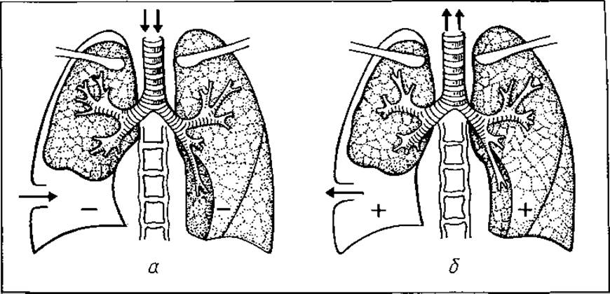
Fig. 50. Open pneumothorax: (a) during inspiration; (b) during expiration
Hemothorax is characterized by the accumulation of blood in the pleural cavity, which may originate from ruptured Vessels of the chest wall and the lung. Initially, the blood clots, but later it liquefies due to Fibrinolysis. Bleeding into the pleural cavity continues until the pressure equalizes with that of the pulmonary vessels due to the accumulated blood. Once this occurs, a unique tamponade effect forms, compressing the lung and bleeding vessels.
Depending on the volume of blood extravasated into the pleural cavity, hemothorax is classified as mild (up to 500 ml of blood in the pleural cavity), moderate (500 to 1000 ml), and massive (exceeding 1000 ml of blood).
If a vascular rupture is accompanied by bronchial injury, blood traces may appear in the sputum.
The clinical presentation of hemothorax includes rapid, weak pulse, low blood pressure, tachypnea, pallor of the skin and mucous membranes, and cold sweats. Patients complain of pain in the injured chest area and coughing. Percussion reveals dullness due to blood accumulation in the pleural cavity. Auscultation reveals absent breath sounds. The heart is displaced toward the healthy side. Anemia develops. Radiography reveals opacification of the corresponding hemithorax.
Hemothorax is frequently combined with pneumothorax, which significantly worsens the clinical course of the injury, promoting blood infection and the development of purulent pleurisy.
During the first 3–6 days, body temperature rises due to blood resorption. A prolonged elevation in body temperature (up to 38–39 °C) may indicate infection of the pleural cavity.
Minor bleeding into the pleural cavity (up to 500 ml) requires no special treatment, as the blood is resorbed within 7–10 days.
For moderate and massive hemothorax, thoracentesis (pleural puncture) is indicated with aspiration of blood and administration of Antibiotics. Puncture is recommended no earlier than the 3rd to 4th day (the time required for vessel thrombosis and secure clot stabilization within the vascular lumen). Early puncture may provoke recurrent bleeding. During this Procedure, care must be taken to prevent air from entering the pleural cavity. For this purpose, a rubber tube is attached to the cannula of the needle used for chest puncture, which is clamped after removing the syringe.
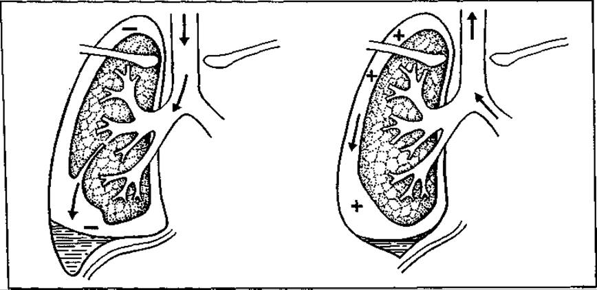
Fig. 51. Tension pneumothorax
Local treatment of hemothorax is combined with general therapy, primarily aimed at combating anemia (blood transfusions, packed red Blood Cells).
Subcutaneous emphysema (emphysema subcutaneum). Injury to the lung, visceral and parietal pleura, and intercostal muscles (occurring when closed chest trauma is combined with rib fractures) can cause air from the pleural cavity to be aspirated into the subcutaneous tissue. This occurs more frequently in tension pneumothorax, when air entering the pleural cavity under pressure seeks an escape route. The clinical presentation includes both local and general manifestations. Air accumulates in the subcutaneous tissue, which is detected during physical examination. Palpation reveals a characteristic crunching sensation or crepitation (presence of air bubbles). Percussion reveals a tympanic sound, while auscultation shows diminished breath sounds.
Subcutaneously, air accumulates most frequently in the region of the affected hemithorax. Only in severe cases of tension pneumothorax may it spread to the neck, face, and the opposite side of the chest. In such instances, the patient's appearance becomes distorted, resembling an inflated rubber toy.
The patient's general condition deteriorates: dyspnea and skin cyanosis develop, and the pulse accelerates.
Mild subcutaneous emphysema does not require special treatment. The patient is prescribed bed rest, analgesics, antitussives, and cardiovascular medications. After a few days, the air begins to be reabsorbed. If air accumulates rapidly in the subcutaneous tissue and spreads to adjacent areas, several small skin incisions are made to release it.
Mediastinal emphysema (emphysema mediastini) occurs as a result of chest trauma with concomitant injury to the Trachea or bronchi. In such cases, air from the respiratory tract penetrates through the surrounding Connective Tissue into the anterior or posterior mediastinum.
Diagnosing mediastinal emphysema (especially of the posterior mediastinal space) in the absence of subcutaneous emphysema is difficult.
Air that has entered the mediastinum can compress the Airways and large blood vessels, causing dyspnea, cyanosis, and a rapid, low-volume pulse. Emphysema develops in the neck region and progresses rapidly. The VEINS OF THE neck and upper extremities are engorged. The patients are agitated. Radiological examinations are of great help in diagnosing mediastinal emphysema. The radiographs reveal widening of the mediastinal shadow and accumulation of air within it. In severe cases of mediastinal emphysema, surgical intervention—mediastinotomy or plastic closure of the injury site—may be required. When the trachea or bronchi are injured, treatment is facilitated by tracheal intubation. Forced artificial ventilation is contraindicated, as it may cause the emphysema to progress.
Chest compression (compressio thoracis). This type of injury most commonly occurs when the chest is squeezed between two hard objects (such as railway car buffers, or during rockfalls and earthquakes). This results in a sudden outflow of blood from the lungs and congestion in the SUPERIOR VENA CAVA system (facilitated by the absence of Valves in the veins of the upper half of the body). All of this leads to traumatic asphyxia syndrome. Hemorrhages (petechiae, ecchymoses) appear on the head, neck, upper chest, mucous membranes, soft palate, conjunctiva, sclerae, external auditory canal, and tympanic membrane. These are the result of ruptured small venules. The reflex spasm of the glottis that accompanies this type of injury exacerbates the congestive phenomena.
The clinical presentation is characteristic. With A large number of hemorrhages, the patient's skin becomes covered with characteristic spots, and when these hemorrhages merge, the entire upper half of the body becomes cyanotic. Interestingly, the cyanotic spots do not extend below the shoulder girdle. Edema of the neck and face frequently occurs. The patient breaks out in a cold sweat; severe dyspnea, tachycardia, and lowered blood pressure are observed.
Treatment consists of ensuring rest for the patient, and administering analgesics, cardiovascular drugs, and general tonics.
Closed chest trauma can be complicated by "shock lung" and "wet lung." In "shock lung," intravascular coagulation of blood in the lungs causes tachycardia, dyspnea, skin cyanosis, and a drop in blood pressure. Fluoroscopy or radiography of the chest reveals decreased transparency of the lung tissue and enhanced pulmonary markings.
The pathogenesis of "wet lung" is based on hypersecretion of the bronchial glands and impaired pulmonary ventilation due to mucus accumulation in the bronchi. The clinical picture is characterized by severe dyspnea; breathing is shallow and noisy. The pulse is rapid. Auscultation reveals numerous moist rales in the lungs.
Patients with "shock lung" are prescribed anticoagulants, while those with "wet lung" undergo bronchial aspiration and artificial pulmonary ventilation.
Rib fracture (fractura costae) occurs from a direct blow, fall, or chest compression. It is observed in 67% of cases of closed chest injuries and is frequently combined with Other types of trauma (concussion, compression, contusion of the chest). Rib fractures can be single or multiple, unilateral or bilateral. A double fracture of several ribs is particularly dangerous. This results in a flail "rib segment" with paradoxical motion: when the patient inspires and the chest expands, the segment, having lost its connection with the chest wall, is drawn inward; conversely, when the patient expires and the chest collapses, the "rib segment" bulges outward (Fig. 52). Such paradoxical Movements of the segment impair respiration.
A displaced rib fracture is frequently accompanied by injury to the parietal and visceral pleura, lungs, and blood vessels, which causes pneumohemothorax and subcutaneous emphysema. Tension pneumothorax may also develop.
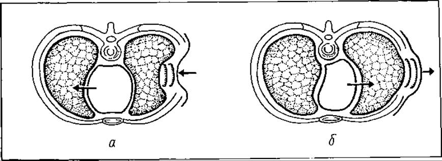
Fig. 52. Displacement of the mediastinum and the "rib segment" in a double rib fracture: a — during inspiration; b — during expiration
The clinical presentation of a rib fracture is characterized by sharp pain, which worsens during deep inspiration, palpation, and coughing. The chest lags behind in the act of respiration, and deformation is noted in multiple fractures. Palpation sometimes reveals the symptom of crepitus (grating of one rib fragment against another), which can also be elicited by gentle chest compression. Edema and ecchymosis may appear in the fracture area.
Multiple rib fractures are accompanied by severe respiratory and circulatory disorders, up to pleuropulmonary shock. Radiological examination plays a crucial role in diagnosing rib fractures, although it should be borne in mind that the fracture site is not always visualized. In such cases, clinical examination becomes of primary importance.
Previously, rib fractures were immobilized using soft and hard bandages. Nowadays, they are rarely used, as they further compromise chest excursion.
Immobilization or surgical intervention is employed only for multiple rib fractures with flail chest wall segments.
In cases of rib fracture, repeated intercostal alcohol-procaine blockades (1% procaine solution and 96% ethyl alcohol) are prescribed. By relieving pain, they improve chest excursion. For multiple rib fractures, paravertebral or vagosympathetic blockade may be used. Patients should remain at rest or in a semi-sitting position for 4–5 days. Antitussive and expectorant drugs, respiratory exercises, and oxygen therapy are prescribed.
A fractured rib knits within 3–4 weeks.
CLOSED INJURIES OF THE Abdominal cavity AND RETROPERITONEAL SPACE
Closed abdominal injuries occur due to The impact of a blunt object against the abdominal wall or from a fall on the abdomen. Such trauma can result not only in a contusion of the anterior abdominal wall but may also be accompanied by injuries to internal organs (parenchymatous or hollow).
The severity of the injury depends on A number of factors: the force of the traumatic agent, the physiological state of the anterior abdominal wall at the moment of injury, and the Anatomical and physiological state of the injured organ. The greater the force of the traumatic agent, the more significant the changes that occur in the body. When the anterior abdominal wall is relaxed at the moment of impact, the likelihood of internal organ injury increases. Injuries to hollow organs (Stomach, intestines, Urinary Bladder) occur more frequently when they are distended at the time of trauma, whereas injuries to parenchymatous organs occur when they are pathologically altered. In such cases, organs can be damaged even by minor trauma. The anatomical Location OF THE organs within the abdominal cavity is also significant (The Liver and Spleen are injured more frequently, while the more deeply situated Pancreas is injured less often).
A severe blunt blow to the epigastric region can cause shock due to injury of the solar plexus, even in the absence of internal organ damage.
The clinical presentation of a closed abdominal injury depends on the severity of the damage. If only the anterior abdominal wall is contused, the patient will experience pain at the impact site, which worsens with movement. A slight swelling may be observed, and muscle rigidity develops in a localized area of the anterior abdominal wall.
Isolated injuries to the abdominal wall can be accompanied by partial or complete rupture of muscles to a greater or lesser extent, along with subcutaneous hematomas. Through the defect in the torn muscle, the Peritoneum protrudes, resulting in a traumatic hernia.
Severe injuries to the abdominal wall are also accompanied by systemic symptoms: nausea, vomiting, accelerated pulse and respiration, hiccup, and flatulence. It is often difficult to make a Differential Diagnosis between an anterior abdominal wall injury and internal organ damage; therefore, such patients must be hospitalized promptly and placed under observation.
Contusions of the abdominal wall usually do not require special treatment. With the initial application of cold followed by heat, the signs of trauma disappear within a few days.
In cases of internal organ injury, the clinical presentation will depend on whether the affected organ is parenchymal or hollow. The former results in varying degrees of hemorrhage, while the latter leads to inflammation of the peritoneum (Peritonitis). Hemorrhage occurs following ruptures of the liver, spleen, pancreas, or mesenteric vessels. Peritonitis is caused by injuries to The Stomach, intestines, or urinary bladder. Sometimes, even minor damage to these organs can trigger severe, fatal peritonitis.
The most severe clinical course is observed in trauma with simultaneous damage to both parenchymal and hollow organs, where hemorrhage is combined with peritonitis.
Diagnosing intra-abdominal injuries is sometimes difficult. Consequently, all patients with abdominal trauma must be hospitalized. Diagnosis is further complicated by the fact that even in cases of severe closed abdominal trauma, a lucid interval may occur immediately after the injury. Therefore, administering painkillers to the patient at the pre-hospital stage is not recommended.
Initially, every closed abdominal injury is characterized by the clinical picture of shock, and only after some time do the symptoms specific to the injury of a particular organ become apparent.
The symptomatology of parenchymal organ ruptures depends on the size of the injured organ and the severity of the hemorrhage. With minor ruptures and minimal bleeding that quickly stops due to vascular thrombosis, the patient's general condition may not be significantly altered. In cases of massive hemorrhage, a typical clinical picture develops. The patient complains of generalized weakness, dizziness, and spots before the eyes. They are pale, covered in cold sweat, with a rapid pulse of low volume and tension, and lowered blood pressure. Nausea and vomiting are observed. The anterior abdominal wall lags behind during respiration, is moderately tense upon palpation, and is acutely tender. Blumberg's sign is positive. Percussion reveals dullness in the flanks (indicating the presence of fluid). Digital rectal examination reveals bulging of the anterior rectal wall due to blood accumulation in the pelvis.
Among Laboratory tests, blood analysis is of primary importance (revealing a decrease in red blood Cell count, Hemoglobin levels, etc.). Blood parameters should be monitored dynamically.
The clinical picture described above indicates internal hemorrhage resulting from the rupture of a parenchymal organ or mesenteric vessels. It is more difficult to determine precisely which organ is damaged. An analysis of The Mechanism of injury and the presence of bruising or abrasions on the skin of a specific abdominal wall region often helps in this regard.
The spleen is the organ most frequently injured in closed abdominal trauma, particularly when it is pathologically altered. Spleen injuries can range from a minor fissure to complete fragmentation or avulsion from the vascular pedicle. Splenic rupture can occur in a "two-stage" manner. At the moment of trauma, the splenic parenchyma ruptures while the capsule remains intact, forming a subcapsular hematoma. After some time, as the hematoma enlarges, the capsule ruptures, and blood pours into the free peritoneal cavity. In such cases of subcapsular hematoma, ultrasound plays a crucial diagnostic role.
Treatment for splenic injuries depends on the extent of destruction. In cases of fragmentation, splenectomy is indicated. For minor ruptures, partial splenectomy or suturing of small defects is currently preferred. In recent years, polyglycolic acid (dexon) mesh wrapping has been used to treat splenic ruptures.
Hematomas within the liver tissue are diagnosed using ultrasound or computed tomography scans. Open liver injuries with intraperitoneal bleeding require surgical intervention, which involves suturing and ligating ruptured blood vessels and Bile ducts. Packing a ruptured liver is unreliable. Severe liver damage may be an indication for partial hepatic resection.
When a hollow organ ruptures, peritonitis develops rapidly. Patients complain of generalized, progressively worsening abdominal pain, nausea, and vomiting. The skin is pale and covered in cold sweat; the pulse is rapid, weak in volume, and tense. The anterior abdominal wall does not participate in respiration. Its muscles are rigid ("board-like"), the abdomen is acutely tender, Blumberg's sign is positive, and hepatic percussion on the right reveals a tympanic sound instead of dullness (indicating free gas under the Diaphragm).
During a plain fluoroscopy or radiography of the abdomen, gas escaping from the injured hollow organ can be observed over the right or left hepatic dome, beneath the diaphragm, rising upward According to the laws of physics ("free intraperitoneal gas").
Without urgent surgical intervention, peritonitis progresses: the abdomen distends, passage of stool and gas ceases, vomiting becomes more frequent, the Tongue is dry, the pulse is rapid and thready, blood pressure drops, the patient's general condition deteriorates, facial features sharpen, and the eyeballs sink ("Hippocratic facies"). As cardiovascular failure worsens, the patient dies.
Important diagnostic techniques in the assessment of closed intra-abdominal injuries include abdominal radiography, diagnostic peritoneal lavage (DPL), and laparoscopy.
The presence of free gas under the diaphragmatic domes on a radiograph indicates a hollow organ rupture; in the retroperitoneal space, it suggests injury to the duodenum, colon, or rectum. Obliteration of the psoas muscle contours (m. ileopsoas) points to a retroperitoneal hematoma, fractured lower ribs may indicate a liver or splenic rupture, and loops of bowel in the thoracic cavity suggest a diaphragmatic rupture.
Abdominal paracentesis involves puncturing the peritoneal cavity with a large-bore needle. The puncture is performed under local anesthesia along the linea alba, 3–4 cm below the umbilicus. The aspiration of blood through the needle indicates hemorrhage. If no blood flows out, a thin polyvinyl catheter—the so-called exploratory catheter—is advanced 15–20 cm into the peritoneal cavity through the needle. It is sequentially directed into the right and left hypochondriac regions and the right and left iliac fossae. If blood is present in the peritoneal cavity, it begins to drip from the catheter. If blood still fails to flow out (which happens when there is little blood in the peritoneal cavity or it has not yet accumulated), the needle is removed, while the catheter is left in place for 24–48 hours.
Peritoneal lavage involves puncturing the peritoneal cavity with a needle (similar to paracentesis) and instilling 1000 mL of warm isotonic sodium chloride solution. The fluid is then drained by lowering the tubing below body level. The presence of blood, bile, gastric, or intestinal contents in the returned fluid indicates The Need for surgical intervention.
Laparoscopy (examination of the peritoneal cavity using a specialized optical instrument) makes it possible not only to detect the presence of blood in the peritoneal cavity but also to identify The Nature of the injured organ. However, some authors question the diagnostic value of this research method.
If the diagnosis of internal organ injury is confirmed or suspected, the patient undergoes immediate surgery. Exploratory laparotomy is performed under endotracheal anesthesia. Blood is aspirated, the peritoneal cavity is cleared and dried, and hemorrhage is controlled.
In cases of hollow organ injury, the peritoneal cavity is cleared and lavaged (flushed with an antiseptic solution). The defect is repaired, and the peritoneal cavity is washed once again with an antiseptic solution. Following such surgery, the anterior abdominal wall is not closed tightly; rather, it is drained in several places using rubber strips and polyvinyl tubing.
Traumatic injuries to retroperitoneal organs require separate consideration.
Among retroperitoneal organs, the pancreas, Kidneys, and urinary bladder are the most frequently injured.
Pancreatic injuries are relatively uncommon and are rarely isolated. As a rule, trauma to the pancreas is concurrently accompanied by injuries to the duodenum, stomach, liver, spleen, etc.
The primary danger of pancreatic trauma lies in the extravasation of both blood and pancreatic juice into the abdominal cavity or retroperitoneal space. Once released, the activated Enzymes cause autodigestion of the pancreas itself as well as the surrounding soft Tissues.
Diagnosing pancreatic injuries is challenging. Patients present with pallor and cold sweat. Tachycardia, hypotension, rigidity of the anterior abdominal wall muscles in the PROJECTION OF THE pancreas, elevation of the left dome of the diaphragm, and tenderness upon palpation in the left costovertebral angle are observed. Blood tests reveal elevated pancreatic enzyme levels and, occasionally, hyperglycemia.
Treatment for pancreatic injuries is exclusively surgical.
Renal injuries resulting from blunt trauma are quite common and may manifest as contusions, lacerations, or complete destruction of renal tissue with avulsion of the renal pedicle.
A renal contusion is accompanied by mild lumbar pain and microhematuria (a trace amount of blood in the urine). More severe renal trauma is characterized by a classical triad: pain in the corresponding lumbar region, localized swelling, and Hematuria.
Renal injuries are classified into extraperitoneal and intraperitoneal.
The triad described above is characteristic of extraperitoneal renal lacerations. In cases of intraperitoneal rupture, where urine leaks into the peritoneal cavity, peritonitis develops.
Laboratory and radiological diagnostic methods play a crucial role in evaluating renal trauma. Blood tests reveal anemia, while urinalysis indicates hematuria. Intravenous urography (intravenous administration of a contrast agent) demonstrates extravasation of urine beyond the Urinary Tract on radiographs.
Renal contusions and minor extraperitoneal lacerations are managed conservatively (bed rest, cold compresses to the lumbar region, hemostatic agents, antibiotics, and infusion therapy).
Indications for surgical intervention include rapidly expanding retroperitoneal hematomas or signs of progressive peritonitis. Prior to surgery, it is mandatory to verify the presence of a contralateral Kidney, as nephrectomy of the injured kidney is sometimes unavoidable.
Bladder ruptures most commonly occur due to trauma to the lower abdomen accompanied by pelvic bone fractures in The Setting of a distended bladder. Similar to renal injuries, bladder ruptures are classified as extraperitoneal and intraperitoneal. Extraperitoneal rupture presents with acute tenderness of the anterior abdominal wall above the Pubic Symphysis, localized swelling, and doughy tissue consistency in this area.
In cases of intraperitoneal bladder rupture, signs of peritonitis develop rapidly.
Both types of rupture present with gross hematuria and urinary urgency.
Retrograde cystography (where a contrast medium is introduced into the urinary bladder via a catheter) AIDS in diagnosing bladder ruptures. It allows for the detection of contrast extravasation into the perivesical tissue or the peritoneal cavity.
The treatment of urinary bladder ruptures is surgical. For intraperitoneal rupture, indications include laparotomy, suturing of the bladder wall defect, aspiration and lavage of the peritoneal cavity, its drainage, and the establishment of a suprapubic cystostomy (for urinary diversion over 7–10 days to reduce intravesical pressure).
In extraperitoneal bladder ruptures, an incision of the skin and aponeurosis is made along the linea alba above the pubic symphysis. The defect in the bladder is identified and sutured. A suprapubic tube is placed, and the surrounding paravesical tissue is drained.
In surgical practice, clinicians frequently encounter patients presenting with multiple, associated, and combined injuries.
Trauma to individual systems and organs is divided into isolated (monotrauma) and polytrauma. An isolated injury refers to trauma affecting a single organ (e.g., traumatic brain injury, liver laceration, bladder rupture). The term "polytrauma" is a collective category that encompasses multiple, associated, and combined injuries.
Multiple mechanical traumas involve injuries to two or more internal organs within a single body cavity (e.g., concurrent injury to the liver and bowel).
Associated injuries refer to trauma affecting internal organs in two or more Body Cavities, or simultaneous injuries to internal organs and The Musculoskeletal System (compression of the chest and fra
cture of the Femur; splenic injury combined with chest contusion; traumatic brain injury associated with pelvic bone fractures).
Combined injuries are defined as those caused by various pathogenic agents of different nature: mechanical, thermal, or radiation (e.g., fracture of the humerus combined with a shoulder burn, closed traumatic brain injury associated with radiation exposure, etc.).
From the perspective of clinical presentation, diagnosis, and treatment, polytrauma has several distinctive features.
First, polytrauma is characterized by the so-called burden syndrome, where hemorrhage accompanying traumatic shock exacerbates the course of injury and worsens the prognosis.
Second, the combination of injuries can sometimes render therapies mutually incompatible; for instance, a humeral fracture combined with suspected closed abdominal trauma and internal organ rupture precludes the administration of narcotics and analgesics.
Third, there is an increased likelihood of severe complications such as hemorrhage, shock, toxemia, renal failure, thromboembolism, and others.
Fourth, certain types of polytrauma can mask the clinical presentation of individual injuries—for instance, when a traumatic brain injury is combined with closed abdominal trauma, the clinical signs of the latter are obscured, frequently leading to diagnostic errors.
Polytrauma is typically caused by major accidents, such as motor vehicle or railway crashes, or falls from a height.
Treating patients with polytrauma is complex and requires a multidisciplinary team involving intensivists, surgeons, traumatologists, internists, and other specialists. Treatment must be comprehensive and pathogenetic. The priority therapeutic measures for polytrauma are outlined below.
First. Eliminate any immediate threats to life:
a) secure a patent airway;
b) maintain adequate ventilation (relieve tension pneumothorax, manage closed and open chest wounds; provide assisted ventilation);
c) support circulation (stop bleeding; relieve cardiac tamponade; monitor patient status and treat shock);
d) halt the progression of elevated intracranial pressure.
Second. Address conditions that pose delayed threats to life or directly impair vital functions:
a) debride cerebral wounds;
b) repair gastrointestinal perforations;
c) explore abdominal and thoracic wounds;
d) relieve Spinal Cord compression;
e) repair vascular injuries;
f) reduce, debride, and immobilize complex fractures.
Third. Manage non-life-threatening conditions and those that do not immediately impair function:
a) minimize bone fragment displacement;
b) reduce closed fractures;
c) debride soft tissue wounds;
d) correct peripheral nerve injuries.
Last update: 08/08/2026
Editorial and Educational Adaptation: This material has been compiled based on the primary/original source text. The project team performed an editorial review, corrected technical inaccuracies, structured sections, and adapted the content for an educational format.
What was processed:
- elimination of formatting defects (OCR errors, structural breaks, corrupted characters);
- editorial organization of content;
- standardization of terminology in accordance with academic sources;
- verification of factual statements against the original source text.
All mentions of the author, publication year, and origin of the primary text have been preserved in accordance with the source.