Military Surgery with Emergency Surgery - V.Ya. Bilyi 2004
Traumatic shock
Introduction. Traumatic Shock (TS) is characterized by acute-phase, interdependent disorders of critical bodily Functions, leading to severe perfusion deficits, Hypoxia, and impaired function of vital Organs.
Among critically ill casualties, clinical signs of TS are observed in two-thirds of patients, with a mortality rate reaching up to 40% of cases.
Among those wounded by firearms during the Great Patriotic War, TS occurred in 8–10% of cases, whereas in modern warfare its incidence reaches 25–30% of the total number of wounded.
Recent studies demonstrate that mechanical trauma-induced impairments of vital functions are prolonged and phased in nature, possess a specific Pathogenesis and defined clinical forms, and can no longer be framed solely within the traditional doctrine of TS.
Due to the severe clinical course of TS, a multitude of early and late complications arising from severe trauma formed the basis in the 1980s for introducing a new concept: traumatic disease. Thus, METABOLISM/2.html">THE CONCEPT OF "shock organs"—such as shock lung, shock Kidneys, shock Liver, and shock gut—emerged as manifestations of multiple organ dysfunction syndrome.
Traumatic disease highlights the inextricable link between shock and the future development of complications, thereby offering Prospects for developing a differentiated approach to treating complications based on prognostication. In traumatic shock, functional disorders develop in an avalanche-like, autocatalytic manner, and preventing adverse outcomes is possible only through early prognostication and timely, differentiated Treatment.
The clinical course of traumatic disease comprises the following periods:
- shock period — acute response to trauma (duration up to 1–2 days);
- period of early manifestations — risk of early complications (duration up to 14 days);
- period of late manifestations — risk of late complications (duration several weeks);
- period of final recovery and rehabilitation (duration ranging from several weeks to many months or even years).
Thus, traumatic disease encompasses the entire course of an Organism's life from the moment of injury to recovery or death. It is characterized by trauma-induced local and systemic pathological processes, wherein traumatic shock is considered the initial period of traumatic disease.
Evolution of the doctrine of traumatic shock. The general Development of the doctrine of TS comprises three periods: descriptive, theoretical-experimental, and modern, which is associated with studying the clinical course of vital organ disorders in casualties.
In his work "Principles of General Field Surgery" (1865), N.I. Pirogov provided a classical Description of the Clinical presentation of TS, which remains relevant today due to its comprehensiveness, precision, and vividness.
The second historical period in The Study of TS began with experimental investigations of its pathogenesis in animals. The main theories of TS were formulated: neurogenic, Blood-plasma loss, and toxemia. However, despite a profound experimental research base, these theories had shortcomings: the absolutization of a single pathogenetic factor of shock and a clear underestimation of others.
Consequently, none of the theories could fully explain the pathogenesis of shock resulting from severe trauma, as the latter exhibits various clinical courses and is caused by a combination of multiple factors. At the same time, it was proven that most cases of TS can be explained from the perspective of the blood-plasma loss theory, given that Hemorrhage is the primary and most frequent cause of shock.
In-depth clinical study of TS revealed that in each case it possesses specific features depending on the localization and Nature of the injury, volume of blood loss, impaired external Respiration, degree of vital organ damage, and other complications.
The current stage of TS research is characterized by further investigation of pathogenesis aimed at objectifying the severity of the clinical course, predicting various complications and outcomes, and ultimately improving surgical and critical care.
Etiology AND PATHOGENESIS of traumatic shock. Modern views on TS are based on its mixed etiology, where the primary factors are excessive pain signaling, blood loss, soft tissue injury, long bone fractures, and trauma to vital organs.
Disorders of Central Nervous system function during trauma are caused by the pain response and associated emotional-neural shocks (the eretile phase of shock).
In the dynamics of the pathogenetic mechanisms of the torpid phase of shock, D.M. Sherman (1987) distinguishes three periods: early, stabilization, and late. The early period is characterized by the mobilization of all compensatory pathways of pathogenesis. The stabilization period reflects the maximal recruitment of protective compensatory mechanisms, which depends on the severity of trauma, the individual CHARACTERISTICS OF THE organism, and the quality of antishock treatment. The late period is characterized by the decompensation of vital functions and the transition to a terminal state.
The formation of core somatic responses to trauma occurs in the subcortical structures of the Brain—specifically within the Cells of the reticular formation—which leads to vasodilation, reduced preload, decreased Cardiac Output, and hypotension, subsequently resulting in severe functional impairment of individual organs and systems. Progressing hypotension and the associated hypoxia create conditions that further impair CNS function, which is highly sensitive to oxygen deprivation.
The reduction in circulating blood volume (CBV) in TS is caused by external or internal bleeding. During hemorrhage, catecholamines stimulate alpha-receptors in peripheral Blood Vessels, leading to vasoconstriction, while stimulation of cardiac beta-receptors increases myocardial contractility.
Constriction of the vascular lumen in the Skin and skeletal musculature serves to conserve blood flow for vital organs, primarily the brain and heart. A "centralization" of Circulation occurs, with blood flowing mainly through arteriovenous anastomoses while bypassing the capillary bed.
Hemorrhage leads to hemodilution. The decrease in CBV is compensated by the organism through the influx of fluid and dissolved Proteins from the interstitial space. Concurrently, the pituitary-adrenal cortex axis is activated, increasing the secretion of aldosterone, which enhances sodium reabsorption in the proximal renal tubules. Sodium retention leads to increased Water reabsorption in the tubules and reduced urine output. The process of autohemodilution develops accordingly.
Subsequently, As a result of sympathoadrenal system exhaustion, increasing metabolic acidosis, and the accumulation of vasoparasitic substances in Tissues (such as histamine, ferritin, beta-hydroxybutyric acid, lactic acid, and other metabolic products), the spasm of precapillary arterioles gradually gives way to paresis and subsequently paralysis, leading to the "decentralization" of Blood Circulation.
In congested capillaries, pressure rises and, due to the predominance of hydrostatic pressure, plasma leakage and hemoconcentration develop. Slowed blood flow and increased coagulation lead to spontaneous blood clotting within the capillaries, resulting in disseminated microthrombosis (the "microcirculatory crisis"). Thereafter, Blood Coagulation decreases, circulatory hypoxia increases, circulating blood volume (CBV) drops significantly, blood flow velocity slows down, and central venous pressure (CVP) decreases. Clinically, this state corresponds to the torpid phase of shock.
Arterial and venous hypotension, Impaired blood supply to The Heart, a drop in CVP, and Metabolic Disorders lead to acute dystrophic Changes in the myocardium and a significant depression of Cardiac Activity. In traumatic shock (TS), this may also be caused by direct cardiac injury (contusion or wound). The aforementioned factors reduce myocardial contractility and deepen the shock.
In the kidneys during shock, blood circulation is redistributed from the cortex to the medulla via a juxtamedullary shunt-like mechanism. When blood pressure drops to 60 mm Hg, renal blood flow decreases by 30%, while a drop in blood pressure below 40 mm Hg halts urine production altogether. Renal blood flow decreases in proportion to the reduction in cardiac output, resulting in ACUTE RENAL FAILURE.
Massive soft tissue and parenchymal organ injuries lead to traumatic toxicosis. This intoxication is caused by The entry of Myoglobin and tissue breakdown products into the bloodstream. These components increase blood viscosity and impair microcirculation, whereas myoglobin in the acidic environment of the renal tubules turns into insoluble acid hematemesis (methemalbumin/acid hematin), causing tubular epithelial necrosis and The Development of Acute Kidney Injury (AKI).
Liver hypoxia during TS leads to impaired antitoxic function. Glycogen and phosphorus compound reserves are depleted in hepatic cells, and prothrombin production drops. Lipid Metabolism, detoxification, and antimicrobial functions are impaired, and plasma bilirubin levels increase.
Hypoxia of the intestinal tissues leads to an increased penetration of toxic metabolic products and aggressive intestinal Bacteria into the bloodstream, causing endogenous toxemia and bacteremia.
Respiratory disorders slow down the venous return to the heart, cause congestion in the Pulmonary Circulation, and lead to hypersecretion in the Trachea and Bronchi. Simultaneously, the cough reflex is suppressed. All of this results in progressively impaired pulmonary gas exchange: Hypoxic hypoxia worsens, and shock lung develops. Circulatory hypoxia joins the hypoxic hypoxia, leading to metabolic disturbances, microthromboembolism, and pulmonary edema.
In shock lung, Acute Respiratory Failure develops, accompanied by venous blood shunting in the pulmonary circulation and a decrease in arterial oxygen partial pressure. Consequently, the ventilation-perfusion ratio is disrupted, which is pathognomonic for shock lung. The final stage of development in shock lung is the decompensation of cardiopulmonary function.
Airway obstruction, multiple rib and sternal fractures, impaired respiratory biomechanics, and pneumothorax lead to a decrease in arterial blood oxygen saturation (SaO2) and the development of hypoxic hypoxia.
In addition to reduced CBV, hemorrhage results in a direct loss of Hemoglobin, causing hemic hypoxia. The circulation of toxic substances—the products of histolysis—in the blood deepens histotoxic hypoxia.
Further progression of TS is characterized by the exhaustion of the Adrenal Glands, the anterior pituitary, and The Thyroid Gland. Corticosteroid deficiency develops. Ion metabolism is disrupted: sodium and chloride concentrations decrease, while plasma potassium levels rise. Protein, carbohydrate, and vitamin metabolism disorders, as well as metabolic acidosis, progress. Blood sugar levels rise, residual nitrogen concentration increases, the alkaline reserve of blood decreases, acetone body excretion increases, and globulin concentration drops against the Background of a general decrease in Plasma Proteins.
Impairment of Vital organ functions during TS is accompanied by disorders in all metabolic processes. In the initial stages, these disturbances are almost always reversible, but with a prolonged course of the torpid phase of shock, they become more severe and sometimes irreversible.
The preservation of Cell viability in vital organs depends on the varying sensitivity of different tissues to oxygen deprivation. Histotoxic changes occur first in the kidneys, followed by the Lungs, liver, heart, and brain.
Most injuries are characterized by the development of endogenous intoxication. Products of tissue crushing and autolysis, enhanced Catabolism of tissue proteins, vasoactive substances, and bacterial endotoxins trigger progressive endotoxicosis. In severe mechanical injuries, endotoxicosis, along with blood loss and other pathogenetic factors of TS, largely determines the severity of TS. Overall, the etiopathogenesis of traumatic shock is illustrated in Figure 2.
Class="center">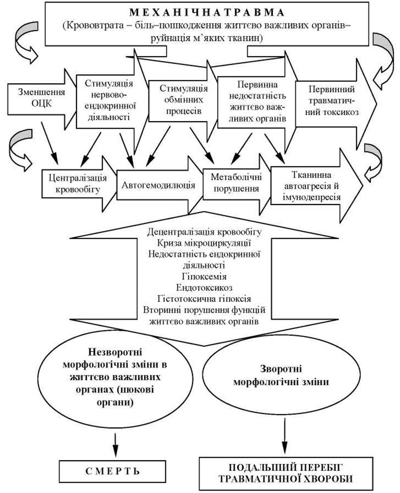
Fig. 2. Etiopathogenesis of traumatic shock.
Thus, TS is an acute critical condition that occurs in victims of severe mechanical injuries, accompanied by mutually dependent Homeostasis disorders in the form of neuroendocrine disorders, hemodynamic, respiratory, and metabolic impairments, dysfunction of vital organs, and altered immunological status, with clinical manifestations depending on the severity and localization of the trauma.
It should also be noted that the following factors play a significant role in the etiopathogenesis of TS:
1. Delayed and inadequate pre-medical care.
2. Re-traumatization during evacuation.
3. Repeated, even minor, blood loss.
4. Hypothermia or overheating of the body.
5. Professional and environmental stress syndrome (physical and emotional exhaustion; prolonged fasting, hypovitaminosis, etc.).
Classification and clinical presentation of TS. Clinically, Two phases of TS are distinguished: erethetic (excitative) and torpid.
During the erethistic phase, consciousness remains intact or impaired, with patients complaining of pain while simultaneously underestimating the severity of their condition. The injured person's voice is "dry," speech is fragmented, and their gaze is restless. The skin is pale, occasionally flushed, and cold sweats appear. General hyperesthesia and hyperreflexia are frequently observed. The pupils react uniformly and briskly to light. Muscular hypertonia and increased tendon Reflexes are noted. The pulse has a satisfactory volume, and tachycardia sometimes occurs.
The second, torpid phase of shock is marked by the depression of all vital functions of organs and systems, manifested by a drop in blood pressure, bradycardia, oliguria, dyspnea, slowed metabolic processes, decreased sensitivity and body Temperature, altered mental status, pallor, cyanosis, dry mucous membranes, and hypodynamia.
Depending on the severity of hemodynamic disorders, the degree of drop in systolic blood pressure, and pulse rate, clinically we distinguish three severity degrees of this shock phase:
1. Grade I shock (mild).
2. Grade II shock (moderate to severe).
3. Grade III shock (extremely severe).
Terminal states are distinguished separately.
Grade I shock frequently occurs in isolated moderate injuries with blood loss up to 20% of BV. The patient's general condition is satisfactory or moderately severe, consciousness is preserved, with moderate motor and mental inhibition, and skin pallor. Maximal BP remains within 90-95 mm Hg, pulse is 100-120 bpm with a satisfactory volume. Respiratory rate is up to 25 bpm. With timely medical care, the prognosis is favorable.
Grade II shock is observed in severe trauma with blood loss of up to 30% of BV. The patient's general condition is severe, consciousness is preserved. Pronounced skin pallor, motor and mental inhibition are noted. BP is 75-90 mm Hg, pulse is 120-140 bpm, of weak volume. Respiration is shallow, up to 30 bpm. Oliguria is present. The prognosis is doubtful.
Grade III shock occurs in severe trauma involving vital organ damage and blood loss exceeding 30% of BV. The wounded person's general condition is extremely severe, consciousness is impaired, and coma sometimes develops. The skin is pale and cyanotic with cold sweat; hypodynamia and hyporeflexia are present, along with impaired renal excretory function (oliguria, anuria). BP is 50-75 mm Hg. Pulse is 140-160 bpm, of weak volume. Dyspnea is up to 40 bpm. The prognosis is unfavorable.
Terminal states are divided into: the preagonal phase, agony, and the state of clinical death.
The preagonal state of the casualty is extremely severe; breathing is shallow and infrequent. The skin is bluish-pale, covered with cold sweat. The pulse cannot be detected in peripheral Arteries, while it is weak in the carotid and femoral arteries. The pupils are dilated and react sluggishly to light. BP is below 50 mm Hg.
The agonal state is characterized by unconsciousness and areflexia (deep coma). Respiratory movements are barely noticeable. Sphincters are relaxed. The pulse on central arteries is thready and poorly defined. Systolic BP is unmeasurable, heart sounds are muffled. In the absence of effective resuscitation measures, clinical death ensues—a state where external Signs of Life are absent (cessation of respiration and cardiac activity, pupil dilation, and absence of the corneal reflex).
The state of clinical death, if effective resuscitation measures are not performed, lasts for 5-7 minutes, after which irreversible changes develop in vital organs, primarily the central nervous system, liver, kidneys, and heart, leading to biological death—the demise of the organism.
In the clinical course of the torpid phase of traumatic shock, it is advisable to distinguish three periods, the duration of which depends on the underlying cause and severity of the shock:
1. The period of compensated reversible shock.
2. The period of decompensated reversible shock.
3. The period of decompensated irreversible shock.
Compensated reversible (early) shock is characterized by the functioning of compensatory mechanisms, as a result of which failure of vital organ functions does not occur. If the primary cause of shock is successfully eliminated during this period, complete recovery ensues with minimal consequences.
During the second period—decompensated reversible shock—microcirculatory and cellular disorders in vital organs worsen and become prolonged, leading to the failure of one or more Organ Systems, though prompt therapeutic measures can prevent death.
In the third period—decompensated irreversible shock (late shock)—irreversible disorders of vital organs occur (multiple organ dysfunction syndrome). Even aggressive intensive therapy cannot prevent death.
The classification approach distinguishing the periods of compensated and decompensated reversible shock, as well as decompensated irreversible shock in the course of the torpid phase of traumatic shock, is of practical importance for prognosis and the Selection of optimal tactics for intensive care and surgical interventions.
Diagnosis of TS. The diagnosis and determination of TS severity are primarily based on general status indicators: the patient's appearance, respiratory rate, and Cardiovascular system function (systolic BP and pulse rate), which on the one hand are the main indicators of overall circulatory function, and on the other hand, reflect the functional state of other organs and systems. The lower the BP, the deeper the Circulatory Disorders and impairments of the body's major vital functions.
Central venous pressure (CVP) is also a quite important hemodynamic indicator of TS.
CVP depends on blood volume and characterizes the venous return of blood to the right side of the heart, as well as the heart's ability to cope with this return. CVP is directly proportional to blood volume and inversely proportional to the capacity of the systemic circulation vessels and cardiac output. A CVP of less than 50 mm H2O is a sign of absolute or relative hypovolemia. In such cases, the blood volume deficit must be replenished immediately. With a CVP ranging from 50 to 100 mm H2O, an adequate volume of blood returns to the heart.
In diagnosing the severity of traumatic shock (TS), determining the extent of blood loss and hypovolemia is of paramount importance.
External bleeding is almost always obvious. An examination of clothing, dressings, the accident scene, vomitus, stool, urine, and gastric contents (obtained via a nasogastric tube) allows for a rough estimation of blood loss volume.
Intrathoracic bleeding can occur in both penetrating wounds and blunt chest trauma. Chest radiography confirms the magnitude of the hemothorax (Fig. 3).
Intra-abdominal bleeding should be ruled out in cases of blunt chest trauma, lower rib fractures, and any penetrating abdominal wound. Clinical signs include abdominal distension, abdominal pain, and the presence of blood in the peritoneal cavity during diagnostic peritoneal lavage. Ultrasound and computed tomography confirm the presence of retro- or intraperitoneal hemorrhage.
Abdominal injuries are frequently complicated by pancreatitis, Peritonitis, and intestinal obstruction, leading to fluid sequestration in the "third space" (atonic bowel, peritoneal cavity, traumatized tissues, and retroperitoneal space).
Pelvic fractures can result in retroperitoneal hemorrhage (with blood loss ranging from 1.5 to 3.0 L).
Long bone fractures are often accompanied by concealed blood loss, amounting to 500–1000 mL.
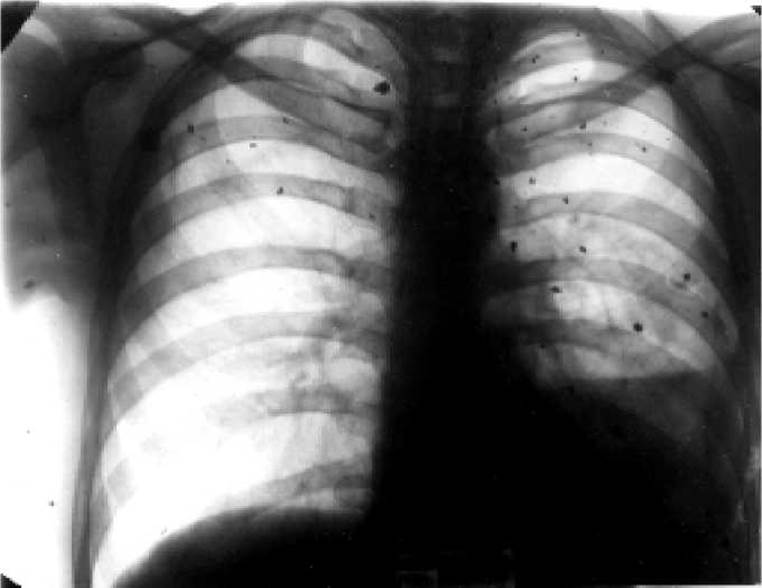
Fig. 3. Radiograph. Multiple shrapnel penetrating chest wounds with injury to the left lung. Small hemothorax.
Soft tissue crush injuries are accompanied by blood infiltration of the traumatized tissues, tissue edema, and additional fluid sequestration.
Intoxication by tissue breakdown products and myoglobin resulting from massive soft tissue and internal organ injuries, coupled with hypovolemia, precipitates acute renal failure (ARF) in TS. In some cases, oliguria and anuria—even in the presence of adequate blood pressure—serve as indicators of TS severity.
The most objective indicator of blood loss is the hematocrit, with optimal values ranging between 30–35%.
The relative volume of blood loss can be estimated using the Allgöwer shock index (The ratio of heart rate to systolic blood pressure). A higher shock index indicates greater blood loss.
Hemoglobin and hematocrit measurements are straightforward laboratory Methods for Assessing blood loss volume. Following hemorrhage and subsequent hemodilution, hemoglobin and hematocrit levels progressively decline.
The diagnosis and severity of shock are established based on the volume of blood loss, blood pressure, heart rate, respiratory rate, hourly diuresis, and mental status (Table 1).
The clinical presentation of traumatic shock is frequently compounded by impairments in the specific Functions of the injured organ.
Disorders of vital organs and systems in TS (shock organs)
Shock lung. Clinically, such patients present with acute dyspnea and a cough productive of blood-tinged sputum. Acrocyanosis and tachycardia are observed, and Auscultation reveals moist rales. Systolic blood pressure may be either elevated or decreased depending on shock severity. Key radiological findings include: accentuation of the pulmonary vascular pattern, evidence of pulmonary edema, and discoid or segmental Atelectasis. Microembolism is characteristically marked by the appearance of disseminated areas of opacification resembling a "snowstorm."
Heart. Tachycardia shortens the time available for diastolic ventricular filling, resulting in diminished coronary perfusion. Impaired coronary blood flow, the effects of cardiodepressive cytokines, acidosis, and toxemia suppress myocardial contractility and provoke arrhythmias. Acute post-traumatic myocardial dystrophy develops.
Liver. Shock liver is characterized by a mild elevation in plasma bilirubin and alkaline phosphatase concentrations, alongside a transient increase in transaminase levels. Transaminase levels return to baseline within 3–12 days following the resolution of shock. Occasionally, massive focal hepatic necrosis occurs, manifesting as acute Liver failure.
Gastrointestinal tract. Splanchnic ischemia triggers various complications: erosive gastritis, acute gastrointestinal ulcers, pancreatitis, and a compromise of the gastrointestinal barrier function, leading to subsequent bacterial translocation from the intestinal lumen into the systemic circulation. Impairment of the intestinal barrier (shock bowel) frequently contributes to the progression of multiple organ dysfunction syndrome.
Kidneys. Shock Kidney is characterized by oliguria and the onset of ARF. Patients experience lower back pain. The urine is lacquer-red or dark brown (due to a high myoglobin content). Blood tests reveal anemia, leukocytosis, hyperkalemia, and metabolic acidosis. Plasma creatinine levels rise to 0.8 mmol/L, and urea to 40 mmol/L. Creatinine clearance drops below 30 mL/min.
Central nervous system. Early shock is characterized by agitation. As shock deepens, lethargy sets in, progressing to stupor and coma. CNS ischemia contributes to the development of irreversible shock or acute post-traumatic encephalopathy.
Metabolism. The activation of anaerobic Glycolysis leads to the accumulation of lactic acid and the development of metabolic acidosis. Oxygen deprivation and lowered blood pH impair cellular membrane function. Metabolic acidosis worsens, myocardial contractility is suppressed, and blood pressure drops.
BLOOD COAGULATION SYSTEM. Traumatic shock is frequently complicated by disseminated intravascular coagulation (DIC). In cases of irreversible shock, the coagulation system remains suppressed for a prolonged period.
Immune system. Numerous changes in The Immune System include: the appearance of functionally active suppressor cells; impaired cytokine production; and an increased likelihood of Sepsis.
Post-traumatic systemic inflammatory response syndrome (SIRS). Severe trauma contributes to leukocyte hyperactivity, leading to microcirculatory disturbances, increased capillary permeability, and vasoplegia. Along with hypoperfusion, these changes play a pivotal role in shock decompensation, the development of multiple organ dysfunction syndrome (MODS), and the early period of traumatic disease. MODS encompasses: acute cardiopulmonary failure, acute renal failure, acute liver failure, and acute encephalopathic disorders.
Features of the clinical course of traumatic shock in localized injuries. Injuries to vital organs (lungs, heart, brain, Spinal Cord, liver, kidneys) exacerbate traumatic shock.
In cranial wounds involving brain damage, the severity of shock is determined primarily by The Nature of the structural brain damage, respiratory distress, and hemodynamic disorders. Traumatic shock is typically masked by symptoms of direct CNS injury and dysfunction, resulting in an atypical clinical course. The causes of circulatory disorders in craniocerebral trauma may include damage to Brainstem and spinal structures involved in the Regulation of cardiovascular activity and respiration. Bradycardia (heart rate of 40–50 bpm) and elevated blood pressure in the Cytology/cytology/16.html">Early stages of progressive intracranial Hypertension can mask the signs of traumatic shock.
In open and closed chest trauma, the clinical signs of traumatic shock are determined by injuries to vital organs such as the heart, lungs, major bronchi, and blood vessels, accompanied by simultaneous respiratory and circulatory disorders that inevitably lead to acute respiratory failure. Penetrating chest wounds typically result in hemothorax, open or tension pneumothorax with mediastinal flutter, paradoxical respiration, and severe pain syndrome. In the first hours following chest trauma, elevated blood pressure may be observed due to impaired external respiration and progressive hypoxia. In severe closed chest trauma, cardiac contusion occurs in 25% of injured patients.
Penetrating wounds and closed abdominal trauma are among the most severe and life-threatening injuries, accompanied by shock in 65–75% of cases. The main pathogenetic factors are massive blood loss, hollow viscus perforation, and irritation of the extensive peritoneal receptor zone by intestinal contents, leading to early peritonitis and systemic intoxication caused by intestinal flora metabolites resulting from gastrointestinal tract paresis.
Wounds and closed INJURIES OF THE pelvis and pelvic organs are likewise characterized by life-threatening profuse bleeding from the vessels of fractured bones, numerous Branches of the internal and external iliac arteries, and venous plexuses. Pelvic innervation is the source of severe pain which, compounded by intoxication, aggravates the patient's condition. Disruption of pelvic bone structures increases the risk of secondary soft tissue injury and heightens the danger of endogenous intoxication due to pelvic organ and soft tissue damage, as well as the presence of interfascial hematomas.
In gunshot wounds and massive closed extremity injuries, the primary cause of traumatic shock is blood loss of up to 1.5–2.0 L, neurogenic/pain factors, and toxemia. Clinical signs of traumatic shock are observed in 60% of such casualties.
The incidence of traumatic shock increases in combined radiation injuries. This is due to the severity of the Combined Trauma, as well as the specific Pathophysiology of ionizing radiation at doses that cause acute radiation sickness.
Radiation injuries exceeding 1 Gy invariably exert a negative impact on the course of traumatic shock in both its early and late stages. Furthermore, the clinical course of traumatic shock will depend on the timing of the injury relative to the period of acute radiation sickness. When wounded at the height of radiation sickness—when the body's compensatory reserves are sharply depleted, oxidation-reduction processes are impaired, vascular membrane permeability is increased, and cardiovascular function is compromised—traumatic shock develops even from mild injuries and follows a severe course with a poorer prognosis. In such cases, the erectile phase of shock is markedly abbreviated or absent.
Thus, The Essence of the mutual exacerbation phenomenon in traumatic shock is that each individual injury increases the severity of the overall condition, and in combined or Polytrauma cases, each specific injury evolves more severely and carries a higher risk of infectious complications than it would as an isolated injury.
The clinical presentation of mutual exacerbation in polytrauma depends on the severity of the dominant injury.
Accordingly, when CNS organs are the dominant site of injury, disorders in the regulation and coordination of vital functions predominate, leading to their rapid decompensation. This significantly increases the incidence of purulent-septic complications in the post-traumatic period, promotes the development of Pneumonia, suppresses Immunity, and diminishes the body's overall nonspecific resistance.
Prevention and treatment of traumatic shock at medical evacuation stages (MES). In accordance with modern requirements of military medical doctrine, the prevention and treatment of traumatic shock must be given top priority across all levels of medical care.
First aid is administered on the battlefield and in areas of mass casualties through self- and buddy-aid by personnel, unit medics and medical instructors, and junior and intermediate medical staff when integrated into peacetime rescue teams.
First medical aid includes: eliminating the action of the traumatic shock-inducing factor; temporary arrest of external bleeding (pressure dressings, tourniquets; wounded individuals after their application or with signs of internal trauma must be evacuated rapidly to the stage of qualified care); pain relief via injections of narcotic or non-narcotic analgesics; application of aseptic dressings to open wounds and burn surfaces; immobilization of fractures using improvised means or standard transport immobilization devices; correction of acute respiratory failure (ensuring upper airway patency, sealing chest wall defects with an occlusive dressing in open pneumothorax using the outer rubberized shell of an individual dressing packet); and urgent evacuation of severely wounded patients in shock.
During pre-medical care, anti-shock measures are carried out by intermediate medical personnel. These measures are largely similar to those performed during first aid, but are executed with a higher level of proficiency.
Pre-medical care: correction of acute respiratory failure (toilet of the Oral Cavity and nasopharynx; relief of Tongue fallback; insertion and securing of an airway); checking or applying an occlusive dressing to an open chest wall defect; oxygen inhalation via a KI-4M inhaler in open pneumothorax; artificial lung ventilation using a DP-10 respirator; and intramuscular administration of 4 mL of cordiamine.
Ensuring blood circulation: arresting external bleeding by applying a tourniquet or pressure dressing. An incorrectly applied tourniquet must be reapplied properly as close to the wound as possible, and improvised tourniquets should be replaced with standard ones. To support cardiovascular function: 1 mL of 20% caffeine is administered intramuscularly; in severe shock, 400 mL of Ringer's solution, saline, or crystalloid solutions (rheosorbilact, rheopolyglucukin, sorbilact) are administered via intravenous bolus; along with intravenous administration of 1–2 mL of 2% promedol solution, 20–40 mL of 40% glucose solution, 5 mL of 2.4% euphylline solution, 4 mL of cordiamine, and 1 mL of caffeine. In cases of hypotension (BP<70 mmHg) or severe TBI, non-narcotic analgesics (2 mL of 50% analgin) and 2.0 mL of 1% diphenhydramine are administered. Closed-chest cardiac massage is performed if the patient develops asystole or cardiac arrest/weakness.
Performing analgesia via intramuscular administration of 1 mL of 2% promedol for stage I traumatic shock.
Application of aseptic dressings to wounds and burn surfaces.
Transport immobilization using standard equipment for bone fractures, extensive soft tissue injuries, thermal trauma, and injuries to major blood vessels and nerve trunks. Correcting previously improperly applied splints.
Administration of hot tea in the absence of contraindications (penetrating wounds and closed abdominal injuries).
Urgent, cautious evacuation of wounded individuals at risk of developing traumatic shock.
First physician's aid. The primary objective of first physician's aid for casualties in a state of traumatic shock is the prompt execution of emergency measures to reduce the risks associated with transporting the wounded to the stage of qualified medical care. Medical triage of wounded patients presenting with shock symptoms and the segregation of a priority evacuation group are of paramount importance.
There are three triage and evacuation groups for casualties in a state of traumatic shock. The first group comprises casualties with internal organ trauma, acute respiratory failure, uncontrolled internal hemorrhage, as well as those with major vascular injuries and traumatic limb amputations. They require emergency life-saving surgical interventions. These casualties are evacuated as a matter of top priority.
The second group consists of casualties in a state of shock who do not require immediate surgical intervention. These are patients with bone fractures showing no signs of major vascular injury or bleeding. They are evacuated In the second priority queue.
The third group includes casualties in a terminal state with injuries incompatible with life (agonal state). Such casualties must be isolated and provided with palliative supportive care following unsuccessful intensive resuscitation attempts (external chest compression, artificial lung ventilation).
First aid for casualties in the First and Second groups suffering from traumatic shock includes the following life-saving emergency measures:
1) Maintenance of adequate gas exchange. Securing upper airway patency; in cases of severe facial trauma, injuries to the Pharynx, Larynx, trachea, or laryngeal Burns with signs of progressive acute respiratory failure, conicotomy or tracheostomy is performed; oxygen inhalation using the KI-4M apparatus; sealing chest wall defects with an occlusive dressing in open pneumothorax according to Banaitis; in tension pneumothorax, pleural puncture is performed in the 2nd intercostal space along the midclavicular line using a wide-bore needle (such as a Dufault needle) or thoracentesis with pleural cavity drainage using Bülau's method; mechanical ventilation using the DP-9 or DP-10 respirator. Mechanical ventilation is delivered via a face mask or endotracheal tube.
2) Ensuring circulatory support. Hemostasis control—application of a ligature or vascular clamp to the injured vessel within the wound, tight wound packing; tourniquet control—if a tourniquet was applied incorrectly but indications for its use persist, it should be reapplied as close to the wound as possible; for grade II-III traumatic shock, intravenous infusion therapy is administered. Ringer's solution (1000 ml) or normal saline (1000 ml) along with sorbilact (rheosorbilact, rheopolyglucin) (500 ml) are infused rapidly into the peripheral Veins (ideally two). Intravenous administration includes 20–40 ml of 40% glucose solution, 10 ml of 10% calcium chloride solution, 10 ml of 10% NaCl solution, 5 ml of 2.4% euphylline solution, 4 ml of cordiamine, and 1 ml of caffeine. If hypotension—pathogenetically caused by vasodilation—is the leading factor in traumatic shock, then following adequate analgesia and against the background of infusion therapy, low doses of vasopressors are used (100 mcg of mesaton or 0.1 mcg/kg of norepinephrine administered as an IV bolus or via continuous infusion at doses of 0.25–1 mcg/kg/min and 20 mcg/min, respectively). However, it should be kept in mind that prolonged administration of these drugs delays the recovery of microcirculation. Closed chest compressions are performed if the patient develops asystole or cardiac arrest.
3) Adequate analgesia. For grade I traumatic shock, 1 ml of 2% promedol is administered intramuscularly along with 0.2 ml of 0.1% atropine sulfate. For grade II-III traumatic shock, narcotic analgesics are administered intravenously. Narcotic analgesics are contraindicated in cases of severe traumatic brain injury. Pain management relies on non-narcotic analgesics (2 ml of 50% analgin) combined with 1.0 ml of 1% diphenhydramine (dimedrol).
For bone fractures and grade I traumatic shock, novocaine blocks are effective. The volume of novocaine is reduced to 40 ml of a 0.5% solution due to the risk of a drop in blood pressure. Novocaine is injected into the hematoma surrounding the fracture site, whereas for pelvic fractures, the Shkolnikov-Selivanov block is used. Regional nerve blocks (administration of novocaine into the area of major nerve trunks) are performed for open limb bone fractures.
For grade II-III traumatic shock, novocaine blocks are strictly contraindicated.
4) Correction or execution of transport immobilization.
5) Warming the injured patient.
Organization and delivery of qualified anti-shock therapy. At the stage of qualified medical care, comprehensive and differentiated anti-shock therapy is administered in full to all injured patients. To fulfill this objective, a specialized shock ward is established at the qualified care level. It is staffed by 2–3 physicians (anesthesiologists-resuscitators) and 6 nurse anesthetists, who shoulder the primary workload of providing intensive shock therapy, which can be delivered simultaneously to 15–16 casualties. Administering the full range of anti-shock measures takes an average of about 2 hours per casualty. Over a 24-hour operational cycle, anti-shock care is provided to 50–60 casualties, accounting for a double-to-triple turnover of beds in the shock wards.
The diagnostic and treatment algorithm comprises the following objectives:
1. Rapid recognition of traumatic shock.
2. Early initiation of primary resuscitation measures prior to identifying the etiological factors of shock (restoring airway patency, ensuring adequate gas exchange, cardiopulmonary resuscitation, emergency hemostasis, maintaining circulating blood volume, fracture immobilization, analgesia, positioning the patient rationally).
3. Determination of the leading etiological factors of shock.
4. Comprehensive intensive therapy for traumatic shock (conservative and surgical interventions).
5. Treatment of shock complications (acute renal failure, etc.).
Determining the leading etiological factors of traumatic shock includes:
1) Examination of the casualty (assessment of respiratory function, circulatory status, neurological status).
2) Assessment of the severity and nature of the trauma in order to provide an early prognosis of shock.
3) Review of laboratory test results (determination of blood group, Rh factor, hematocrit [Ht] and hemoglobin [Hb] levels).
4) Urinary catheterization and diuresis monitoring (urinalysis with microscopic sediment analysis is performed). Urine output serves as an indicator of tissue perfusion.
5) Insertion of a nasogastric tube.
6) Performance of an ECG and chest radiography.
7) If necessary, perform thoracentesis, pleural puncture, or laparocentesis.
Medical triage. During medical triage, top priority must be given to wounded soldiers with severe traumatic shock (TS) who require immediate surgical intervention—those with acute respiratory distress, uncontrolled internal hemorrhage, abdominal wounds with Evisceration, etc. After receiving emergency care in the triage ward, such casualties are immediately transferred to the operating room for simultaneous resuscitation and surgical Procedures.
The second group comprises wounded individuals in a state of shock whose injury patterns do not require immediate surgical intervention. This includes casualties with open pneumothorax and an applied occlusive dressing, pelvic injuries without intra-abdominal organ damage, chemical or radiation contamination, etc. Surgical intervention for this group of wounded should be delayed only as long as necessary to achieve hemodynamic and respiratory stabilization through 1–2 hours of intensive anti-shock management.
The third group consists of wounded patients with signs of TS who have no indications for emergency surgery and can be operated on after emerging from shock (closed and open extremity injuries without signs of arterial bleeding, etc.).
The goal of emergency TS treatment at the stage of qualified surgical care is to eliminate immediate threats to life, diagnose all injuries, and provide comprehensive anti-shock treatment concurrently with urgent surgical interventions: definitive control of external and internal bleeding, repair of vital organ damage, and relief of any asphyxia.
Thus, comprehensive and differential therapy for TS is delivered at the stage of qualified surgical care.
Comprehensive and differential therapy for TS.
1. Analgesia. Novocaine blockades, inhalation of methoxyflurane in concentrations with analgesic effects, oral administration of an alcohol-morphine mixture, intravenous administration of 2 ml of promedol, or injection of a mixture consisting of 4 ml of a 50% analgin solution and 2 ml of a 1% diphenhydramine solution, neuroleptanalgesia (using droperidol and fentanyl), dissociative anesthesia (using ketamine or ketalar) significantly reduce pain. Among commonly used psychotropic medications, seduxen (relanium) has a positive effect. Epidural anesthesia (EA) is used for abdominal and pelvic injuries, and particularly for multiple rib fractures. EA is also indicated in the intensive management of peritonitis, as it simultaneously stimulates gastrointestinal motility.
For anesthesia, a combination of neuroleptanalgesia (NLA) with nitrous oxide and oxygen inhalation (2:1) is frequently used. NLA, seduxen, and epidural anesthesia are applied only after blood pressure stabilization and infusion therapy.
Efforts are made to correct central nervous system and neuroendocrine regulation disorders, and to stop shockogenic impulses using protectors and activators of antinociceptive systems (electro-opioid-neuro-analgesia, diazepines, neuroleptics, central analgesics, ketamine, etc.) and antihistamines (diphenhydramine, pipolfen).
2. Management of acute respiratory failure. In cases of fluid accumulation in the lungs or pulmonary edema, digitalis preparations must be administered along with oxygen inhalation passed through 96% ethyl alcohol, coupled with the simultaneous aspiration of mucus from the bronchial tree and active Oxygen therapy.
Tracheal intubation is performed for severe HEAD, facial, and neck injuries, as well as chest trauma. Through the endotracheal tube, blood, mucus, and vomitus are suctioned from the trachea and bronchi using an electric suction unit.
For casualties with multiple rib fractures, alongside oxygen inhalation, paravertebral intercostal nerve blocks are indicated. For anterolateral flail chest fractures, surgical restoration of the chest wall framework is indicated.
3. Hemodynamic stabilization. To eliminate the causes of cardiovascular dysfunction and hemodynamic disorders, bleeding must be definitively stopped as soon as possible, and circulating blood volume (CBV) must be restored. Anti-shock therapy can only be successful in the absence of hemorrhage.
For rapid blood pressure correction and the elimination of clinical signs of microcirculatory disorders, the initial phase of infusion therapy involves the administration of 400 ml of sorbilact or 400 ml of rheosorbilact, or a hypertonic (5–7.5%) sodium chloride solution combined with sorbilact (rheosorbilact or rheopolyglucukin) at a 1:1 ratio, at a volume of 2 ml per kilogram of the patient's body weight using the oligovolemic resuscitation method.
Restoration of CBV in TS without significant blood loss (up to 20% of CBV) is performed primarily by transfusing crystalloids with the correction of electrolyte imbalances.
Colloids (albumin, fresh frozen plasma, aminol, hydroxyethyl starch) and gelatin derivatives (gelofusine) also effectively increase CBV. Modern colloids (hydroxyethyl starch), unlike dextrans, do not cause endothelial edema or Swelling and thus do not provoke microcirculatory disorders. Hydroxyethyl starch preparations (10% HAES-steril solution) possess anti-adhesive properties: they reduce the concentration of adhesive molecules in plasma and thereby mitigate microcirculatory disturbances during shock.
Blood transfusion (packed red Blood Cells, washed red blood cells) is performed when blood loss exceeds 30% of CBV. Reinfusion of blood shed into the pleural or abdominal cavities is carried out. Blood loss of up to 20% can in most cases be compensated for by the body itself.
The total fluid volume for intensive anti-shock therapy must be calculated as follows:
1) For blood loss up to 20% of CBV (up to 1000 ml), the total fluid volume should be 100–200% of the blood loss volume, with a crystalloid-to-colloid ratio of 1:1.
2) For moderate blood loss—20–30% of CBV (1000–1500 ml)—the total infusion volume is 200–250% of the blood loss. The ratio of saline to colloidal solutions is 1:1.
3) For massive blood loss—30–40% of CBV (1500–2000 ml)—the total infusion volume is at least 300% of the blood loss. Blood transfusion (packed red blood cells) is administered in a volume equal to 50% of the blood loss. The ratio of saline to colloidal solutions is 1:2. It is advisable to administer 15 ml/kg of body weight of perftoran, a 10% submicron emulsion based on perfluoroorganic compounds with oxygen-carrying capacity.
4) For massive blood losses—40–60% of CBV (2000–3000 ml)—the total infusion volume is 300% of the blood loss. The volume of blood transfusion (packed red blood cells and whole blood) is 60% of the blood loss. Saline and colloidal solutions are used in a ratio of 1:3. Intravenous administration of 15 ml/kg of body weight of perftoran is mandatory.
Thus, targeted infusion-Transfusion Therapy is one of the most crucial methods for treating and preventing traumatic shock (see insert, Fig. 4). The greater the blood loss, the larger the volume of infusion therapy required, the higher The amount of protein preparations, and the lower the necessity for transfusing artificial colloidal plasma expanders.
Glucocorticoids (methylprednisolone 5–8 mg/kg, dexazone 1 mg/kg) are used in decompensated TS. They enhance myocardial contractility and relieve peripheral vasospasm.
After stabilizing BCC and BP, restoration of transcapillary exchange (elimination of peripheral vascular spasm) is performed. A drip infusion of nitroglycerin (5-10 mcg/min) is established in combination with a dopamine infusion at inotropic doses (5-10 mcg/kg/min).
Adequate capillary perfusion, which ultimately determines shock compensation, is indicated by the following parameters: BP should be maintained at no less than 80-100 mmHg, CVP within 80-90 mmH2O, diuresis greater than 30 ml per kg of body weight, and the temperature difference between the inguinal crease and the rectum up to 0.5 °C, indicating the relief of arteriolar spasm.
If bradycardia appears, infusion of isadrine and atropine is prescribed.
4. Correction of metabolic acidosis, metabolic therapy. Oxygen inhalation, mechanical ventilation, and infusion therapy (Ringer's solution, sorbilact, rheosorbilact, rheopolyglucin) restore physiological compensatory mechanisms and in most cases eliminate acidosis. In severe metabolic acidosis (pH<7.25), 4% sodium bicarbonate solution is transfused at a dose of 150-300 ml. This may cause metabolic alkalosis, hypokalemia, and arrhythmias.
To correct impaired metabolic processes, intravenous administration of a 3-5% sodium chloride solution, 10% sodium lactate solution, and trisamine is indicated.
Vitamins are no less active biological regulators of metabolism. In traumatic shock, it is necessary to administer vitamins C, PP, B, as well as ATP. Potassium ion compensation can be carried out only with a daily diuresis of at least 500-700 ml. Potassium administration is mandatory if a wounded patient in a state of shock has been prescribed the hormonal drug "Hydrocortisone". In these cases, 5 g of potassium chloride per day is administered.
5. Restoration of liver functions. In case of liver dysfunction, it is necessary to administer large doses of glucose (800 ml of a 20% glucose solution with Insulin, calculated as 1 IU of insulin per 3-5 g of glucose), 400 ml of sorbilact or 400 ml of rheosorbilact, and 400 ml of aminol.
6. Administration of protease inhibitors. To neutralize the action of pathological kinins, 30-60 thousand IU of contrykal or 100-200 thousand IU of trasylol are used.
7. Restoration of microcirculation. DIC therapy includes The Use of sorbilact, rheosorbilact, rheopolyglucin, and neohemodes. Antiadhesive therapy (hydroxyethyl starch, Monoclonal Antibodies) ensures a reduction in the number and activity of adhesion molecules, which decreases leukostasis and unblocks microcirculation. Heparin therapy is carried out after the cessation of internal and external bleeding.
8. Correction of hypoproteinemia. This is performed via infusion of aminol, cryoplasma, and albumin.
9. Use of broad-spectrum Antibiotics, since shock leads to suppression of the immune system.
10. Treatment of fat embolism includes: lipostabil 20 ml intravenously every 6 hours, complamin, rheopolyglucin, sorbilact, rheosorbilact, clofibrate.
11. Elimination of tissue hypoxia. Antihypoxants are prescribed: cytochrome C, riboxin, nicotinamide; antioxidants: ceruloplasmin, vitamins C, E.
12. Maintenance of adequate diuresis, treatment of acute renal failure. Diuretics are not prescribed until BCC is restored. Oliguria generally indicates insufficient BCC restoration. Diuresis is maintained at a level of 30-50 ml/h.
Treatment of hepatorenal failure and other organ disorders includes: diuretic agents; hemodialysis; enterooxygenation; portal vein arterialization; electrochemical oxidation.
13. Combating progressive endotoxicosis using extracorporeal detoxification methods—the removal of toxic substances and excess fluid from the body. Detoxification includes Forced diuresis, hemoperfusion, and peritoneal dialysis.
14. Warming the patient due to impaired heat exchange and increased heat loss. The temperature in the anti-shock room should be within 20-24 °C. Wet clothing must be removed from the casualty and replaced with dry clothing; hot tea should be given if there are no contraindications.
15. Parenteral Nutrition is carried out against the background of complete stabilization of the patient's condition: amino acid mixtures are used (aminol, infesol, aminosteril). Energy Requirements are met by administering a 30% glucose solution with insulin (1 IU per 3-5 g of dry substance). Fat emulsions are contraindicated in shock due to the threat of impaired microcirculation and the risk of fat embolism.
16. Corrective surgical interventions: osteosynthesis, primary surgical debridement of soft tissue wounds without signs of ongoing bleeding; limb amputations are performed after the patient has completely recovered from traumatic shock. Surgical interventions aimed at relieving progressive brain compression should be performed as early as possible.
Control of therapy effectiveness and monitoring. Monitoring of patients with traumatic shock includes control of the level of consciousness, BP, HR, CVP, body temperature, Hb, Ht, leukogram, blood acid-base balance, control of diuresis and urine composition, biochemical blood analysis (plasma concentration of urea, creatinine, bilirubin, transaminases, alkaline phosphatase, protein, electrolytes), and control of the hemostasis system (platelet count, coagulogram). ECG monitoring is advisable.
Criteria for the effectiveness of anti-shock measures are a reduction in pain syndrome and tachycardia, an increase in BP, and an increase in hourly diuresis and CVP.
A fundamental achievement in the treatment of severe injuries is the development and Structure/175.html">Implementation of a new concept in the treatment of traumatic shock—surgical resuscitation, which is closely linked to the treatment of multiple organ dysfunction syndrome (MODS). Thus, for example, it is considered an established fact that fractures of long bones or pelvic bones are a source of endogenous intoxication for the lungs and kidneys. Therefore, rigid surgical fixation of musculoskeletal and chest fractures is one of the methods for preventing multiple organ failure.
In extreme conditions, predicting injury severity based on the Anatomical and physiological parameters that characterize it becomes relevant. The morphological component of an injury is the result of the interaction of the damaging factor with the victim's body and remains unchanged throughout treatment until the final outcome is formed. However, the anatomical component correlates with the severity of blood loss. Thus, the severity of traumatic shock can be predicted based on the anatomical component.
In severe, especially polytrauma cases, traditional gradations of injury and traumatic shock severity often do not coincide. And indeed they do not, because the severity of shock is largely determined by the specific compensatory capabilities of the body, as well as the timeliness and quality of anti-shock measures.
In this case, it is necessary to characterize injury severity as an integral concept: anatomical and functional assessment of traumatic shock severity, life prognosis, and the prediction of early complications of traumatic shock and traumatic disease.
Conclusion. Military conflicts involving modern weaponry, natural disasters, and catastrophes accompanied by a high volume of casualties—primarily due to severe trauma—will be complicated by traumatic shock (TS) in 30–50% of cases.
TS is characterized by acute, interdependent phase disorders of vital bodily functions, manifested by the development of cardiovascular, respiratory, renal, hepatic, gastrointestinal, and immune system failures. The clinical manifestations of these disorders depend on the severity and localization of the injury.
The current stage of TS research is marked by the development of assessment and forecasting systems for shock trauma, which are applied in clinical practice to guide differentiated treatment strategies.
Last update: 08/08/2026
Editorial and Educational Adaptation: This material has been compiled based on the primary/original source text. The project team performed an editorial review, corrected technical inaccuracies, structured sections, and adapted the content for an educational format.
What was processed:
- elimination of formatting defects (OCR errors, structural breaks, corrupted characters);
- editorial organization of content;
- standardization of terminology in accordance with academic sources;
- verification of factual statements against the original source text.
All mentions of the author, publication year, and origin of the primary text have been preserved in accordance with the source.