Antibiotics (Properties, Application, Interactions) - M.P. Cherenko 1999
Transfusion Therapy
Blood TRANSFUSION
The history of BLOOD TRANSFUSION
The entire history of using blood for therapeutic purposes can be divided into three periods: the first spans from ancient times until Harvey's discovery of Blood Circulation (1628); the second runs from 1628 to Landsteiner's discovery of Blood Groups (1901); and the third extends from 1901 to the present day.
Historical sources indicate that blood was used for therapeutic purposes long before our era. For instance, records of blood being used for Treatment are found in Ancient Egyptian artifacts dating back to 2000–3000 BC. However, these early experiments often had a somewhat curious character.
Hippocrates also used blood as a therapeutic remedy, recommending it to psychiatric patients. According to his teachings, healthy blood contained a healthy soul.
Four hundred years ago, the prevailing belief was that a person could become brave and magnanimous by receiving blood from someone who possessed those qualities.
Following the publication of Harvey's book on blood circulation, the issue of blood transfusion emerged with renewed vigor.
Initially, practitioners experimented with animal-to-animal transfusions. This was first accomplished in February 1666 by the London anatomist Richard Lower. The transfusion proved effective, using blood taken from a lamb. The technique was quite primitive: the volume of transfused blood was calculated based on the reduction in the lamb's body weight.
Later, attempts were made to transfuse animal blood into humans. On June 15, 1667, the French philosopher and statistician Jean-Baptiste Denys, assisted by the surgeon Emmerez, infused a small amount of blood directly from a lamb's carotid artery into the arm vein of a patient who was suffering from fever and severely weakened after twenty bleedings. Miraculously, the experiment ended in triumph. It is hard to say today whether the patient's recovery was due to the small doses of transfused blood or other factors; one thing, however, is certain—the transfused blood did them no harm. Despite this successful experiment, no subsequent patients were willing to undergo this method of treatment. Denys then decided on a different approach: he offered a financial reward to anyone willing to receive a blood transfusion.
A 45-year-old resident of a poor Parisian neighborhood (whose name remains unknown) agreed to receive 20 ounces of lamb blood. Completely healthy and unbothered by the prospect, he decided to risk his life not for the sake of science, but for financial gain. Once again, much like in the first instance, he tolerated the Procedure well and offered his own blood to others. Thus, an unknown Parisian porter became the first donor in human history.
Overall, the outcomes of these early transfusions offered a glimmer of hope. Given what we know today about blood transfusions, things could have turned out much worse.
Before long, they did. Following the unsuccessful transfusions performed on the well-known Baron Mauroy, legal proceedings were initiated against Denys. The case was referred to the Faculty of Medicine of Paris for expert review, and the faculty did everything in its power to prevent blood transfusion from gaining widespread acceptance.
Gradually, physicians lost enthusiasm for this treatment method, and the French parliament soon banned it altogether.
Joking about the procedure, opponents of blood transfusion remarked: "This operation requires three sheep: one from whom the blood is taken, one to whom the blood is given, and one who performs the transfusion."
The history of modern transfusion science truly began in the mid-19th century, when the foundational principles of blood transfusion were established.
In 1830, the Russian Military-Medical Journal published an article by V. Khotovitsky, in which he stated that blood transfusion was arguably the only life-saving measure for women suffering from severe postpartum Hemorrhage. This was the first mention of blood transfusion in Russian literature. Just two years later, in 1832, the first blood transfusion in Russia was performed. It was carried out by Dr. Wolf to save the life of a woman who had lost a massive amount of blood during childbirth. The transfusion was successful, and the patient recovered. However, Wolf's subsequent transfusions ended tragically, with the patients dying. Analyzing the causes of death, Wolf already then hypothesized about blood group incompatibility.
Nevertheless, as late as 1845, the Russian surgeon N.I. Buyalsky remarked on the future of blood transfusion: "I believe, and shall remain of the opinion, that the operation of blood transfusion will sooner or later become one of the indispensable practical tools, and that based on accumulated experience, it will eventually take its rightful place alongside other emergency surgical Procedures."
In 1848, Professor Alexei Filomafitsky of Moscow University published a book titled *Treatise on Blood Transfusion as the Only Means to Save Fading Life, Compiled from Historical, Physiological, and Surgical Perspectives*. Through his experiments, Filomafitsky came remarkably close to understanding the dangers of blood clotting during transfusion.
In numerous laboratories across various countries, physicians and biologists began investigating The properties of blood. Their work aimed to answer the most crucial question: why did human-to-human blood transfusions have beneficial effects in some cases, yet proved fatal in others?
At the dawn of the 20th century, the secret of blood was finally unraveled. In 1901, a previously unknown property of blood was discovered. Viennese bacteriologist K. Landsteiner identified a specific pattern in agglutination reactions among healthy individuals and classified blood into three groups. Additionally, he noted the existence of a small group of people whose serum did not agglutinate any red Blood Cells. He considered this an exception and did not attach much significance to it.
The work initiated by K. Landsteiner was completed in 1906 by the Czech scientist Jan Janský, and in 1910 by Moss. Janský identified a fourth blood group and designated them with Roman numerals in the order that is universally recognized today.
These landmark works gave rise to a new understanding of blood group compatibility and incompatibility among individuals. It became clear that hemolytic Shock and fatal transfusion complications occurred when blood of an incompatible group was administered.
Building upon these findings, Crile laid the groundwork for the clinical application of blood group science. He proposed using agglutination testing when selecting Donors and recipients.
A major breakthrough in blood transfusion occurred in 1914, when Hustin recommended adding sodium citrate to blood to prevent clotting. This discovery played a pivotal role in The Development of the indirect method of blood transfusion.
Following the identification of blood groups, blood transfusion found widespread application in clinical practice.
In the USSR, the first scientifically grounded human blood transfusion was performed in 1919 by V. Shamov.
O. Bogdanov also worked on the problems of blood transfusion; he later became the founder and first director of the Central Institute of Blood Transfusion, opened in Moscow in 1926.
During the war with Nazi Germany, about 5 million blood transfusions were performed on soldiers of the Red Army, and later the Soviet Army. Hundreds of thousands of wounded soldiers were saved thanks to the exceptional Organization OF THE blood transfusion service.
Today, blood transfusion is one of the essential therapeutic Methods. In most developed countries, blood, its components, and products are freely available on the market, just like any other medication. However, blood is a material that requires the utmost responsibility.
HUMAN BLOOD GROUPS
Isohemagglutination. A blood group is a genetically determined biological trait defined by the specific set of Antigens present in the formed elements of the blood (erythrocytes, leukocytes, platelets) and Plasma Proteins of a given individual. Consequently, it can be stated that blood groups are not specific to any particular human race.
There are four "classic" blood groups, designated by the numerals I, II, III, IV or, respectively, the letters 0, A, B, AB.
In Europe, 44% of people have blood group II (A), 39% have I (0), 12% have III (B), and only 4–5% have IV (AB).
To date, numerous scientific studies have identified about 300 antigens in human blood, which form several dozen systems. The combination of these antigens across different individuals results in 1.5 million or more possible blood group variants. It is quite possible that every person has their own unique blood group, and identical twins are the only ones who share the exact same profile.
It should be noted that In addition to the ABO system, other systems exist as well. For instance, the M and P factors were discovered in 1927, followed by the Rh factor in 1941 (Landsteiner and Wiener). This factor is found in the erythrocytes of the rhesus macaque (Macacus rhesus). When erythrocytes from this monkey are injected into a rabbit, anti-rh Antibodies are produced in the rabbit's serum. The serum of such rabbits agglutinates the erythrocytes of certain humans. From this, it was concluded that the erythrocytes of such individuals contain a corresponding antigen, which was named the Rhesus (Rh) factor.
The Rhesus factor is hereditary and independent of blood group and sex.
Rh status is determined by several antigens. According to the nomenclature of Fisher and Race, they are designated by the letters D, C, E, d, c, e. These antigens are found not only in erythrocytes but also in leukocytes, platelets, Body Fluids, and Amniotic Fluid.
The antigenic properties are expressed in the following order: D, C, c, E, e. No cases of immunization against the d antigen have been detected.
Since immunization during blood transfusion is most commonly caused by the D antigen, transfusions must avoid introducing this antigen into a recipient whose blood lacks it. Therefore, all recipients are divided into Rh-positive (85% of the population; this group includes everyone whose blood cells carry the antigen) and Rh-negative (15%). With this Classification, other antigens of the Rh system are not taken into account.
Individuals who are Rh(D)-negative do not have anti-Rh(D) antibodies in their plasma unless they have been immunized through a previous blood transfusion or Pregnancy.
The Significance of the Rhesus factor in transfusiology is immense. If Rh-positive blood is transfused to a patient who lacks the Rhesus factor, antibodies are produced in their body. With repeated transfusions, the number of these antibodies progressively increases until it reaches a threshold where an Rh conflict arises—a post-transfusion reaction that can sometimes be fatal. The Rhesus factor also plays a crucial role in obstetrics. It turns out that a child born to an Rh-negative mother and an Rh-positive father may inherit the Rh-positive factor. As a result, the fetus can immunize the mother, and typically by the second or third pregnancy, specific antibodies appear in her maternal blood. These antibodies cross into the Fetal circulation, causing erythrocyte agglutination and destruction, which leads to hemolytic disease of the newborn (also known as newborn jaundice). If an Rh-negative woman has previously received a transfusion of Rh-positive blood, Rhesus antibodies may appear even before her first pregnancy.
A mother may receive Rh(D)-positive fetal cells as a result of fetomaternal hemorrhages, which can accompany normal pregnancy, childbirth, or amniocentesis procedures. To prevent The production of maternal anti-Rh(D) antibodies, all Rh(D)-negative women should be administered anti-Rh(D) immunoglobulin immediately after obstetrical procedures, abortion, antepartum hemorrhage, or the delivery of an Rh(D)-positive infant. This clears Rh(D)-positive fetal cells from the circulation, thereby preventing the mother's body from producing anti-Rh(D).
The A antigen also has several variants that occur with varying frequency in individuals with blood group A. The A1 antigen is observed in 88% of people, and the A2 antigen in 12%. Other variants (A3, A4, A5, etc.) are rare.
There are also reports of Variants of the B antigen; however, these variants are of little practical significance.
People with blood group 0 possess the H antigen, which exhibits recessive properties. As a tribute to Landsteiner, the pioneer of blood groups, the term «0-antigen» has been retained in the modern nomenclature of the system.
Not only erythrocytes but also leukocytes and platelets can carry antigens, located on their Cell surfaces. These antigens are typically disregarded in standard compatibility tests, but in some patients, they can cause complications during blood transfusions due to The formation of corresponding antibodies.
Antibodies to ABO antigens can form not only as a result of prior blood transfusions but are also produced in response to intestinal microflora that harbor antigens similar to blood groups A and B.
Although current blood transfusions focus primarily on the ABO system and the Rhesus factor as the most clinically active ones, clinical practice still encounters antigen variants and their potential complications.
Blood Group Antigens and antibodies are also referred to as agglutinogens and agglutinins. In a dissolved state, they may also be present in Blood Plasma, saliva, gastric juice, and urine.
The antigen molecule consists of 75% CARBOHYDRATES and 15% Amino Acids. The peptide portion is identical across all three antigens, with their Specificity determined by the carbohydrate moiety.
Antibodies are gamma-globulin molecules.
Human blood contains only mismatched agglutinins and agglutinogens, or lacks some of them entirely. Accordingly, there are 4 main blood groups:
I (0) — contains only aß agglutinins and lacks agglutinogens;
II (A) — contains agglutinogen A and ß agglutinin;
III (B) — contains agglutinogen B and a agglutinin;
IV (AB) — contains AB agglutinogens and lacks agglutinins.
Agglutinogens and agglutinins remain unchanged throughout a person's life.
Blood transfusions are guided by Ottenberg's rule, according to which only the donor's erythrocyte agglutination is taken into account during hemotransfusion, rather than the recipient's. This is because the volume of transfused blood contains very few agglutinins, which are therefore unable to agglutinate the recipient's erythrocytes.
It follows from this rule that blood group I (0) can be transfused to patients with groups II (A), III (B), and IV (AB), but not vice versa. Therefore, group I (0) is considered universal. Conversely, group II (A) can be transfused to individuals with blood groups II (A) and IV (AB), and group III (B) to those with groups III (B) and IV (AB). Individuals with blood group IV (AB) are universal recipients.
Exceptions to this rule include cases where a patient receives a large volume of blood (1–2 L).
The reaction between serum and erythrocytes of the same animal species that leads to erythrocyte clumping is called isoagglutination. The clumping of erythrocytes of one animal species by serum from a different species is called heteroagglutination.
Methods of blood typing. Blood groups are determined at an ambient Temperature of 18–20 °C.
Blood typing can be performed using standard sera and standard erythrocytes (simple and double, or cross-matching, reactions).
It is preferable to have 4 groups of standard sera, although only 3 are commonly used. Sera are supplied in 1 ml ampoules. Each ampoule is labeled with the blood group, titer, batch number, and expiration date. In addition, each serum group has its own color coding: I (0) is colorless, II (A) is blue, III (B) is red, and IV (AB) is yellow. Sera are prepared from human blood of the corresponding group.
The titer of a serum is its highest dilution at which agglutination still occurs. The higher the titer, the more reliable the serum. For blood typing, it is recommended to use sera with a titer of at least 64.
The batch number identifies sera prepared from a single individual's blood. The shelf life of standard sera is 4 months.
Blood typing is performed using two batches of standard sera or erythrocytes (for control purposes).
As mentioned earlier, blood typing is more frequently performed using 3 serum groups: I (0), II (A), and III (B). On a special tile (or a regular white plate if unavailable), write the surname of the patient whose blood is being tested, and mark the blood groups 3–4 cm apart. Next to each label, place two drops (from two separate batches) of the corresponding serum group. Clean the recipient's finger (usually the 2nd or 4th finger of the left hand) with a cotton pad soaked in 96% ethanol and let it dry. Prune the finger with a sterile injection needle or lancet until a drop of blood appears. Using the tip of a Glass rod or a corner of a glass slide, pick up a small amount of blood and mix it into the corresponding drop of serum. A separate glass rod or a clean corner of a glass slide must be used for each serum group. The blood-to-serum ratio should be 1:10. Gently rock the tile for 5 minutes, then evaluate the reaction (Fig. 27). If agglutination occurs with group I (0) and III (B) sera but not with group II (A) serum, the blood belongs to group II (A). If agglutination occurs with group I (0) and II (A) sera but not with group III (B) serum, it is blood group III (B). If no agglutination is observed in any of the three drops, it is blood group I (0).
Blood typing using standard erythrocytes involves erythrocytes from 2 batches and 3 groups (or 4): I (0), II (A), and III (B). The method consists of placing a small drop of standard erythrocytes from all 3 groups onto a correspondingly marked tile. Using a Pasteur pipette, apply 2 drops of the test blood serum onto each drop of standard erythrocytes. Then, mix the drops of sera with the corresponding erythrocyte drops. If agglutination occurs with group II (A) erythrocytes and is absent with group I (0) and III (B) erythrocytes, the patient has blood group III (B). Agglutination with group III (B) erythrocytes and its absence with group I (0) and II (A) erythrocytes indicates blood group II (A). Agglutination with both group II (A) and III (B) and its absence with group I (0) indicates blood group I (0), while the absence of agglutination in all drops indicates blood group IV (AB).
Class="center">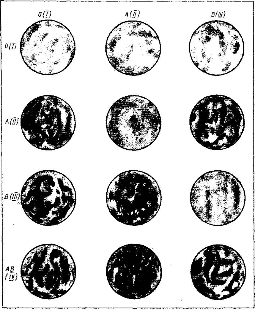
Fig. 27. Evaluation of the hemagglutination reaction with three standard sera across different blood groups
Each of the group antigens (especially A) can induce the formation of corresponding antibodies (a1, a2, a3, etc.) during repeated transfusions, leading to post-transfusion reactions. Therefore, It is important to select identical blood for transfusion not only by the main blood group antigen, but also by subgroup antigens (A1, A2, A3, etc.). This requires special sera containing Monoclonal Antibodies (a1, a2, a3, etc.). In such cases, the compatibility of recipient and donor blood can be precisely determined for both group and subgroup antigens, thereby preventing post-transfusion complications.
In recent years, a method for blood typing using monoclonal antibodies against the A and B antigens of the ABO system—known as цeликлони (celiclones)—has been developed. These agents are obtained from mouse ascitic fluid produced by hybridomas. These hybridomas are generated experimentally by fusing mouse lymphocytes with mouse myeloma cells. Different hybridomas produce monoclonal antibodies against the A and B antigens. The ascitic fluid of mouse hybridomas contains IgM against the A and B antigens, which forms The basis of celiclones. As specific agents, monoclonal antibodies against A and B antigens in celiclones provide a faster and more distinct agglutination reaction compared to standard sera.
Celiclones are supplied as a liquid (diluted ascitic fluid) colored red for anti-A and blue for anti-B antigens, packaged in ampoules or vials. The reaction is carried out at a temperature of 15–25 °C. Celiclones are applied in large drops onto a porcelain plate or tile with appropriate labels. Next to each drop of celiclone, place a drop of test blood that is 10 times smaller, and mix the drops together. Gently rock the plate or tile. Evaluate the reaction after 2.5–3 minutes. The agglutination reaction typically develops very rapidly—within 3–5 seconds—and appears as small red erythrocyte aggregates that subsequently form flakes when celiclones encounter their corresponding antigens. There are 4 possible reaction outcomes:
1) agglutination with anti-A and anti-B celiclones is absent, indicating a lack of antigens A and B; the test blood belongs to group I (0);
2) agglutination occurred in the sample with anti-A Cliclon, indicating the presence of antigen A in the blood; the blood sample belongs to blood group II (A);
3) agglutination is observed in the sample with anti-B Cliclon, indicating the presence of antigen B in the blood; the blood sample belongs to blood group III (B);
4) agglutination occurred in the samples with both Cliclons (anti-A and anti-B), indicating the presence of both antigens A and B in the blood; the blood sample belongs to blood group IV (AB).
If the blood sample under investigation yields an agglutination reaction with both Cliclons (group IV AB), a control test with isotonic sodium chloride solution must be performed. 0.1 mL of the solution is mixed with a drop of the test blood. If no agglutination occurs, the blood is classified as group IV (AB); if agglutination does occur, testing with washed standard erythrocytes must be carried out.
The Rhesus factor is determined using specialized standard anti-Rh sera. They are supplied in two-series kits and stored in a refrigerator at a temperature of 4–6 °C. In addition to Rhesus antibodies, standard anti-Rh sera also contain group agglutinins; therefore, the ABO blood group must be taken into account during testing.
Standard anti-Rh sera are produced from the blood of Rh-negative women immunized by the Rhesus factor. Serum can also be obtained by immunizing guinea pigs with erythrocytes of Macacus rhesus monkeys.
The Rhesus factor is determined using the following method. Three drops of standard anti-Rh serum are placed in two rows on a Petri dish. The erythrocytes of the test blood are added to the first two drops (of two series); standard, predetermined Rh-positive erythrocytes are added to the second two drops (as a control); and standard, predetermined Rh-negative erythrocytes are added to the third two drops (as a control). The Petri dish is placed in a Water bath for 10 minutes at a water temperature of 40–42 °C. If the blood is Rh-positive, agglutination occurs in the First and Second drops; if the blood is Rh-negative, agglutination occurs In the second drops but is absent in the first and third drops.
Genetics of blood groups. Human blood groups, like all other traits, are inherited according to the classical laws of genetics, meaning they are determined by the set of genes received from the maternal and paternal Chromosomes. Consequently, an individual can only possess blood antigens that are present in their parents. Knowing the blood groups of both parents makes it easy to predict the possible blood groups of their children. For example, if both parents have blood group I (0), all their children will also have blood group I (0). However, if the parents have blood group III (B), their offspring may have not only this group but also I (0). This is because every individual receives one chromosome from each homozygous pair from their parents. If one chromosome carries allele B and the other carries allele 0, the genotype of such an individual can be represented as B0. Their blood will always exhibit group B because, during Gene interaction, the B allele (as dominant) suppresses the 0 allele. If two individuals with B0 genotypes marry, some of their offspring may receive a chromosome with the 0 allele from each parent, in which case the children will have the 00 blood group.
Given the above, the genotype of the offspring depending on the parental genotypes can be as follows:
|
Parental |
Possible genotypes |
|
genotypes |
of offspring |
|
00x00 |
00 |
|
00хАА |
А0 |
|
00хА0 |
00, А0 |
|
00хВВ |
В0 |
|
00хВ0 |
00, В0 |
|
ААхАА |
AA |
|
ААхВВ |
AB |
|
А0хВ0 |
AB, А0, В0, 00 |
|
А0хАА |
AA, А0 |
|
А0хА0 |
AA, А0, 00 |
|
ВВхВВ |
ВВ |
|
В0хВ0 |
ВВ, В0, 00 |
|
АВхАВ |
АА, AB, ВВ |
|
АВх00 |
А0, В0 |
|
АВхАА |
AB, АА |
|
АВхА0 |
АА, А0, AB, В0 |
|
АВхВВ |
AB, ВВ, |
|
АВхВ0 |
ВВ, В0, AB, А0 |
Antigen O (H, or the 'silent' antigen) is a weak antigen that can only be detected using specialized sera and has minor practical significance.
Errors during blood group determination can be caused by three main factors.
1. Blood-typing technique: incorrect placement of standard sera or erythrocytes on the tile; mixing of sera or erythrocytes from different groups; incorrect quantitative ratio of serum to erythrocytes; premature reading of results; rinsing pipettes with tap or distilled water instead of isotonic sodium chloride solution; mistaking erythrocyte clumping along the edges of a drying drop for true agglutination.
2. Substandard quality of standard sera: expiration of shelf life; a titer lower than 1:32; use of infected or drying sera.
3. Biological Properties of the test blood: presence of a weak variant of antigen A (A3, A4, etc.) or B in the test erythrocytes, or decreased activity of antigens A and B due to certain pathological conditions (e.g., Sepsis, leukemia).
Errors in blood group determination can be divided into two categories:
1) agglutination is overlooked where it is actually present;
2) agglutination is falsely recorded when it is absent.
Agglutination may be missed when the reaction starts late or is weakly expressed (premature evaluation of the reaction). The reaction may also fail to appear if a larger drop of blood than plasma is used, or in high ambient temperatures.
Agglutination occurs when it should not in the following cases:
1) pseudoagglutination (false agglutination) — the stacking of erythrocytes into coin-like rouleaux. To eliminate this, a drop of isotonic sodium chloride solution is added to the drop where agglutination has occurred;
2) panagglutination (autoagglutination, cold agglutination) is caused by cold panagglutinins present in the serum of most individuals, which are active at temperatures below 15–17 °C. In this case, the serum agglutinates erythrocytes of all blood groups, including its own.
TYPES, METHODS, AND TECHNIQUE OF BLOOD TRANSFUSION
When considering the clinical aspects of blood transfusion, one must first address three key questions: what blood can be transfused, where should it be transfused, and how should it be transfused?
What blood can actually be transfused?
In clinical practice, stored (preserved) blood is most commonly used. This is blood drawn from a donor and mixed with a preservative solution in a 4:1 ratio, or subjected to low temperatures. Preservative solutions perform a triple function:
1) they stabilize the blood in a liquid state (preventing it from clotting);
2) they prevent premature destruction of blood formed elements, thereby ensuring its preservation in a biologically viable state;
3) they prevent the blood from spoiling.
Today, A number of preservatives have been proposed. Their components include: a stabilizer (to prevent blood clotting), an agent that prevents premature hemolysis, and antibacterial drugs.
One such preservative is a glucose-citrate solution. Its composition includes: 2 g of acid citrate (stabilizer); 3 g of glucose (preservative); 0.5 g of sodium sulfacyl (antibacterial agent); 0.01 g of levomycetin (antibacterial agent); and up to 100 ml of redistilled water. The shelf life is up to 3 months. Blood prepared with this solution can be stored in a refrigerator at 4–6 °C for 10–14 days, although it retains its properties for up to 3 weeks.
A preservative containing anticoagulant and nutrient substances (citrate, phosphate, glucose, adenine) that support erythrocyte METABOLISM makes it possible to store blood at 2–6 °C for up to 35 days.
In addition to sodium citrate, cation-exchange resin (which binds Calcium Ions), hirudin, and heparin can be used to stabilize blood.
Currently, the preservation and storage of blood using cold is widely practiced.
Biological freezing makes it possible to preserve cells, Tissues, Organs, and organisms in a viable state. The lower the ambient temperature, the slower the metabolic processes in living cells.
Since temperatures below 0 °C cause ice crystals to form inside cells, leading to their death, biological freezing is performed by adding special substances (cryoprotectants) to the blood to keep the cells intact. Glycerol is one such cryoprotectant. Freezing is carried out rapidly in a liquid nitrogen atmosphere (— 196 °C). After thawing, glycerol is removed from the blood.
Biologically frozen blood can be stored for 15–16 years. To this end, blood transfusion stations have established specialized departments known as long-term blood banks. Such banks exist at the Kiev, Moscow, St. Petersburg, Tbilisi, and Yerevan Institutes of Hematology and Blood Transfusion, as well as at major blood transfusion stations.
However, during prolonged cold storage, certain changes occur in the blood due to erythrocyte metabolism and Aging, the loss of platelet hemostatic activity, and a decrease in The activity of clotting factors V and VIII.
Fresh citrated blood is blood drawn from a donor and mixed with a 3.5–4% sodium citrate solution (1:9). It can be stored in a refrigerator for several hours.
Heparinized blood. Heparin, glucose, and levomycetin are added to it as a stabilizer and preservative. It has a shelf life of up to 24 hours and is used to prime cardiopulmonary bypass circuits.
Salvaged therapeutic blood. This is obtained during therapeutic phlebotomies (for Hypertension or Eclampsia). The blood is collected under sterile conditions, preserved, and transfused after testing.
Placental blood. This is obtained from the Placenta after the umbilical cord is ligated, observing all aseptic rules. It is collected into a 100–200 cm3 container holding a stabilizer. A separate sample is taken to determine the blood group and perform serological tests. A characteristic feature of placental blood is that agglutinogens are clearly expressed, whereas agglutinins cannot be detected by standard methods. Although placental blood yields good therapeutic effects, it is used less frequently than stored blood.
Autologous blood (autoblood) is blood that has extravasated during trauma into the pleural, pericardial, or peritoneal cavities and is not contaminated by the Contents of the large Bronchi or abdominal hollow organs. Mixed with a stabilizer and filtered, it can be reinfused into the same patient. The transfusion must be performed no later than 12 hours after the hemorrhage.
In recent years, it has become common practice to use blood harvested from the patient in advance. This blood is drawn before surgery (400–600 ml), preserved, and transfused to the patient during surgery or in the postoperative period.
Cadaveric blood (post-mortem, fibrinolytic). The idea of its use belongs to V. Shamov, who in 1928, together with M. Kostyukov, conducted experiments on animals and proved the viability of transfused erythrocytes and whole cadaveric blood.
The pioneer of clinical fibrinolytic blood transfusion is S. Yudin, who in 1930 at the Sklifosovsky Institute in Moscow was the first to transfuse cadaveric blood to a human.
Fresh cadaveric blood differs very little from regular donor blood. Its only distinctive feature is that 1–4 hours after death, it no longer clots and does not require The addition of a stabilizer. Cadaveric blood is harvested from patients who died suddenly (from myocardial infarction, electrocution, or other closed trauma not accompanied by damage to the Skin, Blood Vessels, or hollow organs, asphyxia, or signs of agony). Contraindications for harvesting cadaveric blood include malignant tumors, blood and hematopoietic disorders, malaria, tuberculosis, Syphilis, extensive wounds, ulcers, infections, Burns, etc.
Under sterile conditions, 4 liters of blood are collected from the jugular vein of a cadaver within the first 6 hours after death. Its shelf life is up to 14 days. Due to the specific conditions required for harvesting fibrinolytic blood, its use is limited.
Artificial blood belongs more properly to blood substitutes. It performs one of the primary Functions OF BLOOD—Oxygen transport—which no other blood substitute can do.
In recent years, scientists have focused on developing a blood substitute capable of transporting oxygen. Many approaches have been proposed to achieve this goal. Successes in this
Japanese scientists have achieved a breakthrough in this field by developing a blood substitute that appears as a milky-white emulsion, where microscopic fluorocarbon droplets play The Role of red blood cells. Animal experiments have confirmed the feasibility of administering it to humans.
In the spring of 1979, a 60-year-old patient with prostate Cancer underwent surgery at a hospital in Fukushima, Japan. During the operation, he suffered massive blood loss. The hospital lacked the necessary blood supply at the time, so artificial blood was transfused instead. The patient survived.
By 1980, the preparation had been transfused to 70 patients in Japan and 3 in the USA. Subsequently, the artificial blood preparation synthesized in Russia began to be successfully used in clinical practice in both Russia and Ukraine (L.V. Usenko). This novel blood substitute offers certain advantages over donor blood: it can be transfused without blood type matching, it absorbs 3 times more oxygen from the air than red blood cells, and it can be stored in a refrigerator for up to 3 years.
Research on the production of artificial blood is ongoing.
The second question is: where is the best site to infuse blood? There are several routes for administering blood into the body. The most common is the intravenous route: percutaneous or via venesection into superficial Veins (of the antecubital fossa, forearm, hand, or FOOT). Its drawback is that if prolonged transfusion is required, the veins may thrombose and develop inflammation.
Conditions for transfusion therapy are optimized by catheterization of a major venous vessel with a high volumetric blood flow rate. The subclavian vein is most frequently used for this purpose.
Intra-arterial blood transfusion is performed under specific indications. The main difference between this transfusion method and the intravenous approach is the pronounced reflex stimulation of Cardiac Activity resulting from the irritation of angioceptors and the restoration of Coronary Circulation. Intra-arterial blood is transfused under a pressure of 180—200 mm Hg. Indications for this method include cardiac arrest in clinical death caused by massive uncompensated blood loss, and terminal states. Blood can be administered into various Arteries, preferably of a larger caliber. It is transfused from an ampoule with a manometer connected to the system.
Intra-arterial blood transfusion is performed during Operations on the abdominal or thoracic organs, in cases of clinical death against the Background of deep and prolonged hypotension caused by significant uncompensated blood loss, as well as during massive hemorrhage in abdominal or thoracic surgeries.
Intra-arterial [intra-osseous] blood transfusion is used very rarely, mainly in patients with extensive burn surface areas (into the Sternum, calcaneus, ilium, or the epiphyses of long tubular bones).
Other routes of blood administration (into the fontanelles of infants) are used even less frequently. Since the intravenous method of blood transfusion is the most commonly used, its technique will be described in detail.
The ampoule of blood is removed from the refrigerator and warmed in a water bath to a temperature of 18—20 °С. The transfusion is carried out using a disposable administration set (Fig. 28). The disposable set is centrally sterilized using gamma rays.
The patient lies on a bed or dressing table, with the arm positioned alongside the body and slightly abducted. The pouch is opened, and the administration set is removed under sterile conditions. The metal seal is removed from the blood ampoule, and the rubber stopper is wiped with 96% ethyl alcohol. It is then punctured with a needle attached to one end of the set, proximal to the filter. The blood ampoule is inverted. By raising and lowering it relative to the other end of the set, the tubes are filled with blood, and air is expelled from them (to equalize the pressure inside the ampoule with atmospheric pressure, an additional long needle is inserted through the stopper, with its tip positioned above the blood level). After the set is filled and blood begins to flow from its other end, the tube is clamped with a special clamp attached to the set. The drip chamber with the filter should be half-filled with blood. The patient's arm is compressed in the shoulder area with a rubber tubing, and the patient is instructed to clenching and unclenching their fist several times. This causes the arm veins to fill with blood and become more prominent. The skin in the antecubital fossa is disinfected with 96% ethyl alcohol, and the superficial vein is punctured with a needle that was also kept in the plastic pouch (Fig. 29, a).
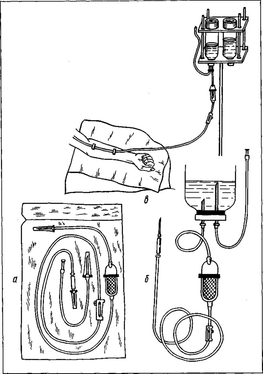
Fig. 28. Disposable blood transfusion set: a — in a sterile plastic pouch; b — assembled; c — blood transfusion into a forearm vein
When blood appears from the needle, the rubber tubing is removed from the shoulder, and the needle is connected to the infusion set. Using the clamp that compresses the tubing, the flow of blood into the vein can be regulated (drip or jet). The rate of blood flow into the vein is monitored via the drip chamber.
A blood transfusion is never carried out to absolute completion; a few milliliters are left in the tubing system and the ampoule. Upon completion of the transfusion, the tubing is clamped, and the needle is removed from the vein. The puncture site is pressed for 2—3 minutes with a cotton ball soaked in 96% ethyl alcohol.
If the subcutaneous veins are poorly contoured and very difficult to puncture, venesection is performed. The procedure is conducted under aseptic conditions. It is preferable to perform it in an operating room or a dressing room, or in the patient's ward on the bed if the patient is non-transportable (Fig. 29, b).
The surgeon scrubs in as for surgery, prepares and drapes the operative field (usually the antecubital vein, although any superficial vein may be used). Local anesthesia is administered using a 0.5% novocaine solution. The skin and subcutaneous tissue are incised parallel or perpendicular to the vein at a distance of 2—3 cm. A segment of the vein is mobilized. Two silk ligatures are passed beneath it. The distal ligature is used to ligate the vein. At 0.5—1 cm proximally, at a distance of 1 mm, the anterior wall of the vein is incised transversely, and a thin polyvinyl catheter with a cannula at the end is inserted into it. The proximally placed ligature is tied around the vein and the catheter (taking care not to occlude the lumen of the vein), thereby securing the catheter in the vein. When blood begins to flow from the catheter, the infusion set is connected to the cannula. The skin over the vein is sutured, and the tubing is secured to the skin with adhesive plaster. After the transfusion is completed, various approaches can be taken. If no further transfusions are planned in the near future, the catheter is removed, and the vein is compressed for several minutes with a tight bandage.
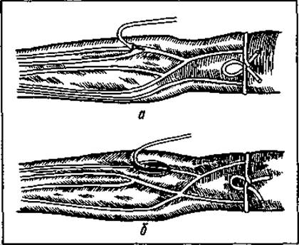
Fig. 29. Intravenous blood transfusion:
a — venipuncture; b — venesection
If repeated infusions are necessary, 0.5 ml of heparin is injected into the catheter (to prevent catheter thrombosis), and its tip is sealed with a special plug. During subsequent transfusions, the plug is removed, and the infusion set is reconnected to the catheter.
How is blood transfused? The following methods of blood transfusion are distinguished: direct, indirect, exchange transfusion, and retrograde.
Direct transfusion involves the immediate transfer of blood from a donor to a recipient (without prior stabilization or conservation). The main advantage of this method is that the transfused blood maximally retains its properties. At the same time, direct transfusion has its drawbacks. It is more complex and requires specialized equipment. With direct transfusion, it is impossible to administer blood by drop infusion. During the hemotransfusion, the donor lies next to the patient, which poses a risk if the patient has an infectious disease.
In view of the above, the indications for direct blood transfusion are quite limited. These include: ongoing hemorrhage that cannot be stopped by other methods; hypocoagulation in blood system disorders; stage III Traumatic shock combined with a blood loss exceeding 45—50% of the circulating blood volume (CBV); and deep burns covering more than 20% of the body surface area.
Contraindications to direct blood transfusion include pulmonary and cardiac insufficiency, pulmonary edema, hypercoagulation, as well as oncological and infectious diseases in the recipient.
Technically, a direct blood transfusion is performed as follows. The donor and recipient are positioned on two parallel operating tables (since direct transfusion is a surgical procedure, it is usually carried out in an operating room). A small table is placed between them at table height to support the arms of both the donor and the recipient. The transfusion process itself can be performed using either syringes or specialized apparatuses. The simplest method (now rarely used) is direct transfusion using syringes. The VEINS OF THE donor and recipient are punctured with needles fitted with attached tubing (the donor's upper arm is constricted with a tourniquet). Blood is drawn from the donor's vein using a 20 ml syringe. Afterward, the tube is clamped, and the blood is infused into the recipient's vein through a second tube. By alternately drawing blood from the donor and infusing it into the recipient, 200–400 ml of blood is transfused to the patient. This method is labor-intensive and is currently used only when no other options for direct transfusion are available and the transfusion is critically necessary.
The second method of direct blood transfusion is more sophisticated. Three rubber or polyvinyl chloride tubes are attached to a metal three-way stopcock mounted on a special stand. The end of the first tube is connected to a needle inserted into the donor's vein, and the end of the second tube is connected to a needle inserted into the recipient's vein. A 20 ml syringe is inserted into the third tube (the Ttsang-Braitsev apparatus). By alternately turning the stopcock toward the donor's or the recipient's vein, blood is drawn from one into the syringe and infused into the other. To prevent the tubing from clotting, it is periodically flushed with sodium citrate.
Even more advanced are devices that operate on the displacement pumping principle (the Jouvalier apparatus, the PKPU universal apparatus). Blood is pumped through a polyvinyl chloride tube using a continuous-action device equipped with a roller or finger pump and a manual drive.
Indirect blood transfusion. The donor and recipient are completely separated both in time and in space. Blood is initially collected from the donor into a special container holding sodium citrate or a blood preservative, and is subsequently used at various times.
Indirect blood transfusion is performed using a closed system from the same container (glass bottle or plastic bag) in which the blood was originally harvested.
Transfusion can be administered by the drip method (50–60 drops per minute), the jet (rapid) method, or under pressure (intra-arterially or intra-aortically).
Exchange blood transfusion. This method combines simultaneous phlebotomy (blood removal) and blood transfusion. It is used primarily for detoxification purposes. During phlebotomy, toxic substances adsorbed onto erythrocytes or dissolved in the plasma are removed along with the blood, after which the lost blood volume is replaced with donor blood. Recently, with the advent of newer and more effective Methods of Detoxification Therapy (hemodialysis, peritoneal dialysis, hemo- and lymphosorption), exchange blood transfusion has largely lost its practical significance.
Absolute indications for exchange blood transfusion include hemolytic disease of the newborn and poisoning with substances that exert a direct toxic effect on the blood.
Reverse blood transfusion (autologous blood transfusion, reinfusion). In recent years, surgeons have become increasingly interested in autologous blood transfusion—that is, the transfusion of blood previously collected from the patient themselves. This method's popularity stems from the fact that major surgeries require large amounts of stored blood for transfusion, and finding suitable donors is often challenging. Furthermore, massive transfusions of donor blood frequently lead to severe complications and adverse reactions.
Most often, autologous transfusion is used to compensate for intraoperative blood loss exceeding 500–800 ml (10–15% of the circulating blood volume).
The technique of autologous blood transfusion involves exfusion (withdrawal) of 400–800 ml of blood from the patient 1–2 days prior to surgery, with simultaneous partial replacement (by 1/3 or 1/2) using a suspension of cryopreserved erythrocytes or other blood substitutes. During surgery, if bleeding occurs, the patient loses this diluted blood. The blood collected prior to surgery is then returned to the patient during the postoperative period.
During autologous blood transfusion, care must be taken to ensure that the exfusion does not exert a negative impact on the patient's body, and that the stored blood used for transfusion during surgery has a minimal storage age—meaning it retains its oxygen-transport function and provides maximum therapeutic efficacy.
Reinfusion is the transfusion of a patient's own blood that has accumulated in serous cavities, provided there is no hemolysis, infection, or other contraindications. This occurs in cases of closed abdominal trauma with rupture of the Liver or Spleen, ruptured Ectopic Pregnancy, closed hematothorax, etc. Under sterile conditions, the blood is collected into a special container, filtered through 8 layers of gauze, mixed with a preservative, and returned to the patient.
Assessment of blood suitability for transfusion. Stored or fresh citrated blood is evaluated based on its quality, seal integrity, storage period, proper documentation, and macroscopic inspection.
Blood assessment should be performed immediately after removing it from the refrigerator, without agitation. Normally, such blood should separate into three layers: a layer of erythrocytes at the bottom, a thin layer of leukocytes above it, and a layer of transparent, yellowish plasma at the top.
Indications that blood is unsuitable for transfusion include:
1) erythrocyte hemolysis (in which case the plasma has a pinkish hue);
2) presence of blood clots (if the blood container is tilted, the clots float to The surface of the plasma);
3) blood contamination (appearance of flakes, films, or turbidity in the plasma).
It is essential to verify that the storage time limits have been respected (checking the manufacturing date) and to confirm the blood group and Rh factor.
Pre-transfusion testing.
Before performing a direct or indirect donor blood transfusion, several tests must be carried out.
1. Determine the blood groups of the donor and recipient according to the ABO system and Rh factor (as discussed previously).
2. Perform an individual compatibility test for the ABO system at room temperature. This test is performed using the patient's blood serum. For this purpose, 2–4 ml of blood without an anticoagulant is drawn from the recipient's vein (as anticoagulants can negatively affect the test result). Once a clot has formed in the test tube, a drop of serum is collected from the bottom using a pipette and placed on a dry white porcelain plate or Petri dish. A volume of donor blood (taken from the ampoule) that is 10 times smaller than the serum drop is added and mixed. After waiting 5 minutes, the test result is evaluated. The absence of agglutination indicates that the donor blood from that particular ampoule and the recipient's blood are compatible.
An individual compatibility test using the ABO system can also be performed in a water bath. It is carried out similarly to the previous one, except after mixing the donor's blood with the recipient's blood serum. The Petri dish is kept for 10 min in a water bath at a temperature of 46—48 °С. This test prevents the transfusion of Rhesus-incompatible blood.
During blood transfusion, a biological test is performed. 20—25 ml of donor blood is transfused in a jet stream three times at 3-minute intervals. During the intervals, the tubing system is clamped or the blood is administered drop by drop, very slowly, to prevent needle clogging. The absence of subjective or objective signs of incompatibility (which will be discussed in subsequent chapters) indicates that the transfusion can be continued.
The biological test prevents the transfusion of blood that is incompatible exclusively according to the ABO system.
It should be noted that blood transfusion is a critical procedure. It may only be performed by a physician. It must be carried out taking into account possible contraindications and observing all technical rules. Otherwise, a whole range of complications, sometimes fatal, may occur.
MECHANISM OF ACTION OF TRANSFUSED BLOOD
To what does blood owe its therapeutic properties? To answer this question, it is necessary to characterize the functions that blood performs in the body. Speaking of these, it must be emphasized that blood plays the primary role in ensuring the vital activity of the Organism. Like no other biological environment, it features a rich chemical composition. More than 200 different substances are contained within it. Blood fulfills its purpose with great precision: it ensures the RESPIRATORY FUNCTION OF the body, facilitates The excretion of Metabolic waste products, participates in numerous chemical processes within the organism, regulates Water and Electrolyte balance as well as temperature, protects the body against microbes and various harmful factors, and maintains the stability of all organs and systems by integrating them into a single whole.
Based on the foregoing, it can be concluded that blood performs numerous functions in the body.
The Respiratory Function of Blood lies in the fact that, passing through the alveolar capillaries, it captures oxygen from them and delivers it to all body tissues. It picks up carbon dioxide from the tissues and carries it to the Lungs, from where it is eliminated from the body via the alveoli.
The substitution function is particularly noticeable when blood is transfused following blood loss. It replenishes the total circulating blood volume, increases the number of red blood cells in the vascular bed—thereby improving oxygen Transport from the lungs to the tissues—and stimulates the release of blood from depots, incorporating it into the general circulation. In the patient's body, the transfused blood finds normal conditions for survival. This is confirmed by the fact that the transfused donor erythrocytes retain their physiological properties in the recipient's body for up to 30 days, playing an important role in oxygen metabolism. Leukocytes also preserve their physiological properties, participating in phagocytosis.
The hemostatic function of transfused blood is associated with the presence of factors that actively participate in Blood Coagulation. The best effect in this regard is achieved by direct blood transfusion.
The stimulating action is directed toward a number of important bodily functions. It is the most complex and, therefore, perhaps the least understood. Transfused blood improves the functions of The Cardiovascular system, the Central Nervous system and its peripheral divisions, as well as hemodynamics; it activates external and internal Respiration and increases the body's resistance. It is believed that transfused blood plasma, which contains
abundant colloids, proteins, breakdown products of formed elements, salts, etc., irritates the Bone Marrow and the reticuloendothelial apparatus, thereby achieving its therapeutic effect.
The detoxifying function of blood consists in a significant reduction of intoxication in cases of exogenous or endogenous poisoning of the organism. This is presumably related to the enhanced adsorption of toxins by transfused erythrocytes and proteins, their dissolution in the blood, and their transport to excretory organs. The detoxifying effect of transfused blood is particularly pronounced in poisonings where Hemoglobin converts into methemoglobin or carboxyhemoglobin (which occurs in intoxications with phenol, potassium chlorate, Carbon Monoxide and illuminating gas, hemolytic poisons including arsenic, snake venom, mushrooms, etc.).
The immunobiological function of transfused blood is associated with: the possibility of transferring immune bodies during transfusion, which stimulates the development of passive Immunity in the recipient's body; an increase in the phagocytic activity of leukocytes; The stimulation of antibody formation; and the normalization of the body's reactivity. To enhance the immunostimulating effect, physicians sometimes use blood from donors who have immunity to a specific infection.
The trophic function of blood transfusion is based on the fact that transfused blood contains a number of energetic and plastic components (proteins, fats, carbohydrates, Vitamins, electrolytes, water, etc.). Introduced into the recipient's body along with the blood, they become integrated into the general metabolism.
The thermoregulatory function consists in the fact that blood transfers heat from energy-intensive organs and warms up organs that are losing it.
The humoral function involves The transport of Hormones and other biologically active compounds by the blood from the cells where they are produced to other organs and tissues. This ensures chemical interaction among all PARTS OF THE organism.
When examining The Mechanism of action of transfused blood on the organism, it should be noted that in recent years, some of its individual functions have been called into question. For instance, certain researchers believe that transfused blood not only lacks a hemostatic effect but, conversely, provokes bleeding; the nutritional effect is exerted not by the blood itself, but solely by albumin, and even then it is assimilated by the body only after 2 weeks; furthermore, blood transfusion exhibits no stimulating influence and instead inhibits hematopoiesis. Only a single function of blood remains beyond doubt—its respiratory function.
INDICATIONS AND CONTRAINDICATIONS FOR BLOOD TRANSFUSION
When, in what cases, and under what circumstances is blood transfusion indicated? Proceeding from what is known about its functions, until recently blood was widely used in clinical practice even when one could have managed without it.
Currently, the transfusion of whole preserved blood is significantly restricted. It is transfused only when absolutely necessary. Otherwise, it is replaced by components or blood substitutes.
From a practical standpoint, all indications and contraindications for blood transfusion can be divided into relative and absolute. Absolute indications require blood transfusion when it is necessary to save a human life and the patient would inevitably die without it. These include fatal hemorrhages, stage III and IV shock, and clinical death. Such indications must be clearly justified. Often, clinical signs alone are insufficient, but a drop in systolic pressure below 100 mm Hg following blood loss, indicating a 30% deficit in circulating blood volume, provides a grounds for transfusion. In such cases, whole blood should not be used immediately. One should start with the infusion of crystalloid solutions, later switch to packed red blood cells, and then, if necessary, transfuse whole blood over the course of the day.
Under relative indications, the patient can manage without a blood transfusion. It serves merely as one of the components of complex therapy and is not of decisive importance. It is preferable to transfuse blood, but if contraindications exist, it can be dispensed with.
Relative indications for blood transfusion include minor parenchymatous and capillary hemorrhages, stage I and II shock, leukopenia, hypoproteinemia, endogenous and exogenous intoxications, sepsis, acute and chronic purulent processes, sluggish inflammatory processes, delayed wound healing, and intoxication with poisons that convert hemoglobin into carboxyhemoglobin (carbon monoxide) and methemoglobin (phenol, aniline), as well as preoperative preparation.
In recent years, relative indications for whole blood transfusion have been notably narrowed. Absolute contraindications to blood transfusion include acute cerebral hemorrhages, thrombosis of cerebral vessels, and pulmonary edema.
Relative contraindications to blood transfusion include the following:
1) cardiovascular pathology (acute septic endocarditis, myocarditis, heart defects, stage II and III Circulatory Disorders, stage II hypertension, pronounced cerebral arteriosclerosis, thromboembolism);
2) lung diseases (active and Disseminated Pulmonary Tuberculosis);
3) Kidney and liver diseases accompanied by impaired function (since conserved blood introduces a significant amount of protein breakdown products, this places high demands on the organs responsible for neutralizing them; therefore, relative contraindications to blood transfusion may include Renal Amyloidosis, acute Glomerulonephritis, nephrosclerosis, and acute and chronic hepatitis);.
4) acute rheumatism;
5) allergic diseases (Bronchial Asthma during an acute flare-up);
6) hemorrhagic vasculitis;
7) central nervous system disorders (concussion, contusion, and compression of the Brain).
Thus, the approach to blood transfusion for each patient must be individualized, taking strict account of both indications and contraindications.
COMPLICATIONS DURING BLOOD TRANSFUSION
Blood transfusion is a serious intervention in the body's vital processes. Occasionally, it can cause undesirable severe reactions and complications. Many of these are associated with gross violations of blood transfusion guidelines.
Numerous schemes and classifications have been proposed for transfusion-related reactions and complications. According to the classification proposed in 1973 by V. Filatov, all complications can be divided into 3 main groups: 1) mechanical complications; 2) reactive complications; 3) complications associated with the transmission of infection from the donor's blood.
Mechanical complications are mainly related to violations of instructions regarding blood procurement or transfusion, as well as technical errors. These include acute cardiac dilatation, air embolism, thromboembolism, and thrombosis.
During the transfusion of large volumes of conserved blood in elderly patients with heart disease, acute cardiac dilatation may occur. It is characterized by the onset of difficulty breathing, a feeling of chest tightness, cardiac pain, arrhythmia, tachycardia, and a drop in blood pressure during or at the end of the transfusion. Cardiac arrest may occur during diastole.
To prevent this complication, it is preferable to transfuse red blood cell concentrates in patients with risk factors.
If signs of circulatory overload appear, the transfusion should be stopped, 200–300 ml of blood should be withdrawn, and medications that tone The Heart Muscle should be administered (calcium chloride, cordiamine, glucose with Insulin, and vitamins). In cases of bradycardia, atropine sulfate solution is recommended. If the patient suffers cardiac arrest, general resuscitation measures should be performed to bring them out of clinical death.
A life-threatening complication is air embolism, which occurs when air enters the bloodstream during a blood transfusion. Air entering a vein moves toward the right side of the heart, and from there into the pulmonary artery and its branches, obstructing them. This manifests as sudden fainting, cyanosis, respiratory arrest (apnea), and cardiac arrest (syncope). The face becomes pale and cyanotic, the pulse accelerates and weakens, and blood pressure drops.
First aid involves rapidly lowering the HEAD end of the table or bed, performing artificial pulmonary ventilation, and administering closed-chest cardiac massage. Puncture of the right side of the heart is performed in the VII–IV intercostal space to the right of the sternum, and about 200–250 ml of frothy blood is aspirated.
Sometimes blood transfusion can be complicated by blood clot embolism.
Pulmonary Embolism or thrombosis of its branches occurs most frequently.
This complication can be caused by three factors: improper blood preservation (stabilization), faulty transfusion technique, and the detachment of a thrombus formed in a vein. Thrombi can detach from distant thrombosed veins (most commonly in the lower extremities) due to increased venous pressure caused by the blood transfusion. The occlusion of one of the main Branches of the pulmonary artery or multiple embolisms of smaller branches is accompanied by collapse, facial pallor and cyanosis, and a cold, clammy sweat. Difficulty breathing, retrosternal pain, and a cough appear (initially dry, later producing blood-tinged sputum). After some time, body temperature rises and clinical signs of pulmonary infarction develop.
If this complication occurs, the blood transfusion must be stopped immediately, and analgesics, antispasmodics, and cardiac drugs should be administered. Anticoagulants and sulfonamides are prescribed to prevent Pneumonia and pulmonary abscess.
In the case of Thrombosis of the vein used for transfusion, a dense, painful, cord-like tract may appear along a certain section of the vessel. Treatment consists of local application of warming compresses, anticoagulants, and fibrinolytic drugs (heparin, phenindione, pelentan, fibrinolysin, etc.), along with heparin Electrophoresis.
Among reactive complications, the following can be distinguished: fever, allergic reactions, massive transfusion syndrome, transfusion and citrate shock, and potassium intoxication.
Post-transfusion fever is caused by The entry of pyrogenic substances into the blood—specifically, products of protein breakdown and microorganisms.
Fever is most frequently caused by the interaction of recipient antibodies with transfused leukocytes, platelets, or IMMUNOGLOBULINS.
The reaction typically occurs 1.5–2 hours after transfusion. The patient experiences a sensation of warmth, chills, occasionally a headache, body temperature rises to 38–39 °C, and vomiting may occur. Depending on the severity of manifestations, three degrees of febrile reaction are distinguished.
In mild reactions, the patient experiences general weakness, slight chills, and a minor elevation in body temperature (within 1 °C).
Moderate reactions manifest as chills, weakness, headache, mild abdominal and lower back pain, and a 1.5–2 °C increase in body temperature.
Severe reactions present with pronounced chills, headache, nausea, vomiting, labored breathing, bone and lower back pain, lip cyanosis, and a significant rise in body temperature (more than 2 °C).
Mild and moderate reactions resolve on their own within a few hours. Severe reactions are prolonged, but in most cases end favorably.
Mild and moderate reactions do not require specific therapeutic measures. The patient should be warmed (covered with blankets, a heating pad applied to the feet, given hot tea). In severe reactions, in addition to the measures listed above, the administration of analgesics (morphine hydrochloride, norphine, tramal, promedol), cardiac medications (camphor, caffeine, cordiamine), antihistamines (dimedrol, diazolin, suprastin), corticosteroids (hydrocortisone, prednisolone — 80–100 mg), antipyretics (acetylsalicylic acid, ascophen, amidopyrine) is recommended, along with an intravenous drip of 10% calcium chloride solution (10 ml) and 5% glucose solution (500 ml) with ascorbic acid.
Allergic reactions are associated with the body's sensitization to donor Blood Plasma Proteins. They occur during repeated blood and plasma transfusions, the administration of protein substances and protein preparations, as well as in certain diseases (chronic inflammatory processes, malignant tumors, blood system disorders).
Signs of an allergic reaction appear during the blood transfusion or 15–20 minutes after its completion. Urticaria, allergic edema in various parts of the body, dyspnea, and other symptoms join the characteristic signs of a febrile reaction.
Allergic reactions are usually mild in nature (except for anaphylactic shock) and resolve spontaneously within 30–40 minutes. Hives, joint pain, and itching may persist for 1–2 days.
If an allergic reaction occurs during the blood transfusion, it must be stopped immediately. The patient is administered intravenously 10 ml of a 10% calcium chloride solution and 5–10 ml of a 5% ascorbic acid solution, diazolin, dimedrol or suprastin, and corticosteroids. Caffeine or camphor is given according to clinical indications.
Transfusing a large volume of blood is accompanied by a number of adverse reactions, known as massive transfusion syndrome (homologous blood syndrome). This syndrome is associated with the Introduction of sodium citrate and potassium into the recipient's body via stored donor blood, changes in biochemical constants and formed elements during blood preservation, and the immunological incompatibility of donor and recipient blood regarding erythrocyte, leukocyte, and platelet antigens as well as plasma protein antibodies, which are practically not accounted for during donor blood Selection.
Massive transfusion syndrome is characterized by: vascular spasm, increased recipient blood viscosity, erythrocyte aggregation, capillary plugging, circulatory disorders, petechial hemorrhages in The Liver and Kidneys, and pulmonary blood congestion. The blood fails to clot, leading to a bleeding tendency.
All of this occurs because it is practically impossible to perfectly match the blood of multiple donors with the recipient's blood across all factors and parameters. Numerous minor immune incompatibilities accumulate, with quantity turning into quality.
Homologous blood syndrome occurs more frequently following the transfusion of stored blood with a prolonged shelf life, in patients operated on under conditions of extracorporeal circulation, in states of shock, and with massive blood loss. It is also common in oncological patients.
To prevent this complication, The Use of fresh stored blood is recommended, combined with the intravenous administration of low-molecular-weight plasma-substituting solutions (hemodes, rheopolyglucukin, rheomacrodex, neocompensan, etc.).
A dangerous complication of blood transfusion is transfusion shock. It is caused by the transfusion of blood incompatible according to the ABO and Rh systems, among other factors. It can also be triggered by the transfusion of infected or altered blood, or blood that was accidentally frozen and thawed or heated to temperatures exceeding 40 °C.
The main causes of this complication are the destruction of donor erythrocytes and their hemolysis with the release of toxic breakdown products (histamine, bradykinins, catecholamines, etc.). Hemolysis of the recipient's erythrocytes under The Influence of donor blood agglutinins is also occasionally possible. This occurs during universal donor blood transfusions to recipients with other blood groups.
Ultimately, all of this leads to the development of shock, severe intoxication, blood clotting disorders, and ACUTE RENAL FAILURE.
The course of transfusion shock comprises three periods: I — shock; II — renal failure; III — recovery.
The shock period (the first few hours). It may develop during the blood transfusion itself, after the introduction of 20–40 ml of blood into the bloodstream. The patient becomes restless, complaining of a sensation of warmth, chest tightness, headache, and lower back pain, nausea, and vomiting. The pain is caused by the spasm of cerebral, mesenteric, and renal vessels. The pulse accelerates, and arterial pressure drops. The pain is so severe that patients become agitated and scream. The patient's face first flushes and then turns pale, chills appear, and body temperature rises. Subsequently, involuntary urination and defecation occur. Hemoglobinuria and hemoglobinemia set in. Jaundice develops rapidly.
The renal failure period is characterized by signs of kidney damage. Urine output decreases or ceases entirely. Uremia develops, characterized by headache, nausea, vomiting, loss of appetite, diarrhea, adynamia, drowsiness, chills, and elevated blood pressure. Pallor and yellowish skin tint, pastiness, and edema increase. Body temperature rises to 38 °C or higher. Anemia develops, and blood levels of urea, creatinine, bilirubin, and potassium increase. Protein, leukocytes, erythrocytes, and casts are detected in the urine.
If the disease progresses, patients die between the 3rd and 18th day following the transfusion. With a favorable course, the patient's condition begins to improve starting from the 2nd–3rd week, ushering in the third period — recovery.
The first sign of the recovery period is the restoration of diuresis. It gradually increases and may reach 3–4 liters per day by the 8th–12th day. The patient's general condition gradually improves. However, even after renal function is restored, general weakness, rapid fatigue, and a temporarily reduced concentrating ability of the kidneys may persist for a long time.
The clinical manifestations of transfusion shock in Rh incompatibility share many similarities with the shock developing from an ABO conflict, but it occurs more frequently after blood transfusions and has a less acute course.
The management of transfusion shock caused by ABO or Rh incompatibility is identical and depends on the stage of the complication.
Treatment must be initiated immediately upon the appearance of the initial signs of shock. During the first 12 hours, exchange blood transfusions provide favorable outcomes. From 1000—2000 ml of the patient's blood is exfused and replaced with group-identical donor blood (stored for 3—5 days). Treatment should aim to normalize hemodynamics and eliminate hemolysis products from the body. Subcutaneous administration of 1 ml of morphine hydrochloride, promedol, 0.5—1 ml of atropine sulfate, cordiamine, or caffeine is indicated, alongside intravenous administration of 20 ml of 40 % glucose solution with vitamins B and C, corglycon, strophanthine (0.5 ml), 10 ml of 10 % calcium chloride solution, and hydrocortisone. Subsequently, 400—500 ml of rheopolyglucukin, hemodes, 400—500 ml of 5 % sodium bicarbonate solution, or 200—250 ml of 10 % sodium lactate solution are administered intravenously. To stimulate diuresis, diuretic agents are administered (20 % mannitol solution at a dose of 1 g of dry substance per 1 kg of the patient's body weight). If no effect is observed after 6 hours, the mannitol administration may be repeated. To dilate renal blood vessels and improve renal blood flow, the administration of caffeine-sodium benzoate (10 % — 1 ml) and euphylline solution (2.4 % — 5—10 ml) is recommended.
To prevent intravascular coagulation, it is advisable to administer 5000 IU of heparin.
The management of patients with acute renal failure should be carried out in specialized units equipped to perform hemodialysis using an artificial kidney machine if necessary.
During this period, fluid intake must be restricted. The amount of administered fluid should equal the volume lost by the patient through urine, feces, vomitus, and exhaled air.
In cases of anuria and the absence of other fluid losses, the daily water requirement should not exceed 500—600 ml. The diet must be easily digestible and contain 20—30 g of protein, 50—60 g of fat, and 240—300 g of carbohydrates. Foods with high potassium and sodium content (fruits, vegetables) are excluded from the diet.
Intravenous administration of 20—40 % glucose solution (200—400 ml) with ascorbic acid, thiamine bromide, pyridoxine, cyanocobalamin, and insulin (40—50 IU per 100 g of dry glucose) is recommended. For detoxification purposes, hemodes, rheopolyglucukin, plasma, and solutions of albumin, protein, and albuminate are administered. To reduce Protein Catabolism, anabolic hormones (dianabol, phenobolin, retabolil) are used.
To enhance the elimination of toxic products from the body, patients are prescribed daily gastric lavage, cleansing enemas with sodium bicarbonate solution, and siphon enemas.
Meticulous patient care is essential (for the Prevention of pneumonia, pressure ulcers, and oral infectious complications). If the aforementioned measures yield no effect, hemodialysis is indicated.
During the convalescent period, the intensity of treatment is reduced. The patient requires careful nursing care and an appropriate diet.
Citrate toxicity develops following rapid transfusion of large volumes of blood stabilized with sodium citrate, most commonly occurring in individuals with impaired liver or kidney function. An elevation in serum sodium citrate levels is accompanied by a decrease in ionized calcium, as sodium citrate forms a complex with it, triggering a cascade of reactions: vasospasm of the pulmonary and cardiac vessels, depression of myocardial contractility, and electrolyte imbalance (primarily calcium and potassium ions). All of this leads to impaired hemodynamics and nervous system dysfunction. A toxic dose of sodium citrate is considered to be 10 mg/kg/min, which corresponds to the administration of 2—3 ml of conserved blood per 1 kg of the recipient's body weight per minute.
Clinically, citrate toxicity manifests as restlessness, tachycardia, arrhythmia, hypotension, dyspnea, and convulsions. These signs appear during or at the end of the blood transfusion. Citrate toxicity can be fatal due to cardiac arrest.
To prevent citrate toxicity, it is recommended to administer 10 ml of 10 % calcium chloride or calcium gluconate for every 500 ml of blood.
In the event of citrate toxicity, the blood transfusion must be stopped immediately, followed by the intravenous administration of 10 ml of 10 % calcium chloride or calcium gluconate solution, and subcutaneous administration of caffeine.
The third group of complications is associated with the transmission of infections from the donor to the recipient. Most frequently, blood transfusions can transmit Viral Hepatitis, malaria, syphilis, brucellosis, Toxoplasmosis, trypanosomiasis, and AIDS.
Some of these infections (hepatitis B, AIDS) may manifest many months or even years after the blood transfusion.
Since the CAUSATIVE AGENT OF syphilis (the spirochete) has a short lifespan, blood stored for more than 4 days at 4 °C is no longer infectious.
To prevent HIV transmission, donor blood in most countries undergoes routine antibody screening against the virus. Concurrently, other measures are implemented to exclude donors belonging to high-risk groups for AIDS. The virus is particularly sensitive to high temperatures and is also inactivated during plasma fractionation for the production of immunoglobulin preparations.
The Clinical presentation of specific infectious diseases and their treatment methods differ very little from those acquired via other routes of infection.
Alongside the transmission of a specific infection into the recipient's bloodstream, bacterial contamination of the blood with opportunistic microbes (streptococci, staphylococci, proteus, etc.) may occur during transfusion, potentially leading to sepsis. This is observed when aseptic techniques are breached during blood collection. Gram-negative microbes are particularly dangerous. In addition to anti-shock therapy, the treatment of such complications requires the administration of high doses of Antibiotics intravenously. The risk of transmitting infection is increased by latent, acute, and chronic donor diseases. Therefore, thorough screening of donors prior to blood collection is of paramount importance in preventing infectious complications.
During blood storage, potassium leaches out of red blood cells, which, upon rapid infusion of large volumes of blood, can lead to potassium intoxication. This exerts a detrimental effect on the myocardium. This complication occurs more frequently in patients with renal pathology and traumatic toxicosis.
An analysis of transfusion-related complications indicates that the majority arise from procedural errors during transfusion or non-compliance with current guidelines for the collection and administration of blood products. Consequently, strict adherence to blood collection and transfusion protocols is essential for their prevention.
DONATION
Worldwide, large volumes of blood are transfused annually for therapeutic purposes. This raises the question of its source. Today, the primary supplier of blood is the donor (from the Latin *donare*, meaning "to give"). Blood donation is both a biological and a social phenomenon.
In most Western countries, whole blood is collected from unpaid volunteers. However, the majority of plasma is obtained via plasmapheresis from paid donors, who yield 500—600 ml of plasma weekly or biweekly. Raw Materials for the majority of commercial plasma products (albumin, immunoglobulin, clotting factors, etc.) are obtained in this manner.
Who can be a donor? Any healthy individual aged 18 to 60 who understands Structure/19.html">The Importance of their patriotic duty to society can become a blood donor. Prospective donors sign a written statement confirming their awareness of the liability for concealing a history of syphilis.
Before donating blood, a person undergoes a thorough medical examination, which includes measuring body temperature (blood donation is prohibited if the temperature reaches 37 °C), blood pressure, a physical examination by a general practitioner and a dermatovenerologist, determination of blood hemoglobin and leukocyte levels, ABO and Rh blood typing, the Wassermann serological test for syphilis, blood bilirubin screening, a urinalysis, and chest fluorography.
For repeat blood donations, the procedure is streamlined: a check-up by a general practitioner, body temperature measurement, blood hemoglobin and bilirubin level determination, and the Wassermann test.
In female donors, the hemoglobin level must not be lower than 120 g/L, and in male donors, 124 g/L. The erythrocyte count should be at least 4 ∙ 1012/L for women and 4.2 ∙ 1012/L for men. The leukocyte count ranges from 4,000 to 9,000 per 1 mm3. The ERYTHROCYTE SEDIMENTATION RATE (ESR) must not exceed 16 mm/h in women and 12 mm/h in men.
Blood donation is contraindicated in individuals with the following medical conditions: 1) congenital and acquired syphilis; 2) all forms of tuberculosis; 3) brucellosis; 4) tularemia, toxoplasmosis; 5) malaria with a history of attacks within the last 3 years; 6) infectious hepatitis (Botkin's disease); 7) serum hepatitis; 8) physical exhaustion, avitaminosis; 9) cardiovascular diseases (stage III hypertension, cerebrovascular disorders, atherosclerosis, endarteritis, endocarditis, myocarditis, and heart defects in the stage of subcompensation or decompensation); 10) malignant tumors; 11) bronchial asthma and other allergic conditions; 12) infectious diseases; 13) mental disorders; 14) pregnancy and Lactation periods; 15) drug addiction, alcoholism; 16) HIV infection.
In some countries, the majority of blood collection organizations adhere to rigorous standards during donor recruitment. Special emphasis is placed on safety against infections.
A first-time donor can safely give 450 mL of blood without any adverse health effects. The donated blood volume is restored within 30–35 days.
BLOOD COMPONENTS AND PRODUCTS
Over recent decades, transfusion medicine has achieved significant milestones. Few would dispute the vital importance of blood transfusion in treating a wide range of diseases. However, clinical practice shows that even in cases of massive blood loss, a patient does not necessarily require whole blood transfusions, as blood products can adequately meet these needs. For instance, patients in shock are administered fresh-frozen or native plasma, whereas severe blood loss is treated with packed red blood cells, among other components.
Thanks to advancements in chemistry, biology, and medicine, methods for isolating specific blood fractions have been developed. All blood components and products can be classified into several groups: 1) anti-anemic agents (packed red blood cells); 2) coagulation system correctors (platelet concentrate, fibrinogen, fibrinolysin, antihemophilic plasma, hemostatic sponge, Thrombin, cryoprecipitate); 3) multi-purpose agents (plasma, albumin, protein solutions); 4) immunological agents (immunoglobulins); 5) agents for anti-infective therapy (leukocyte concentrate).
All of these products are widely utilized in clinical practice.
Packed red blood cells (erythrocyte mass) consist of blood from which 60–65% of the plasma has been removed. They can be native, leukocyte-poor, or washed. Native red blood cell mass contains erythrocytes, 35–40% of the original plasma, leukocytes, and platelets. Washed red blood cells lack these cellular elements and are used under specific indications in patients with plasma protein intolerances. Leukocyte-poor red blood cells serve as an intermediate option between native and washed types, containing 20% plasma, 10–20% platelets, and 30–40% leukocytes.
Washed erythrocytes must be used within 6 hours.
The indication for transfusion of packed red blood cells is a significant reduction in the oxygen-carrying capacity of the blood, which occurs in severe anemia (Hypoxia) caused by hemorrhage, impaired hematopoiesis, and other factors leading to decreased erythrocyte counts.
Transfusion of washed erythrocytes is indicated in cases of marked patient sensitization to donor blood plasma factors.
An erythrocyte suspension is obtained by aspirating the plasma from settled blood and replacing it with an equal volume of isotonic sodium chloride solution.
Transfusion of packed red blood cells is contraindicated in hemoconcentration and when the patient's blood is prone to hypercoagulation.
The storage shelf life of packed red blood cells and Suspensions is 14 days.
Erythrocytes can be preserved for extended periods in a frozen state. Although costly, this method is useful for storing very rare blood types. Once thawed, erythrocytes can be stored for no longer than 24 hours and contain only negligible amounts of leukocytes, platelets, and plasma.
Blood plasma is a protein-rich yellowish fluid. Additionally, it contains various electrolytes, carbohydrates, Lipids, hormones, vitamins, and other substances. It is obtained from whole blood by sedimentation or centrifugation. An alternative procedure is "apheresis" (withdrawal). During plasmapheresis, a donor's blood is drawn, erythrocytes are separated from the plasma and returned to the donor, while the plasma is retained for transfusion. Because plasma protein fractions regenerate much faster than red blood cells, large volumes of plasma can be collected from a single donor over an extended period.
Plasma can be native (in liquid form), vacuum heat-dried, or freeze-dried (lyophilized). Freeze-dried plasma can be fresh-frozen or long-term frozen. Fresh-frozen plasma, when stored at —30 °C, retains all blood clotting factors. It is thawed for 60 minutes and must be used within 4 hours, as prolonged storage leads to the loss of factors V and VIII. Fresh-frozen plasma must be ABO-compatible with the recipient's blood.
Fresh-frozen plasma is an exceptionally effective treatment for all types of hemorrhaging. A patient should be transfused with 800 mL (4 donor doses) over 60 minutes. Fresh-frozen plasma can be stored in a standard household refrigerator.
In some Western countries, long-term frozen dry plasma is manufactured. It is produced from multiple donor units (forming a small pool) by mixing blood of various ABO groups, making it universally compatible for patients of any blood type. Freeze-dried long-term plasma can be stored for up to 5 years.
Prior to administration, dried plasma is reconstituted with an isotonic sodium chloride solution. The shelf life of native plasma is 1 day, and for dried plasma, it is 3 years.
Plasma transfusion helps normalize the protein COMPOSITION OF THE blood and reduces vascular permeability. The fluid portion of the blood is retained within the vascular bed, thereby normalizing blood circulation and oxidation-reduction processes.
Plasma transfusion is indicated for patients with traumatic shock accompanied by blood loss and dehydration, patients with Protein deficiency or the absence of certain protein components, and in septic conditions. It is used for hemostatic purposes to promote faster blood clotting. It is not recommended to transfuse large volumes of plasma (exceeding 1.5 L) to prevent complications such as massive transfusion syndrome, as well as the development of homologous blood syndrome due to tissue incompatibility of immunoplasma factors.
By dissolving dry plasma not in 200 ml, but in 50 ml or an even smaller volume, a concentrated plasma preparation (plasmol) can be obtained. Such plasma induces tissue dehydration, which is crucial in the treatment of cerebral edema.
In addition to standard native or dry plasma, specialty plasmas are also produced, including antihemophilic, antistaphylococcal, and antipseudomonal plasmas, among others.
Other blood components are isolated from plasma: albumin, polyglobulin, fibrinogen, у-globulin, prothrombin, and antihemophilic globulin, which are used as therapeutic agents.
Albumin is the primary fraction of plasma from which it is derived. Solutions are available in 5%, 15%, and 25% concentrations. The 5% solution is used for rapid volume expansion, while 15% and 25% solutions are used to correct hypoproteinemia. The high efficacy of the drug is attributed to its high oncotic and rheological properties.
Albumin actively draws tissue fluid into the bloodstream, enhances autohemodilution, restores circulating blood volume (CBV), binds with toxins, and eliminates them from the body.
Albumin is used to treat depleted patients with pronounced hypovolemia and hypoproteinemia, as well as in burn shock and blood loss.
Protein solution is a 4–8% plasma protein solution available in 100 ml and 200 ml doses. It is derived from retroplacental blood. It contains albumin (75–80%) and stable α- and β-globulins (20–25%). Indications for use are the same as those for albumin. It is administered in combination with donor blood or packed red blood cells.
Fibrinogen is one of the proteins of the BLOOD COAGULATION SYSTEM. It is supplied in vials in dry form. Before use, it is reconstituted with sterile water. To achieve a therapeutic effect, the patient must be administered 5–6 g of the dry preparation; 1–2 g of the dry preparation can be obtained from 1 L of donor blood. It is used in pathological conditions accompanied by a decrease in blood fibrinogen levels, in Fibrinolysis following bleeding in obstetric practice, in liver and spleen diseases, and in disseminated intravascular coagulation (DIC) syndrome. To prevent thromboembolism, fibrinogen transfusion must be combined with the administration of low-dose heparin. Fibrinogen is available in standard vials (similar to blood transfusion vials) of 0.2–1 g. The shelf life is 2 years. Fibrinogen is contraindicated in thrombophlebitis, thrombosis, and decompensated heart defects.
Fibrinolyzin is derived from donor plasma or placental blood serum. It is also a white powder, supplied in the same type of vials as fibrinogen. A single vial contains 10,000 to 20,000 to 30,000 IU of specific activity. The shelf life of fibrinolyzin is identical to that of fibrinogen. The drug is administered concurrently with heparin. It is indicated for pulmonary and peripheral arterial thromboembolism, myocardial infarction, and thrombophlebitis.
Fibrinolyzin is frequently administered alongside thrombolytic agents, such as streptase, streptokinase, streptodecah, and urokinase.
Platelet concentrate consists of a suspension of 60–70% platelets in 40 ml of plasma. Achieving hemostasis requires 5–6 donor doses. Platelet transfusion must be performed immediately after harvesting, as it cannot be stored.
Platelets play a crucial role in the blood clotting process. Upon contact with a foreign surface, they disintegrate, releasing substances contained within the platelets that accelerate The conversion of fibrinogen into fibrin.
Platelet concentrate is used for bleeding caused by platelet deficiency (below 20,000 ∙ 109/L). It is also indicated for patients experiencing blood loss following massive transfusions of stored blood. Since platelet concentrate contains a certain amount of red blood cells, matching donor blood according to the ABO system is advisable.
Cryoprecipitate contains at least 200 IU of antihemophilic globulin (coagulation factor VIII) per single dose, along with fibrinogen and small amounts of other proteins, including fibrin-stabilizing factor (factor XIII). It is prepared from a single donor unit. The product is used for the treatment and prevention of bleeding in patients with hemophilia, as well as in hemorrhages of other etiologies characterized by a depletion of blood coagulation factors VIII and XIII.
Hemostatic sponge. It is applied topically as a hemostatic agent. Produced from blood plasma and supplied in a dried form.
Thrombin is an effective local hemostatic agent used for capillary bleeding and other conditions accompanied by minor hemorrhages, as well as during surgical interventions on parenchymal organs.
Leukocyte concentrate is a blood component containing a high concentration of white blood cells from peripheral blood, mixed with platelets, red blood cells, and plasma. Leukocyte concentrate can be obtained through blood sedimentation or leukapheresis. In this process, a donor's blood is collected, differentially centrifuged, the necessary components (leukocytes, platelets) are harvested, and the remaining constituents are returned to the donor.
Leukocyte concentrate is supplied in 50 ml vials. This volume contains a single therapeutic dose of leukocytes (equivalent to the leukocyte count in 500 ml of blood). Transfusion requires accounting for the blood group compatibility of both the donor and the recipient. Leukocyte concentrate should not be stored for longer than 24 hours. It is most commonly used as a component of anti-infective therapy. The mechanism of its therapeutic action relies on introducing deficient, functionally competent neutrophil granulocytes—the primary cellular mediators of antimicrobial defense—into the recipient's body.
Leukocyte concentrate is transfused in courses with intervals of 2–3 days.
According to their mechanism of action, immunological preparations are divided into two groups: nonspecific (nonspecific у-globulin, polyglobulin) and specific. Nonspecific preparations contain a broad spectrum of antibodies against various viral and bacterial pathogens. They are used for passive immunization against conditions such as streptococcal infection, Influenza, and a range of childhood infections. They are manufactured from donor and placental plasma (serum).
Today, targeted immunoglobulins are of paramount importance. Their therapeutic and prophylactic action is directed against specific infections. This is achieved through the active immunization of donors with the corresponding antigen, the collection of immune plasma, and the Isolation of the immunoglobulin fraction. The resulting immunoglobulin preparation exhibits high specific activity against the pathogen whose antigen was used for immunization. Widely used agents of this type include antistaphylococcal у-globulin, antistaphylococcal polyglobulin, tetanus immunoglobulin, anti-Rh (D) immunoglobulin, hepatitis B immunoglobulin, and rabies immunoglobulin.
Targeted immunological preparations are manufactured from plasma with high titers of specific antibodies, convalescent plasma, and plasma from donors actively immunized with the corresponding antigens.
All immunological preparations are supplied in 1–1.5–3 ml ampoules and stored in a refrigerator at temperatures of 2–10 °C. The shelf life is up to 3 years. They are administered intramuscularly.
BLOOD SUBSTITUTES
In many cases, patient treatment can be managed without transfusing whole blood or even its components by utilizing blood substitutes. These are substances that, when administered intravenously, can partially replace donor blood and are widely used in treating various medical conditions.
Today, numerous classifications of blood substitutes have been proposed; however, most of them are cumbersome and often incomplete. The simplest yet sufficiently comprehensive classification is as follows: 1) hemodynamic blood substitutes (antishock agents); 2) detoxification blood substitutes; 3) parenteral Nutrition substitutes; 4) crystalloid (saline) solutions.
Hemodynamic blood substitutes are often referred to as antishock agents because they are primarily used to treat shock (traumatic, burn, or operative) and acute blood loss. Their mechanism of action aims to restore normal hemodynamics. For this purpose, preparations made from dextran, gelatin, and hydroxyethyl starch are utilized.
The therapeutic efficacy of antishock solutions is attributed to their high colloid osmotic pressure and the hydrophilicity of colloids, which helps increase blood volume. Additionally, they reduce or prevent erythrocyte stasis and aggregation.
Among antishock substitutes, dextrans—glucose polymers with high (70,000) or medium (40,000–50,000) molecular weight—stand out in particular. A prime example is polyglucukin, a water-soluble, medium-molecular-weight glucose polymer. Its intravenous administration leads to an increase in circulating fluid volume within the vascular bed, owing to its high colloid osmotic pressure and prolonged persistence in the circulation. It is available in 400 ml vials with a shelf life of 5 years.
Gelatinol is an 8% solution of partially hydrolyzed gelatin, obtained from the Collagen-containing tissues of cattle. It is supplied in 250–400 ml vials with a shelf life of 3 years. The Molecular Weight of gelatinol ranges from 20,000 to 40,000. Dosage depends on the patient's condition, with up to 2 liters administered simultaneously.
Hydroxyethyl starch is a starch molecule with a molecular weight ranging from 10,000 to 2,500,000, used as a 6% solution.
Polyvinol is a 2.5% polyvinyl alcohol solution.
Antishock blood substitutes are administered intravenously or intra-arterially, with the optimal dose determined on an individual basis (up to 2000 ml).
Before administering the aforementioned agents, the recipient's blood type should be determined, because infusing large amounts of hemodynamic blood substitutes can disrupt the blood clotting system and complicate subsequent blood typing.
Detoxification blood substitutes. The therapeutic effect of these solutions relies on their ability to bind toxins and eliminate them through the renal barrier. Furthermore, by resolving erythrocyte stasis in capillaries, they alleviate Organ and tissue hypoxia, enhance renal function, and increase diuresis. Under the influence of detoxification substitutes, intracellular fluid shifts into the vascular bed, which subsequently increases circulating blood volume and improves hemodynamics. This group of substances includes: hemodez, rheopolyglucukin, rheogluman, polydez, enterodez, and neocompensan.
Hemodez is a low-molecular-weight dextran available in 150, 250, and 400 ml vials. Its shelf life is 5 years.
Preparations analogous to hemodez are marketed abroad under the names Neo-Compensan and Periston.
Rheopolyglucukin is also a low-molecular-weight dextran, supplied in 400 ml vials.
Rheogluman (a mixture of rheopolyglucukin and mannitol) is a clear, colorless, odorless liquid, available in 100, 200, and 400 ml vials.
Polydez is a 3% solution of polyvinyl alcohol in isotonic sodium chloride solution. The preparation effectively binds toxins and is rapidly excreted in the urine.
Enterodez is a low-molecular-weight polyvinylpyrrolidone preparation recommended for oral administration at a dose of 5 g 1–3 times daily.
Blood substitutes for parenteral nutrition. These are indicated when a patient is unable to take food for various reasons, or when ingested food is not absorbed within the digestive tract.
Assessing quantitative and qualitative requirements for parenteral nutrition is challenging. Numerous methods exist to detect protein-energy malnutrition—ranging from simple anthropometry to complex radionuclide diagnostic techniques.
The following simpler criteria can be applied: 1) rapid, unexpected body weight loss of 10% or more; 2) body weight less than 80% of normal for height; 3) serum albumin levels dropping below 30 g/l; 4) total lymphocyte count exceeding 1.2 ∙ 109 /l.
Indications for parenteral nutrition are divided into absolute and relative. Absolute indications include: preoperative states for Diseases of the Pharynx, Esophagus, or Stomach involving mechanical obstructions to food passage (tumors, strictures); the first 3–7 days (prior to initiating enteral nutrition) following laryngeal resection and major surgeries on the pharynx, esophagus, or digestive tract; the immediate days after extensive thoracic or retroperitoneal surgeries; severe trauma and purulent-septic conditions; severe postoperative complications (Peritonitis, gastric, intestinal, biliary, or pancreatic fistulas); acute pancreatitis; and terminal states in intensive care practice.
Relative indications for parenteral nutrition comprise: gastrointestinal disorders accompanied by significant impairments in Digestion, absorption, and motility; conditions associated with pronounced protein deficiency or impaired Protein Synthesis (liver cirrhosis, hepatitis, biliary tract pathology); and clearly defined Protein metabolism disorders in surgical patients during the preoperative period.
Under absolute indications, parenteral nutrition must be total (containing all nutritional components); under relative indications (when the enteral route remains functional), it may be partial. The Nature and volume of the transfusion fluid are determined taking into account the severity of Metabolic Disorders.
Preparations used for parenteral nutrition are broadly categorized into two groups: nitrogen sources and Energy Sources.
Preparations that rapidly compensate for the body's nitrogen requirements via parenteral administration include protein hydrolysates (hydrolysin, casein hydrolysate, aminopeptidase, aminosol, amigen, etc.). These consist of mixtures of Amino Acids and simple Peptides.
To ensure nitrogen equilibrium and achieve a positive nitrogen balance, 1.5–2 L of hydrolysates must be administered daily.
Hydrolysates are produced from animal and human blood Serum proteins, which are broken down using Enzymes, bases, or acids.
These preparations are available in 200–400 ml bottles.
Balanced amino acid mixtures containing free L-amino acids (such as poliamine, morianin, aminofusin, phreamin, etc.) are widely used for Parenteral Protein Nutrition.
Amino acid mixtures are superior to protein hydrolysates not only because they are peptide-free, but also because they feature an optimal combination of Essential Amino Acids that are readily utilized by the patient's body.
Amino acid mixtures are administered intravenously at a dose ranging from 400 to 1200 ml daily for 5–10 days.
Energy is required for the proper assimilation of the administered nitrogen; otherwise, nitrogen itself is used to meet energy demands. Carbohydrates, fats, and alcohols can serve as energy sources.
Among carbohydrates, glucose has found widespread application. To prevent circulatory disorders, the administration of 20–50% glucose solutions is recommended.
Fructose is superior to glucose. It has the same caloric value as glucose but is metabolized by the body independently of insulin. When administered intravenously, it is cleared from the bloodstream and taken up by liver cells more rapidly. However, fructose solutions are expensive and consequently have not found widespread use.
Invert sugar solution is a mixture of equal parts of glucose and fructose. It is readily available for parenteral nutrition, causes no complications upon administration, and can be used to meet the body's Energy Requirements.
Although carbohydrates are the primary source of energy in the body, parenteral administration alone is insufficient to fully cover high energy demands.
Alongside carbohydrates, ethyl alcohol, sorbitol, and xylitol can be utilized as energy sources.
In terms of caloric value, ethyl alcohol exceeds glucose by a factor of 1.7. It is administered at a dosage of 1 g of alcohol per 1 kg of body weight. The administration of 7–8 g per hour does not cause adverse reactions or exert a hepatotoxic effect.
Polyhydric alcohols (polyols) — sorbitol and xylitol — represent accessible energy sources. Compared to Monosaccharides, they possess a higher caloric value and can be combined with amino acid solutions (hydrolysates) during sterilization, as alcohols do not interact with amino acids in the absence of carbohydrates. An example of a preparation combining sorbitol and amino acid mixtures is Aminofusin.
Diols, such as 1,3-butanediol and 1,2-propanediol, can also be used as Energy Sources for parenteral administration. They serve as more efficient energy sources than polyols.
The high caloric density of fat allows small quantities to supply the body with significant amounts of energy, which gives fat emulsions a distinct advantage over carbohydrates. Fat emulsions also solve The problem of supplying the body with essential substances such as Fatty acids and Fat-soluble vitamins.
Various plant-based fats, predominantly derived from soybean oil at concentrations of 10–20%, are used to manufacture fat emulsions. One liter of a 20% fat emulsion, together with an emulsifier, provides 2000 kcal (8370 kJ).
A fat emulsion is a milky-white liquid, available in 400 ml bottles. International preparations proposed for parenteral nutrition include Lipomul (USA), Lipofundin (Germany), Fatgen (Japan), Lipofiphisan (France and UK), Intralipid (Sweden), and Lipomayz (Russia). Among these, Intralipid, manufactured in Sweden from soybean oil, is the most effective.
Fat emulsions play merely an ancillary role in parenteral nutrition, covering up to 30% of energy requirements, while the remainder is supplied by carbohydrates and alcohols.
In recent years, preparations designed as optimal energy sources have been introduced for parenteral nutrition. The daily dose of fats, carbohydrates, and proteins is mixed and infused over 12–24 hours (depending on the patient's condition).
Abroad, Triphamin (Fre-Amine E) is used for parenteral nutrition. Its daily dose ranges from 1000 to 1500 ml, with 1 L of Fre-Amine E emulsion yielding 1200 kcal (5020 kJ).
Crystalloid (electrolyte) solutions are of low efficacy as blood substitutes. To increase circulating blood volume (CBV), they must be administered in volumes 3 to 4 times exceeding the blood loss. At the same time, introducing such large amounts of fluid can lead to undesirable consequences. Crystalloid solutions are most commonly used in critical conditions to achieve a rapid, albeit short-lived, effect. They improve blood rheological properties, normalize microcirculation, and help prevent disseminated intravascular coagulation (DIC) syndrome.
Salt-based blood substitutes are more widely employed to correct water-electrolyte balance and acid-base status. Both simple and complex salt solutions are used for this purpose. Simple solutions include isotonic fluids such as 0.9% sodium chloride solution, 1.5% sodium bicarbonate solution, 0.9% ammonium chloride solution, 1.1% potassium chloride solution, and others. Additionally, hypertonic solutions are used clinically: 2%, 3%, and 10% sodium chloride solutions; and 5% and 8.4% sodium bicarbonate solutions. Potassium and sodium chloride solutions are used to treat laboratory-confirmed deficiencies, whereas sodium bicarbonate solutions and 0.3M Tris-Buffer solutions are indicated for acidosis. Ammonium chloride solution is effective in cases of severe alkalosis.
Complex salt solutions are used in various pathological states. They contain several electrolytes, notably sodium, potassium, magnesium, and calcium. Examples include Ringer-Locke, Darrow, Hartmann, and Butler solutions, among others.
Standard ready-to-use electrolyte solutions that are easy to prepare in a hospital pharmacy are widely utilized, including Lactasol, Acesol, Trisol, Disol, Chlosol, and Ionosol.
The transfusion of blood substitutes generally does not require prior testing, with few exceptions. Complications observed during their administration resemble those following blood transfusion:
1) primary toxic reactions caused by the preparation itself; 2) delayed toxic manifestations resulting from poor solution absorption and its accumulation in the body; 3) anaphylactic shock; 4) infectious complications caused by contamination of the solution during its preparation; 5) elevated body temperature; 6) thrombophlebitis of the vein accommodating the catheter tip; 7) septicemia resulting from the introduction of infection into the bloodstream; 8) metabolic disorders (hyperosmolar syndrome, hypo- or hyperglycemia, Deficiency of certain Trace Elements, etc.).
Last update: 08/08/2026
Editorial and Educational Adaptation: This material has been compiled based on the primary/original source text. The project team performed an editorial review, corrected technical inaccuracies, structured sections, and adapted the content for an educational format.
What was processed:
- elimination of formatting defects (OCR errors, structural breaks, corrupted characters);
- editorial organization of content;
- standardization of terminology in accordance with academic sources;
- verification of factual statements against the original source text.
All mentions of the author, publication year, and origin of the primary text have been preserved in accordance with the source.