Medical Radiology - Lazar A.P. 2008
Radiological Examination of the Chest Organs
Pulmonary Tuberculosis
Tuberculosis is an infectious disease caused by Mycobacterium tuberculosis. Recently, Ukraine has seen a rise in the incidence of this disease, driven by adverse ecological and social factors that impair human immune reactivity, particularly among marginalized groups such as drug users, individuals with alcohol dependence, and inmates. The pathogen is most commonly transmitted aerogenically.
Class="center">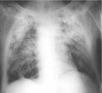
Fig. 115. Pulmonary silicosis on a radiograph.
The following clinico-radiological forms of respiratory tuberculosis are distinguished:
1. Primary tuberculosis complex.
2. Tuberculosis of intrathoracic Lymph Nodes.
3. Disseminated pulmonary tuberculosis.
4. Focal pulmonary tuberculosis.
5. Infiltrative pulmonary tuberculosis.
7. Pulmonary Tuberculoma.
8. Cavernous and Fibrocavernous pulmonary tuberculosis.
9. Cirrhotic pulmonary tuberculosis.
10. Tuberculous Pleurisy (including empyema).
11. Tuberculosis of the Bronchi, Trachea, and Upper Respiratory Tract.
12. Respiratory tuberculosis combined with occupational dust-induced lung diseases (coniotuberculosis).
Clinical forms of respiratory tuberculosis are characterized by localization (specifying the segment, lobe, and lung), presence of bacterial excretion (with detection of Mycobacterium tuberculosis [MTB+] or without [MTB-]), and the phase of the tuberculous process (progressive or regressive). Progression of pulmonary tuberculosis is accompanied by infiltration, destruction, or dissemination, whereas regression is marked by resorption, compaction, scarring, and calcification.
As the disease progresses, one form of tuberculosis can transition into another. For instance, the progression of infiltrative tuberculosis with breakdown within the infiltrated area can lead to The Development of fibrocavernous pulmonary tuberculosis. Over time, almost all forms of pulmonary tuberculosis can evolve into focal tuberculosis.

Fig. 116. Primary tuberculosis complex of the right lung at the compaction stage on an X-ray image.
Primary inhalation of anaerobic Mycobacterium tuberculosis occurs into the segments of the middle and lower lobes of the lung. Subsequently, the pathogen spreads intrapulmonary via Lymphatic vessels and nodes, frequently implanting in areas of the Lungs with higher oxygen partial pressure—namely, the apical segments. With adequate Immune Response, the tubercle bacillus is either destroyed or encapsulated and rendered into an inactive, dormant state. In the case of insufficient Immunity, the infection leads to The formation of the primary tuberculosis complex, which predominantly occurs in children and adolescents, accounting for 5–10% of all clinical forms of tuberculosis.
The primary tuberculosis complex is characterized by specific lesions of: 1) the lung area where the primary focus is formed, 2) the lymphatic vessels (tuberculous lymphangitis), and 3) the regional lymph nodes (tuberculous lymphadenitis). Clinically, the primary tuberculosis complex may be asymptomatic or mimic the course of Influenza, pneumonia, or Chronic Bronchitis. Radiologically, four Stages of the complex are distinguished: infiltration, resorption, compaction, and calcification. In The First stage (infiltration), a composite of pathological shadows is revealed as a single entity: the primary focus at the periphery, a linear shadow of lymphangitis leading to the ROOT, and enlarged regional lymph nodes at the lung hilum. In the second stage (resorption), lasting 2–7 months, partial resorption of the infiltrate results in the bipolarity of the infiltrative lesion, meaning the pulmonary and glandular Components of the primary complex can be identified separately, although connected by the linear shadow of lymphangitis. In the Third Stage (compaction), lasting 7–12 months, the lesion zone becomes denser and decreases in size (Fig. 116). The Fourth Stage (calcification) is characterized by petrification of the primary lesion site due to calcium salt deposition, forming a single calcified Ghon Focus up to 0.5 cm in size. The calcification process begins approximately one year after the onset of the disease and can continue for several years. Treatment during the Cytology/cytology/16.html">Early stages of the primary tuberculosis complex leads to a reduction of the infiltration area within 3–4 weeks and its complete resorption within 3–4 months. During routine fluorographic screenings of the Ukrainian population, the Ghon focus is detected in 10–15% of healthy individuals.
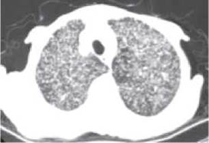
Fig. 117. Disseminated pulmonary tuberculosis on a computed tomography scan.
Tuberculosis of the intrathoracic lymph nodes (or tuberculous bronchoadenitis) is also a primary form of tuberculosis, developing in 35–45% of children and 1–3% of adults who contract tuberculosis. Pathomorphological changes predominantly occur in the bronchopulmonary lymph nodes of the upper lobes of the lungs and the middle lobe of the right lung.
There are two clinical forms of tuberculous bronchoadenitis (intrathoracic lymph node tuberculosis): infiltrative and tumor-like. The infiltrative form is more common than the tumor-like one. It develops when the inflammatory process spreads to the bronchial walls and is radiographically characterized by an enlarged, homogeneous root shadow of the lung with blurred outlines. The tumor-like form of tuberculous bronchoadenitis features larger lymph nodes within the lung root; they have a distinct outer margin but cannot be differentiated individually. During dynamic follow-up, these two forms may transition into one another. Enlargement of the bronchopulmonary and tracheobronchial lymph nodes is best visualized by computed tomography.
Disseminated pulmonary tuberculosis is characterized by multiple scattered tuberculous foci throughout the lungs, resulting from the spread of the disease via lymphatic, hematogenous, and bronchogenic pathways. Depending on its clinical course, disseminated pulmonary tuberculosis can be acute, subacute, or chronic. All forms of disseminated pulmonary tuberculosis typically present with bilateral, symmetrical lesions (Fig. 117).
In acute disseminated tuberculosis, the foci in the lungs are extremely small—about the size of a millet seed—which is why it is also known as "Miliary tuberculosis" (from the Latin milium meaning millet). The Clinical presentation of disseminated tuberculosis (dyspnea, cough, profuse sweating, high fever) is nonspecific and can mimic other conditions such as influenza or acute respiratory viral infections (ARVI); therefore, radiography plays a crucial role in diagnosing acute miliary tuberculosis. Monomorphic foci of individual tuberculous granulomas, 2-3 mm in size, are scattered throughout both lungs across all lung fields, predominantly in the middle and lower zones. The pulmonary vascular pattern is depleted, the transparency of the lung fields is reduced, and the shadows of the lung roots appear unstructured and poorly defined.
Subacute disseminated pulmonary tuberculosis most frequently affects the upper lung zones (segments I, II, and VI). The foci tend to merge, forming infiltrates of varying sizes that are relatively uniform in shape and density. These infiltrates are prone to destruction and may break down to form cavities lacking a thick fibrous capsule (unlike cavities seen in Other forms of tuberculosis). These cavities are surrounded by foci and infiltrates, or occasionally by nearly unchanged lung tissue. Such cavities are referred to as "punched-out" cavities.
If foci are incompletely resolved and treatment is inadequate, subacute disseminated pulmonary tuberculosis progresses into a chronic form. Sometimes, disseminated tuberculosis presents as a chronic disease from the outset. Radiographically, Chronic disseminated tuberculosis manifests as foci of varying density and size, distributed asymmetrically amid fibrotic and emphysematous lung parenchyma, accompanied by destruction cavities and deformed, displaced lung roots.
Focal tuberculosis most commonly develops As a result of an exacerbation of the inflammatory process surrounding the Primary tuberculous complex. In segments I, II, and VI, against the Background of an enhanced pulmonary pattern, one can detect focal shadows of various sizes, round shapes, low density, and blurred contours, with pleural thickening visible at the level of these foci. Specific treatment leads to the resorption of such foci within 2–3 months. In cases of inadequate treatment and disease progression, along with the aforementioned soft "exudative" foci, denser "productive" foci 4–6 mm in size with medium density and sharp contours appear, while the pulmonary pattern becomes coarse and cord-like. Under METABOLISM/18.html">The Influence of treatment, productive foci do not resolve; instead, they become denser, shrink, and undergo calcification.
Infiltrative tuberculosis develops as perifocal inflammation surrounding primary foci, causing them to coalesce and form a tuberculous infiltrate of varying size. Infiltrative tuberculosis is diagnosed in 40–60% of newly detected tuberculosis patients. Clinically, the onset resembles an acute respiratory viral infection or pneumonia: acute beginning, cough, chest pain, and a Temperature rise to 38–39 °C. Radiographically, various types of infiltrates are detected in segments I, II, and VI. A limited (subclavicular) infiltrate appears as a round shadow of low density, heterogeneous Structure, irregular shape, and blurred contours, measuring 2–3 cm, located at or just below the clavicle.
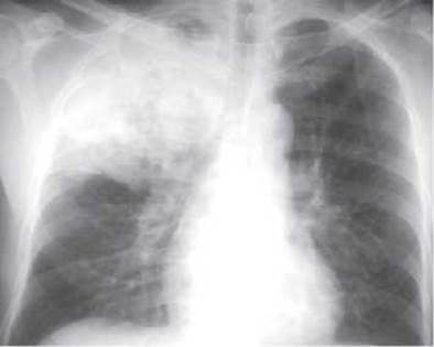
Fig. 118. Infiltrative tuberculosis of the right upper lobe on a radiograph.
A cloud-like infiltrate produces a faint shadow with indistinct borders, occupying an entire segment or a part of it. A lobar infiltrate involves an entire lobe and is termed tuberculous lobitis (Fig. 118). An infiltrate localized along the edge of a lung lobe following the interlobar fissure is known as tuberculous persiscuttitis.
All infiltrates are typically accompanied by a lymphatic tract (lymphangitis) leading toward the widened lung root. Under The Effect of treatment, tuberculous infiltrates resolve and become denser, resulting in fibrous-cirrhotic changes and calcifications at their site. In an unfavorable course, destruction occurs within the infiltrate, leading to caseous necrosis, which may result in cavity formation and bronchogenic dissemination.
Caseous pneumonia is a severe form of pulmonary tuberculosis characterized by the infiltration of an entire lobe and rapid tissue destruction. The contours of the infiltrative shadow and the destruction focus are jagged and blurred. There is a rapid bronchogenic spread of the process accompanied by the appearance of numerous lobular foci that tend to coalesce and break down.
Tuberculoma is a rounded focus of caseous necrosis separated from the surrounding lung tissue by a Fibrous Connective Tissue capsule. Clinically, tuberculoma is often asymptomatic, though tuberculin Skin tests are consistently positive.
Radiologically, a tuberculoma appears as a dense, rounded shadow with distinct yet somewhat irregular margins and a heterogeneous structure, typically measuring 2–4 cm. Tuberculomas are most frequently located subpleurally in the I, II, and VI segments, which accounts for nearby pleuropulmonary scars as well as thickening of the costal and interlobar Pleura. Calcified foci of varying sizes and fibrotic changes are often observed in adjacent or remote areas of the lung. During exacerbation phases, the outer contours of the tuberculoma become blurred due to perifocal inflammation, accompanied by the appearance of new foci in neighboring lung segments, pleural effusion, and lymphangitis pathways.
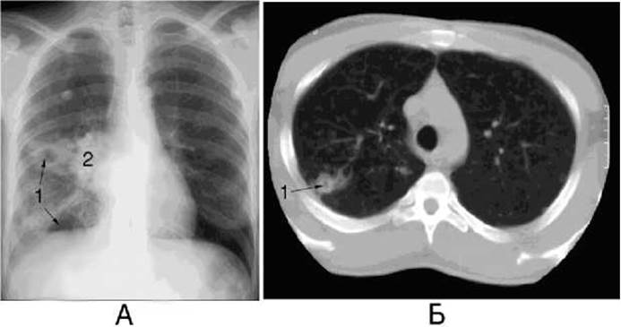
Fig. 119. Breakdown of infiltrative tuberculosis with cavity formation in the right lung on a radiograph (A) and a computed tomogram (B).
1 - cavity formation; 2 - lymphadenopathy.
Cavernous and fibro-cavitary tuberculosis develop from any form of tuberculosis as a result of delayed Diagnosis, late initiation of treatment, specific body reactivity features, and other factors that drive disease progression and lead to caseous-necrotic and destructive Changes in the lung tissue.
Cavernous pulmonary tuberculosis is characterized by the presence of a stable cavity in the lungs, with an absence of perifocal inflammation and fibrotic changes in the surrounding lung tissue. The cavity contains caseous masses, and its thick inner wall is irregular in surface. Radiologically, it presents as a ring-shaped shadow of varying size (most commonly 2–4 cm) with blurred outer and sharp, scalloped inner contours. Computed tomography facilitates the detection of cavities against the background of tuberculous foci and pneumosclerosis areas (Fig. 119). As caseous masses are discharged through the draining bronchus, the cavity wall thickness decreases and its inner surface becomes smoother. In protracted cases, the disease progresses into the fibro-cavitary form.
Fibro-cavitary pulmonary tuberculosis is an active, destructive form of tuberculosis marked by a chronic course featuring alternating periods of exacerbation and remission. Radiomorphologically, this form is characterized by one or more cavities with thick, three-layered walls (an inner caseous layer, a middle granulation layer, and an outer fibrous layer), alongside the development of sclerotic changes in the adjacent lung tissue, dissemination foci of various ages, pleural thickenings,
and varying degrees of pulmonary emphysema. The vascular and bronchial pattern of the affected lobe is enhanced and distorted, and the pulmonary hilum on the ipsilateral side is deformed, displaced toward the sclerotically altered lobe, and frequently widened. The pleura is unevenly thickened, and the costophrenic angles are partially or completely obliterated. This radiographic pattern may remain stable for many months or even years (Fig. 120). Upon disease progression, daughter cavities emerge, and the inflammatory process spreads to new areas, potentially involving an entire lobe or lung. Under the influence of treatment, the cavity heals, leading to the development of cirrhotic tuberculosis.
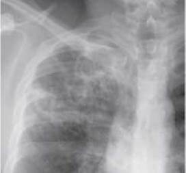
Fig. 120. Fibro-cavitary tuberculosis of the upper lobe of the right lung.
Cirrhotic pulmonary tuberculosis is the terminal stage of tuberculosis, characterized by excessive proliferation of connective tissue in the lungs that replaces the alveolar parenchyma. Cirrhosis is invariably accompanied by contraction and volume reduction of the lung or its lobe. However, caseous foci and residual cavities persist and may trigger disease relapses. Cirrhotic changes are most commonly localized in the upper lobes. Upper lobe cirrhosis presents with the following radiological features: the lobe is significantly reduced in volume, the root is dense and pulled upward, the pulmonary pattern is distorted, areas of emphysematous lung tissue are visible, the parietal pleura is thickened, the mediastinal Organs are shifted toward the lesion, and the intercostal spaces are narrowed.
Tuberculous pleurisy is most frequently a complication of other forms of tuberculosis, although it can also present as a primary condition. It shares the same radiological features as pleurisy of non-tuberculous Etiology and frequently leads to pleural calcification.
Tuberculosis of the upper respiratory tract, trachea, and bronchi develops as a complication of other clinical forms of pulmonary tuberculosis. Radiological signs of tuberculous damage to the Airways are primarily detected using computed tomography and bronchography. These include varying degrees of narrowing of the large bronchi, and occasionally their occlusion. The bronchial walls are unevenly thickened, with irregular inner contours.
Koniotuberculosis refers to respiratory tuberculosis combined with occupational dust-induced lung disease, or Pneumoconiosis. Tuberculosis most frequently coexists with silicosis (silicotuberculosis). The radiological presentation is a combination of features characteristic of both conditions.
Last update: 08/08/2026
Editorial and Educational Adaptation: This material has been compiled based on the primary/original source text. The project team performed an editorial review, corrected technical inaccuracies, structured sections, and adapted the content for an educational format.
What was processed:
- elimination of formatting defects (OCR errors, structural breaks, corrupted characters);
- editorial organization of content;
- standardization of terminology in accordance with academic sources;
- verification of factual statements against the original source text.
All mentions of the author, publication year, and origin of the primary text have been preserved in accordance with the source.