Tuberculosis Study Guide - M.M. Savula 2002
Primary tuberculosis in children
Primary tuberculous complex
Pathogenesis. In the event of massive entry of Mycobacterium tuberculosis into a child's Airways combined with low body resistance, tuberculous Changes in the lung tissue and intrathoracic Lymph Nodes are detected In addition to symptoms of intoxication (a localized form of tuberculosis develops). Around the mycobacteria that have settled in the Lungs, an area of specific inflammation of various sizes forms (ranging from barely noticeable to spreading over an entire lobe). These changes are called the primary pulmonary focus.
From this focus, mycobacteria and the inflammatory process spread via Lymphatic vessels toward the hilum of the lung, affecting the hilar lymph nodes, where specific morphological changes also occur, with a particular tendency toward caseous necrosis. Thus, a localized form of Primary tuberculosis is formed—the primary tuberculous complex, which consists of three components: tuberculous inflammatory changes in the lungs (primary pulmonary focus), inflammation along the lymphatic vessels (lymphangitis), and tuberculous involvement of the hilar lymph nodes.
Clinical presentation. The onset of the disease in the primary complex can vary and depends on the extent of changes in the lung tissue and lymph nodes, the presence and severity of caseous necrosis within them, and any complications.
Sometimes, this form of tuberculosis is detected incidentally during follow-up examinations of children who showed a "tuberculin Skin test conversion" (virage) during mass tuberculin screening. However, a detailed interview with the parents often reveals that the child had experienced mild symptoms of intoxication for some time, which had been attributed to fatigue, a common cold, etc.
In other cases, parents seek medical attention because they have noticed changes in the child's behavior over time, such as lethargy, sweating, poor appetite, weight loss, and a low-grade fever that did not resolve with Antibiotics. Coughing is rare in children, occurring only when complications arise. In the event of a significant enlargement of the intrathoracic lymph nodes, younger children may present with a hacking, pertussis-like cough. Children are usually unable to expectorate sputum.
Quite rarely, the primary tuberculous complex begins acutely with a high (febrile) fever, cough, and shortness of breath, mimicking Pneumonia, for which the child is unsuccessfully treated with antibiotics. It is worth noting that children with tuberculosis, unlike those with pneumonia, tolerate high body Temperature relatively well, remaining active and playful. The disease runs a more severe course in infants and young children.
During history-taking, it is often revealed that There is a family member with Pulmonary Tuberculosis or that the child has not been vaccinated against tuberculosis. Sometimes, an adult tuberculosis patient in the family is only identified during screening of the sick child's household contacts.
During physical examination, it is necessary to check for a post-vaccination scar on the child's left shoulder (unvaccinated children are affected more frequently). Often, children with early forms of tuberculosis appear no different from healthy ones. Sometimes, pallor, decreased skin elasticity, and occasionally Conjunctivitis, blepharitis, or erythema nodosum are noted. On Palpation, some children present with multiple enlarged, soft-elastic peripheral lymph nodes (micropolyadenitis).
Percussion changes in this form of tuberculosis are either absent or mild. Dullness is detected over the area only in the presence of a large pulmonary focus. Dullness near the Sternum or spine is observed with significant enlargement of the intrathoracic lymph nodes.
On Auscultation, breath sounds are usually normal, occasionally diminished or harsh over a localized area of the lungs, and sometimes dry or fine moist crackles can be heard there. Thus, the absence of changes on percussion and auscultation does not rule out a localized form of tuberculosis in a child.
For Diagnosis, the results of tuberculin skin testing (tuberculin conversion or strongly positive reactions to tuberculin) and chest X-ray Examination of the child are of key importance.
In the primary tuberculous complex, the chest radiograph reveals an opacity in the lung tissue, connected by a pathway of lymphangitis to the shadow of enlarged hilar lymph nodes (Fig. 15).
Class="center">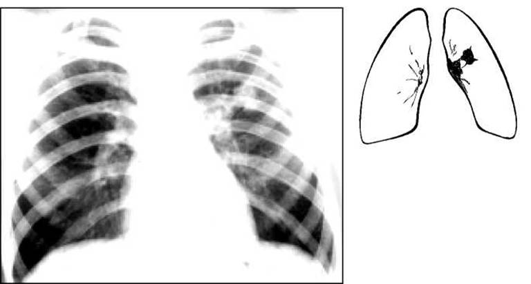
Fig. 15. Chest radiograph. Primary tuberculous complex on the left.
Detecting Mycobacterium tuberculosis in children is difficult because they do not expectorate but rather swallow their sputum. Therefore, repeated searches for MTB in bronchial washings, gastric aspirates, and laryngeal swabs are necessary.
Changes in the peripheral Blood in the primary tuberculous complex are minor: moderate leukocytosis (10.0-13.0-109/L) with a slight left shift, lymphopenia, and an increased ESR of up to 25-35 mm/h.
Course. With timely detection and complete Treatment, primary forms of tuberculosis have a favorable prognosis and a strong tendency to heal. In the past, when mass tuberculin screening was not performed, residual changes from healed tuberculosis were sometimes incidentally discovered on chest X-rays in children who had never been monitored or treated for it. More complete recovery occurs after full-course treatment, during which the symptoms of the disease quickly disappear and body temperature normalizes. However, the healing of changes in the lungs and lymph nodes is slow, so treatment must last at least 6-9 months.
When therapy is initiated in a timely manner, complete resolution of tuberculous changes in the lung tissue and hilum occurs. More commonly, calcium salts are deposited at the site of the residual pulmonary focus, forming the so-called Ghon Focus, while small calcifications remain in the hilar lymph nodes. These changes persist for life and do not harm human health. However, they harbor "dormant" viable MTB, which, under certain conditions many years later, can cause reactivation of the disease and The Development of secondary forms of tuberculosis.
In cases of late detection and incomplete treatment of primary forms of tuberculosis, areas of caseation persist even in partially calcified lymph nodes, serving as a source of prolonged intoxication and causing the process to transition into so-called chronic primary tuberculosis. It is characterized by a discrepancy between minimal, seemingly healed changes in the LYMPH NODES AND a persistent Intoxication syndrome. It is advisable to refer such children to pediatric sanatoriums.
Complications. Timely detection and complete treatment are also crucial because complications can occur in primary forms of tuberculosis, some of which are life-threatening to the child. The most common complication is the extension of tuberculous inflammation from the affected lymph node to a bronchus, which can result in the rupture of caseous material into the bronchial lumen and its occlusion, leading to Atelectasis. This is accompanied by a paroxysmal, uncontrollable cough, shortness of breath, and sometimes fever. Over the area of atelectasis, the percussion note is dull, and breath sounds are severely diminished. The chest radiograph shows a homogeneous opacity with sharp margins, corresponding in shape to the atelectatic area of the lung.
Spread of the inflammatory process to the Pleura leads to the development of pleural effusion (exudative Pleurisy).
If the infection from the affected lymph nodes enters the bloodstream and spreads hematogenously, tuberculous involvement of other Organs may occur, the most formidable of which is inflammation of the Meninges (Tuberculous meningitis). In the case of total dissemination of the lungs and other organs with tiny tuberculous foci, a severe, sometimes fatal form of tuberculosis develops—Miliary tuberculosis.
A rare complication of the primary tuberculous complex is the liquefaction of caseous masses in the center of the primary pulmonary focus, their evacuation through a draining bronchus, and The formation of a cavity—a primary pulmonary cavity.
The primary tuberculous complex must be differentiated from non-specific pneumonia, which runs a more acute course and resolves quickly with antibiotics. In pneumonia, such a significant enlargement of the hilar lymph nodes is not observed, and the Mantoux test is often negative.
Treatment of children with anti-tuberculosis drugs, desensitizing agents, and vitamin preparations is initiated in a hospital Setting. After 3-6 months, it can be continued in a sanatorium or on an outpatient basis.
Last update: 10/08/2026
Editorial and Educational Adaptation: This material has been compiled based on the primary/original source text. The project team performed an editorial review, corrected technical inaccuracies, structured sections, and adapted the content for an educational format.
What was processed:
- elimination of formatting defects (OCR errors, structural breaks, corrupted characters);
- editorial organization of content;
- standardization of terminology in accordance with academic sources;
- verification of factual statements against the original source text.
All mentions of the author, publication year, and origin of the primary text have been preserved in accordance with the source.