Tuberculosis - I.T. Pyatnochka 2005
Primary tuberculosis
Pulmonary tuberculosis combined with occupational dust-induced lung diseases (coniotuberculosis)
Tuberculosis and dust-induced occupational respiratory diseases refer to the combination of a clinical form of tuberculosis and Pneumoconiosis, resulting in a distinct condition known as silicotuberculosis, which features characteristic pathomorphological, radiological, and clinical presentations (Fig. 22).
Tuberculosis combined with occupational dust-related diseases accounts for 0.3–0.5% of newly diagnosed tuberculosis cases, and 1–1.5% of patients under observation in anti-tuberculosis dispensaries.
According to their Etiology, pneumoconioses are classified into five groups: 1) silicosis; 2) silicatosis (asbestosis, cementosis, talcosis, kaolinosis); 3) anthracosis; 4) mixed-type pneumoconiosis (silicosilicatosis, anthracosilicosis, siderosilicosis); and 5) pneumoconioses of other etiologies (siderosis, aluminosis, apatitosis, amidosis).
Tuberculosis develops most frequently in individuals with silicosis.
Silicosis is an occupational disease caused by the inhalation of dust containing free silicon dioxide. There are three stages of silicosis. The prevalence of tuberculosis correlates to some extent with the stage of pneumoconiosis. Tuberculosis is observed in approximately 20% of individuals with stage I silicosis, 40% with stage II, and 80% with stage III.
The Pathogenesis of silicotuberculosis stems from the interaction of two etiological factors: free silicon dioxide and MBT. Tuberculosis typically develops As a result of the endogenous reactivation of residual post-tuberculosis lesions in the Lungs or intrathoracic Lymph Nodes, whereas exogenous superinfection plays a significantly lesser role.
The pathogenesis of silicosis itself is complex and not yet fully understood. Factors contributing to its development include high dust concentrations, the size of dust particles (silicon dioxide) suspended in the air, prolonged exposure duration, and the overall condition of the body (inflammatory processes in the Upper Respiratory Tract and lungs, as well as impaired mucociliary clearance).
The pathological anatomy of silicotuberculosis is polymorphic, determined by the clinical form and phase of tuberculosis, as well as the form and stage of pneumoconiosis. Depending on The Nature and localization of the pathological process, pneumoconioses are classified into interstitial, diffuse, sclerotic, nodular, and tumor-like forms.
Alongside the typical forms of Pulmonary Tuberculosis frequently associated with silicosis (focal, infiltrative, disseminated, and fibro-cavitary), several atypical forms of silicotuberculosis are distinguished: silicotuberculous bronchoadenitis, nodular silicotuberculosis, silicotuberculoma, massive silicotuberculosis, and destructive silicotuberculosis.
The Clinical presentation of silicotuberculosis is multifaceted and determined by the stage and course of silicosis, as well as the form and phase of pulmonary tuberculosis. The clinical course of silicosis can be rapid, gradual, or delayed.
Clinical signs indicating The Development of tuberculosis against the Background of pulmonary silicosis include the onset of Intoxication syndrome and localized moist rales in the lungs, moderate leukocytosis with a shift to the left in the leukogram, lymphopenia, accelerated ESR, and the detection of MBT in sputum.
Class="center">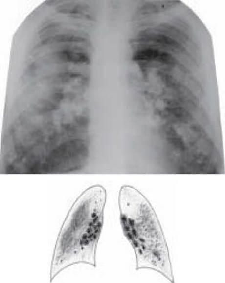
Fig. 22. Silicotuberculosis: stage III silicosis, Disseminated pulmonary tuberculosis, infiltration phase. Plain chest radiograph
Radiologically, stage I silicosis is characterized by enhanced and distorted pulmonary markings, small nodular shadows predominantly in the middle lung zones, widened and dense lung roots, and early signs of emphysema.
Stage II silicosis is typically characterized by an increase in the size and number of nodular shadows, primarily in the middle and lower lung zones, loss of differentiation in pulmonary markings, and progressive bullous-emphysematous changes.
In stage III silicosis, nodular shadows coalesce to form massive conglomerates against a background of fibrosis, the lung roots appear "truncated," and peripheral calcification of the hilar lymph nodes is occasionally observed.
The Diagnosis of silicotuberculosis is based on radiological, clinical, laboratory, and tracheobronchoscopic findings.
When formulating the diagnosis, it is advisable to first indicate "Silicotuberculosis," followed by a detailed Description of the silicosis component and subsequently the tuberculosis process.
For instance: Silicotuberculosis: stage I silicosis (nodular form), newly diagnosed pulmonary tuberculosis (0.2001) of the upper lobe of the left lung (focal) (infiltrative), Dest-, MBT - M-K-HistO, Resist-, respiratory failure grade II, CatI Cog4(2001).
The Treatment of patients with silicotuberculosis must be intensive and prolonged (at least 1 year), utilizing 3–5 antituberculosis drugs, often combined with glucocorticoids for a duration of 2–4 months. In destructive forms of silicotuberculosis, anti-mycobacterial agents are advantageously administered intravenously or endobronchially. For concurrent nonspecific endobronchitis, bronchodilators, mucolytics, and broad-spectrum Antibiotics are used alongside anti-mycobacterial drugs until the process resolves. Surgical intervention (partial lung resection) is performed quite rarely, specifically for silicotuberculomas or fibro-cavitary pulmonary tuberculosis in patients with limited silicosis.
CONTROL QUESTIONS
1. Definition of silicotuberculosis.
2. Pathogenesis and pathomorphology of silicotuberculosis.
3. Clinical presentation of silicotuberculosis.
4. Radiological findings in silicotuberculosis According to the disease stage.
5. Diagnosis of silicotuberculosis.
6. Treatment of patients with silicotuberculosis.
Last update: 10/08/2026
Editorial and Educational Adaptation: This material has been compiled based on the primary/original source text. The project team performed an editorial review, corrected technical inaccuracies, structured sections, and adapted the content for an educational format.
What was processed:
- elimination of formatting defects (OCR errors, structural breaks, corrupted characters);
- editorial organization of content;
- standardization of terminology in accordance with academic sources;
- verification of factual statements against the original source text.
All mentions of the author, publication year, and origin of the primary text have been preserved in accordance with the source.