Critical conditions in pulmonary tuberculosis in adults - N.I. Fomichova 2010
Intoxication syndrome
Intoxication syndrome represents a generalized functional disorder of the entire Organism resulting from The Development of pathological changes. Its mechanism, both in tuberculosis and other pulmonary diseases, involves the poisoning of the body by toxic products of various origins (endo- and exotoxins of tuberculosis Bacteria, as well as products of destructive Changes in the lung tissue). These toxins poison the body's Organs and physiological systems, thereby triggering functional disorders. In some cases, these manifestations are the result of autonomic dystonia, while in others they stem from trophic, dystrophic, or allergic tissue changes. Intoxication syndrome manifests through systemic signs affecting the entire body, symptoms localized in specific physiological systems, and alterations in laboratory parameters.
Pathogenesis. The primary causes of intoxication syndrome in tuberculosis are most frequently Bacterial toxins that migrate from the Lungs into the lymphatic and circulatory systems, exerting a damaging effect on body organs and Tissues. Evidence of this includes the escalation of intoxication at the onset of Treatment for Disseminated Pulmonary Tuberculosis, when patients are prescribed a combination of 4–5 antituberculosis drugs. This leads to the massive destruction of MBT, resulting in an increased production and release of toxic substances into the bloodstream, and consequently, an exacerbation of intoxication symptoms. Intoxication can also be triggered by Protein Denaturation (caseous necrosis) and The breakdown of lung tissue. For instance, it is well known that in certain forms of infiltrative tuberculosis, pronounced intoxication syndrome persists until pulmonary tissue breakdown occurs. Following the rejection of caseous masses and The formation of a cavity, intoxication decreases significantly, creating an illusion of recovery. In some patients, intoxication syndrome develops As a result of neuro-reflex impulses originating from the affected lungs. In pulmonary tuberculosis, intoxication syndrome may have a dual origin, associated not only with the main Clinical forms of tuberculosis, but also with secondary infections and tissue alterations.
Intoxication disrupts the Functions of various organs and systems. Central Nervous system dysfunction leads to fever, changes in respiratory rhythm and depth, and headaches. Autonomic dystonia, states of collapse, myocardial trophic changes, and impaired Functions of the Stomach, Liver, Kidneys, and other organs also occur. Due to Endocrine System dysfunctions—particularly of the Pituitary Gland and Adrenal Glands—patients experience physical deconditioning (hypodynamia), hypotension, and weight loss. Depending on the combination or predominance of specific symptoms, physicians distinguish febrile-septic, Influenza-like, and severe generalized fatigue syndromes. Other manifestations of intoxication syndrome, such as prolonged low-grade fever ("hidden" intoxication) and hypodynamia, do not constitute emergency conditions in phthisiology.
Class="center">DIAGRAM 8. DIAGNOSTIC SCHEME OF INTOXICATION SYNDROME IN RESPIRATORY TUBERCULOSIS
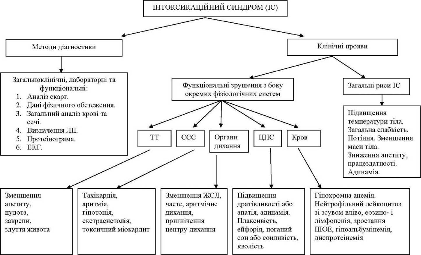
Clinical manifestations (Diagram 8). Five key signs of intoxication syndrome are identified: elevated Temperature, generalized weakness (adynamia), weight loss, sweating, and impaired general development. It can be mild, moderate, or severe. Emergency conditions typically arise precisely in cases of moderate-to-severe intoxication syndrome.
The clinical manifestations of intoxication syndrome may intensify or diminish in sync with the progression of the pathological process, or their severity may vary. For example, a high body temperature may occur without sweating and with only mild adynamia, and conversely, patients with a low-grade fever may exhibit a loss of appetite, weight reduction, and impaired physical development.
Elevated temperature is the primary and main sign of tuberculosis intoxication, determining the stage of the disease and caused by either the underlying condition or a secondary infection.
The elevation of body temperature in Various Forms of tuberculosis can be sudden and rapid (Caseous Pneumonia, Miliary tuberculosis, lobitis, periscisuritis in infiltrative tuberculosis)—developing within a few hours to 1–2 days—or slow and gradual, taking several days to 1–2 weeks to reach peak levels. In certain forms of tuberculosis (caseous pneumonia, miliary tuberculosis), the onset of the disease may present with fever and cold sweats.
Depending on its peak value During the first days of the illness, the temperature level can vary: very high (39.1°–40.0°C and above), high (38.1°–39.0°C), and mildly elevated (37.1°–38.0°C).
Most clinical forms of tuberculosis feature an elevated temperature response. In caseous pneumonia, miliary, and disseminated tuberculosis, body temperature can be high or very high.
The temperature curve pattern in pulmonary tuberculosis also varies. Initially, it is nearly constant (daily fluctuations up to 0.5°); later, fluctuations range from 0.6° to 1.0° or more per day, or present as temperature spikes (hectic fever). For instance, in initial forms of tuberculosis, caseous pneumonia, Pleural Empyema, and the tumorous form of tuberculous bronchial adenitis—when treatment stabilizes the process, or when the draining bronchus becomes obstructed by sputum, caseous masses, or Blood clots—both minor elevations and sharp temperature spikes are observed. Waves of periodically elevated body temperature can be prolonged, lasting 1–2 months or more (e.g., in caseous pneumonia).
Generalized weakness is the second major sign of intoxication syndrome. It is invariably present during the gradual Development of the disease, throughout its full-blown clinical manifestations, and persists even during the regression phase of the illness. Generalized weakness in tuberculosis (as in all pulmonary and extrapulmonary Inflammatory Diseases) is divided into three degrees of severity. In mild cases, patients can perform physical work, but tire quickly and recover their strength slowly. In moderate severity, patients can get out of bed but are unable to engage in occupational activities; this is how most clinical forms of tuberculosis begin in the majority of cases. Severe weakness is so pronounced that patients are forced to remain bedridden. This is the course taken by typhoid-like, meningeal, and pulmonary forms of miliary tuberculosis, caseous pneumonia, and subacute disseminated or advanced fibrous-cavernous pulmonary tuberculosis in their terminal phases.
Profuse sweating accompanied by a hectic body temperature—serving as one of the primary symptoms at disease onset—occurs in miliary tuberculosis and caseous pneumonia. Hectic fever accompanied by daily profuse sweating is encountered in neglected forms of pulmonary tuberculosis. In initial clinical forms of the disease, profuse sweating is rarely observed. Excessive sweating is also a frequent sign of silicosis complicated by tuberculosis.
With the onset of intoxication syndrome in tuberculosis, functional disorders develop. Digestive System disorders are frequent in this disease: reduced appetite progressing to complete anorexia, a feeling of heaviness in the epigastric region, abdominal distension, constipation, and less frequently, nausea. This consequently leads to weight loss. Sometimes this weight loss is rapid, with patients losing 5–8 kg or more within a few days; in other instances, it is gradual, amounting to several kilograms over several months. The most rapid weight loss is observed in acute forms of tuberculosis (caseous pneumonia, miliary tuberculosis, and certain forms of infiltrative tuberculosis). Gradual weight loss is characteristic primarily of mild forms of tuberculosis and chronic, slow-progressing tuberculosis.
Impaired general body development is currently a rare sign of intoxication syndrome in tuberculosis. It is frequently observed in children and young individuals with prolonged tuberculosis intoxication that transitions into secondary forms of tuberculosis. Trophic changes develop in the Skin, and the Development of Muscles and bones is delayed.
Functional changes in The Cardiovascular system induced by tuberculosis intoxication most commonly manifest as tachycardia, and occasionally as a soft pulse or extrasystole.
Signs of intoxication syndrome originating from the Respiratory system are closely linked to local functional disorders: rapid breathing and a reduction in vital lung capacity. Respiratory rate in miliary tuberculosis and caseous pneumonia can reach 40–60 breaths per minute.
In addition to the aforementioned functional deviations, the development of intoxication syndrome in respiratory tuberculosis can be influenced by alterations in the excretory, endocrine, autonomic, and central nervous systems. In caseous pneumonia and severe forms of disseminated tuberculosis, other clinical symptoms typically arise simultaneously with the fever—and sometimes even earlier: increased irritability or, conversely, apathy, listlessness, and an indifferent attitude toward the environment; insomnia and drowsiness; decreased work capacity, tearfulness, and euphoria. All these signs reflect the phase states of the Cerebral Cortex resulting from tuberculosis intoxication. This same cause triggers symptoms of Autonomic nervous system dysfunction, such as sweating (predominantly at night or early in the morning), tachycardia, decreased appetite, and vasomotor and dyspeptic disorders.
The most frequent hemogram sign of intoxication syndrome is various hypochromic anemia states, moderate neutrophilic leukocytosis with a left shift, eosinophilia and lymphopenia, and an elevated ESR. Characteristic features include hypoproteinemia and dysproteinemia with reduced albumin and elevated globulin levels.
To assess the severity of endogenous intoxication in respiratory tuberculosis, the leukocyte intoxication index (LII) is calculated using the following formula:

neutrophils:
my - myelocytes,
ju - juvenile (metamyelocytes),
p - stab neutrophils,
s - segmented neutrophils,
pl. c. - plasma Cells,
mon - monocytes,
e - eosinophils.
Normally, the leukocyte index of intoxication ranges from 0.3 to 1.5. In tuberculosis, this indicator progressively increases in accordance with the disease progression.
Pronounced intoxication syndrome can trigger critical conditions, such as acute respiratory, cardiovascular, or endocrine failure, as well as psychomotor agitation.
EMERGENCY CARE
To rescue a patient from severe intoxication syndrome, general and individualized measures are implemented (Scheme 9).
General measures involve etiotropic rational therapy for tuberculosis tailored to the patient's specific treatment category. First and foremost, intensive, continuous, and combined antimycobacterial Treatment of the underlying tuberculosis and its purulent-inflammatory complications—which serve as the source of endogenous intoxication—is essential. Antimycobacterial therapy is intensified by using exclusively those drugs to which the MBT strains are susceptible, while maximizing all possible administration routes: intravenous, endolymphatic, intracavitary (in cases of pleural obliteration and subcortically located cavities), and intrapleural (for pyopneumothorax and pleural empyema). In case of concomitant purulent endobronchitis, therapeutic bronchoscopy is performed to lavage the tracheobronchial tree.
SCHEME 9 SCHEME OF EMERGENCY CARE FOR INTOXICATION SYNDROME IN TUBERCULOSIS PATIENTS

Etiotropic drugs should preferably be administered parenterally, because severe intoxication syndrome impairs the functions of The Stomach, intestines, and liver, which can negatively affect drug absorption and their delivery to the lungs. Antituberculosis drugs, such as isoniazid and fluoroquinolones, should be administered via intravenous drip in a standard daily dose dissolved in 50–100 ml of isotonic sodium chloride solution, whereas rifampicin is diluted in 250 ml of a 5% glucose solution.
Nonspecific Pathogenetic Therapy should comprise antipyretic, desensitizing, antihistamine, anabolic, detoxicating, and symptomatic agents, as well as drugs that normalize Protein METABOLISM.
As antipyretics, analgin (50% — 2 ml) is administered intramuscularly twice a day, and acetylsalicylic acid is prescribed at 0.5 g three times daily after meals (tablets should be thoroughly crushed and washed down with plenty of fluids). Paracetamol is administered in tablets at 0.5 g two to three times a day, with a maximum daily dose of 1.5 g. Nimesulide, Sigan, Olfen, and similar anti-inflammatory and antipyretic agents are also prescribed (to be taken after meals with a generous amount of Water).
For detoxification purposes, abundant fluid intake is prescribed (mildly mineralized mineral waters, tea, infusions of rosehip, motherwort, and St. John's wort). Concurrently, diaphoretics (infusions of linden flowers, chamomile, elderberry, raspberry tea) and Diuretics (infusions or tinctures of bearberry leaves, birch leaves, field horsetail herb, juniper berries, parsley seeds) are applied. Pharmacological agents include aminophylline (2.4% — 10 ml in 500 ml of isotonic sodium chloride solution), Triampur Compositum, furosemide (40–80 mg in the morning), and Trifas (1.0 g for two to three days).
Reosorbilact and Sorbilact exhibit pronounced detoxicating properties. Both drugs contain essential cations: Na+, K+, Ca++, Mg++, the Cl- anion, an organic lactate anion, and sorbitol. Due to their hyperosmolarity, reosorbilact and sorbilact induce fluid shift from the intercellular space into the vascular bed, thereby improving microcirculatory flow and tissue perfusion, which is the primary objective of detoxification therapy. The fluid shift from the interstitial to the intravascular compartment increases circulating blood volume (CBV) at the expense of plasma volume expansion, i.e., causing hemodilution. Furthermore, owing to the potent specific osmodiuretic effect of sorbitol—stemming from the absence of natural human Mechanisms for the reabsorption of polyhydric alcohols in the proximal renal tubules—a marked diuretic action of both preparations is observed, particularly with sorbilact. Notably, the latter also exerts a nephroprotective effect and enhances diuresis even in the presence of ACUTE RENAL FAILURE. In addition, sorbitol is partially metabolized into fructose, thereby facilitating the normalization of carbohydrate and Energy Metabolism. This has an especially beneficial effect on improving the functional state of hepatocytes by restoring Glycogen stores, which significantly contributes to the intensification of natural detoxification processes. Importantly, the lactate anion contained in both drugs helps correct the acid-base balance of Blood Plasma and, by participating in carbohydrate-energy metabolism reactions, restores and stimulates the functions of cells belonging to the reticuloendothelial system, liver, and kidneys.
It is also recommended to administer a 5% glucose solution and an isotonic sodium chloride solution via intravenous drip concurrently with cardiac and diuretic agents. For this purpose, 1 ml of a 0.06% corglycon solution and 5–10 ml of 2.4% aminophylline are added to the infusion bottle. During and after fluid rehydration, medications that improve renal blood flow are sometimes used (2% papaverine hydrochloride solution, 1–2 ml intravenously; 2% No-spa solution, 2 ml intravenously; or 10% diprophylline solution, 5–10 ml intravenously), along with fast-acting diuretics, such as furosemide (1–2 ml intravenously) and a 15% mannitol solution at a single dose of 0.5–1.5 g/kg intravenously to eliminate toxins from the body (Forced diuresis).
Significant intoxication is frequently accompanied by hypoalbuminemia; therefore, blood protein composition is normalized by administering 5% or 10% albumin solution, dry, native, or fresh-frozen plasma (100–200 ml 1–2 times a week, for a course of 4–5 infusions), or 200–400 ml of protein or lactoprotein solutions. To restore The amino acid profile of the blood, Aminoplasmal 5% E or Polyamine is prescribed via intravenous drip for 5 days or more.
To improve protein metabolism and hematopoiesis, and to stimulate leukocytosis and cellular growth, methyluracil is prescribed at 1 g 3–4 times daily.
Diphenhydramine and heparin possess antihistamine properties and are administered subcutaneously into the rectus abdominis Muscle or intramuscularly (1–2 ml of a 1% solution and 1,000–5,000 IU, respectively). As antiallergic agents, calcium gluconate is used at 1–3 g 2–3 times a day or 5–10 ml of a 10% solution intravenously; loratadine at 0.01 g once daily; Telfast at 180 mg; and tavegil at 2 ml intramuscularly once daily. Glucocorticosteroid preparations and heparin yield the strongest antiallergic effect.
The formation of exudative-destructive changes, cavities, and the exacerbation of intoxication in tuberculosis are promoted by lysosomal Enzymes, particularly proteases. Consequently, protease inhibitors are utilized in severe cases of tuberculosis. Treatment is initiated with natural antifermentative agents: kontrikal (10,000–20,000 IU), trasylol (10,000–20,000 IU), gordox (100,000 IU), pantrypin (12 IU), or ingitril (15–20 IU). These are administered via intravenous drip in physiological sodium chloride solution or 5% glucose twice a week, comprising a total of 3–5 infusions.
Antioxidant therapy aims to suppress Lipid Peroxidation (LPO), for which tocopherol acetate (vitamin E) is prescribed orally or intramuscularly at 0.1–0.3 g daily, in courses lasting 15–20 days.
Below is a clinical case of tuberculosis where the patient's severe condition was characterized by all the hallmarks of intoxication syndrome, the primary ones being persistently high BODY TEMPERATURE AND varying degrees of general weakness corresponding to the developmental stages and clinical course of the disease.
CASE REPORT
Patient R., 24 years old, was admitted to a tuberculosis hospital with suspected miliary pulmonary tuberculosis. He denies any history of contact with tuberculosis patients. In childhood, he suffered from left-sided exudative Pleurisy of unknown Etiology. According to the patient, a week ago he experienced general malaise, headache, and low-grade fever. Five days later, his temperature rose to 40° C, accompanied by profuse sweating, tachypnea up to 40 breaths per minute, tachycardia (pulse 98 bpm), and a dry cough. He was hospitalized in the pulmonology department with a Diagnosis of pneumonia. A chest X-ray showed an enhanced vascular pattern throughout both lungs. Over 5 days of treatment, the patient's condition significantly worsened with increasing symptoms of intoxication, clouded consciousness, hyperesthesia, adynamia, and worsening dyspnea. Abdominal distension, hepatomegaly, a drop in body temperature to 38° C morning and evening, and weight loss of up to 10 kg were observed. Suspecting typhoid fever, the patient was transferred to the infectious diseases department. A repeat chest X-ray revealed small, moderately intense focal shadows without a tendency to confluence in both lung fields, with slightly widened roots. Sputum smear Cell/15.html">Microscopy showed no Mycobacterium tuberculosis. A blood test revealed relative leukocytosis (10.0×10⁹/L), ESR of 20 mm/h, marked lymphopenia, aneosinophilia, and monocytosis.
After analyzing the disease course and examination results, it was concluded that the patient's severe condition was caused by a pronounced intoxication syndrome. Therefore, the patient primarily underwent detoxification therapy (Reosorbilact 400 mL IV drip once daily, 200 mL of 4% sodium bicarbonate solution, 150 mL of albumin IV drip, Contrical 10,000–20,000 IU IV drip in 300 mL of isotonic sodium chloride solution) along with etiotropic treatment: isoniazid 5 mL of a 10% solution intravenously daily, rifampicin 600 mg intravenously daily in 250 mL of 5% glucose solution, streptomycin 1.0 g intramuscularly, ethambutol 1.6 g, and pyrazinamide 2.0 g per day. To reduce the inflammatory response and fever, nimesulide 0.5 was prescribed morning and evening for 7 days, paracetamol 0.5 g three times a day, and 30% sodium thiosulfate solution 5 mL daily. To help reduce intoxication, phytosorbents were recommended, along with frequent fluid intake including tea, rosehip decoction, and mineral waters.
For a month, the patient received calcium gluconate, vitamin E, methyluracil, and prednisolone 20 mg daily in the morning. After the tenth day, the prednisolone dose was reduced by 5 mg weekly until complete withdrawal. Ambroxol and mucaltin were prescribed to suppress the dry cough. The patient also received hepatoprotectors, B-complex Vitamins, and 5% ascorbic acid 2 mL intramuscularly daily. As intoxication decreased and the patient's general condition improved, the pathogenetic therapy was tapered.
Following five months of intensive anti-tuberculosis and pathogenetic therapy, complete resorption of the focal shadows in the lungs was achieved, internal organ functions were restored, and the patient was discharged in satisfactory condition to continue treatment in a sanatorium Setting.
Selection/41.html">Review Questions and TASKS WITH ANSWER KEYS
Case Study. Patient K., 22 years old. Two weeks ago, following hypothermia, she began to feel unwell: her body temperature rose to 37.5 0C, accompanied by a cough with a small amount of mucous sputum, chest pain, weakness, and poor appetite. She treated herself at home for a week, but her condition continued to deteriorate. An emergency doctor hospitalized her in the pulmonology department with suspected community-acquired polysegmental right-sided upper-lobe pleuropneumonia, which allegedly was supported by radiological findings. Intensive antibacterial therapy was administered for a week, but the patient's condition progressively worsened. Her body temperature rose to 39.5 0C and became hectic, breathing became difficult, poor appetite progressed to anorexia, The amount of sputum increased and it became purulent, profuse night sweats and intense headaches appeared, and the patient became bedridden and inactive.
Objective status: the patient's condition is severe, skin is pale, lip cyanosis, acrocyanosis, lies motionless in bed, consciousness is clouded, does not fix gaze, Nutrition is depleted. Respiratory rate is 28/min, accessory muscles are involved in breathing. Pulse rate is 110/min, Heart sounds are muffled. Blood pressure is 100/65 mm Hg. Over the upper lobe of the right lung, Percussion sound is significantly dulled, breathing is bronchial, with numerous resonant moist rales of various calibers that can be heard even at a distance from the patient (chugging rales). Throughout the left lung, percussion sound is clear and pulmonary, breathing is harsh. Abdomen is soft and tender upon Palpation of the right upper quadrant. The liver is enlarged by 3 cm.
A repeat chest X-ray reveals an intense, non-homogeneous opacification of the upper lobe of the right lung with blurred margins, associated with an infiltrated ROOT. Against the Background of the opacification, separate denser foci and areas of radiolucency caused by lung tissue destruction are visible. The cavities are multiple and irregular in shape. In the lower sections of this and the contralateral lung, there are multiple focal shadows of moderate and weak intensity without clear margins.
Blood test: RBC 2.8 T/L, Hb 80 g/L, WBC 18.2 G/L, E 0%, Band 15%, Seg 67%, L 8%, Mo 10%, ESR 45 mm/h. Urine analysis: protein, hyaline casts.
Sputum smear direct microscopy revealed Mycobacterium tuberculosis. The patient was transferred to the tuberculosis dispensary.
Question 1. What clinical form of tuberculosis was diagnosed in the patient?
A. Fibro-cavernous tuberculosis.
B. Caseous pneumonia.
C. Infiltrative tuberculosis.
D. Tuberculous Sepsis.
E. Disseminated tuberculosis.
Question 2. What critical condition is most likely to develop first in this patient if necessary medical care is not provided?
A. Acute Respiratory Failure combined with hemodynamic disorders.
B. Pulmonary Hemorrhage.
C. Cerebral coma.
D. DIC syndrome.
E. Asphyxia.
Question 3. What, in your opinion, is the leading mechanism in the development of severe intoxication syndrome in this patient?
A. Bacterial toxins.
B. Dystrophic and allergic tissue changes.
C. Neuroreflex impulses originating from the affected lungs.
D. Destruction of lung tissue.
E. Protein Denaturation and destruction of lung tissue.
Question 4. What should be the starting point of treatment for this patient?
A. Massive anti-tuberculosis therapy.
B. Massive anti-tuberculosis therapy combined with broad-spectrum Antibiotics.
C. Comprehensive detoxification therapy.
D. Agents that normalize blood protein composition.
E. Anti-inflammatory drugs.
Question 5. Which combination of anti-tuberculosis drugs is most appropriate to prescribe for the patient?
A. Isoniazid, rifampicin, pyrazinamide, streptomycin.
B. Lomefloxacin, isoniazid, rifampicin, streptomycin.
C. Isoniazid, rifampicin, pyrazinamide, streptomycin, prothionamide.
D. Isoniazid, rifampicin, pyrazinamide, ethambutol, streptomycin.
E. Isoniazid, pyrazinamide, cycloserine, ethambutol.
Answers and their rationale.
Answers to the questions: 1 B, 2 A, 3 E, 4 C, 5 D
1. The diagnosis of caseous pneumonia in this patient is indicated, firstly, by the gradual, progressive deterioration of her condition, even against the background of broad-spectrum antibiotic therapy, and the patient's extremely severe condition caused by pronounced intoxication syndrome. This is characteristic of caseous pneumonia, as opposed to infiltrative tuberculosis, in which intoxication is less severe. Secondly, radiological findings—specifically upper-lobe localization, the presence of intense inhomogeneous shadowing with multiple cavities, and dissemination foci in the lung tissue bilaterally—strongly support caseous pneumonia (the absence of "old" cavities and pulmonary fibrosis rules out fibro-cavitary tuberculosis, while the absence of a dissemination syndrome typical of disseminated tuberculosis and tuberculous sepsis excludes the latter). Furthermore, sputum smear microscopy revealed MBT. Auscultation reveals moist, so-called "caseous" rales. Clinical blood analysis shows anemia, marked lymphopenia, and leukocytosis with a significant stab shift, along with a substantially elevated ESR. These findings are also typical of caseous pneumonia, which is frequently complicated by secondary nonspecific infection (the patient notes that her sputum has become purulent), accompanied by severe intoxication syndrome.
2. First and foremost, the patient is at risk of developing acute respiratory failure combined with hemodynamic disorders. Lobar caseous pneumonia, which in this patient most likely developed on the background of a cloud-like infiltrate or lobitis, is accompanied by a pronounced hyperergic tissue reaction with nearly total caseous necrosis that dominates over perifocal inflammation. Liquefaction and subsequent sloughing of caseous masses resulted in the formation of multiple cavities in the lung tissue. All of this is accompanied by oxygen deficiency, which was rapidly compounded by endogenous intoxication resulting from the action of microbial toxins and excessive amounts of intermediate and End products of Metabolism generated in the affected lung tissue. In addition, the manifestation of severe intoxication syndrome in caseous pneumonia is driven by massive breakdown and denaturation of lung tissue Proteins. Neuroreflex impulses originating from the affected lungs also play a significant role in the development of acute respiratory failure and hemodynamic disorders. Therefore, the pronounced intoxication syndrome may primarily lead to suppression of the respiratory center with the development of acute respiratory failure, and to toxic myocarditis resulting in hemodynamic disorders. Critical conditions such as DIC syndrome and cerebral coma are secondary, while pulmonary hemorrhage and asphyxia may not occur at all in this case.
3. The leading mechanism in the development of severe intoxication syndrome in this patient is caseous Necrosis of the lung tissue with its breakdown and protein denaturation, which is pathogenetically and morphologically characteristic of this form of tuberculosis.
4. Treatment must undoubtedly begin with the administration of massive, comprehensive, pathogenetically substantiated detoxification therapy, which will subsequently be supported by a regimen of anti-tuberculosis drugs. The severity of the patient's condition is primarily caused by severe intoxication syndrome, which can lead to vital organ dysfunction and patient death. Initiating combined etiotropic treatment from the very beginning in this case would lead to massive destruction of MBT, further exacerbating the manifestations of intoxication syndrome.
5. The patient requires the administration of 5 first-line anti-tuberculosis drugs. Specifically, these are isoniazid, rifampicin, pyrazinamide, ethambutol, and streptomycin.
Last update: 08/08/2026
Editorial and Educational Adaptation: This material has been compiled based on the primary/original source text. The project team performed an editorial review, corrected technical inaccuracies, structured sections, and adapted the content for an educational format.
What was processed:
- elimination of formatting defects (OCR errors, structural breaks, corrupted characters);
- editorial organization of content;
- standardization of terminology in accordance with academic sources;
- verification of factual statements against the original source text.
All mentions of the author, publication year, and origin of the primary text have been preserved in accordance with the source.