Intensive Care of Emergency Conditions - V. M. Zaporozhan 2006
Acute Respiratory Failure
Respiration, alongside Blood Circulation, is essential for sustaining life, as its primary function is to facilitate METABOLISM by delivering the required amount of oxygen to the Tissues and removing carbon dioxide as a metabolic byproduct. In this process, external respiration ensures The transfer of these gases from the external environment into the blood, which transports them to and from the tissues, whereas tissue respiration integrates oxygen into tissue metabolism. Respiratory failure specifically refers to the failure of external respiration.
Based on the foregoing, respiratory failure is defined as a pathological state in which the Lungs have a limited capacity to maintain the normal gas composition of arterial blood while breathing ambient air.
An emergency condition that requires intensive care is designated as acute respiratory failure (ARF).
It typically develops within a matter of minutes or hours and may either resolve completely or progress to a chronic form. Unlike acute failure, the chronic variant develops gradually over many months and years. It can also acquire acute features during an Exacerbation of chronic disease. However, THE CONCEPT OF acuteness relates solely to the speed of onset and reversibility rather than the severity of the clinical course.
To understand the Pathogenesis of respiratory failure, THE ORIGIN OF its characteristic symptoms, and to confidently navigate the Selection of appropriate intensive care Methods, one must briefly review the Anatomical and physiological foundations of respiration.
The Essence of respiration is to enable a human of average body mass (approx. 70 kg) under basal metabolic conditions to consume 250 mL/min of oxygen and eliminate 200 mL/min of carbon dioxide. The regulation of breathing is carried out neurohumorally. The respiratory center consists of a group of Cells located in the reticular formation (controlling inspiration and expiration) and the Pons (pneumotaxic center). The intrinsic automatism of the center is weak, its sensitivity to changes in pH and pCO2 is unstable, while it is most resistant to pO2 (via the carotid and aortic sinuses) as well as to alveolar pressure.
External respiration is characterized by such factors as the Mechanics of Breathing, ventilation, perfusion, ventilation-perfusion ratios, and diffusion.
The mechanics of breathing refers to the functional interaction between the lungs with their airways and the chest wall with the Respiratory Muscles and Diaphragm, whose coordinated efforts drive the movement of gases during respiration.
The primary effort is expended on overcoming airway resistance. This resistance depends on the diameter of the bronchial lumen and the compliance of the lung tissue. In certain pathological states, this resistance can increase significantly, for instance, by 10- to 30-fold in Bronchial Asthma. Minimal effort is required to open the alveoli; they do not collapse even at low lung volumes because their inner surface is lined with surfactant. Surfactant (from the English 'surface') is a phospholipid capable of altering the surface tension of the alveoli, rendering them non-wettable and preventing them from sticking together.
The mechanical work of the respiratory muscles requires a certain expenditure of energy and oxygen. The percentage of the total body oxygen consumption utilized for inspiration and expiration is referred to as the work of breathing. Under normal conditions, it does not exceed 3%, but in certain pathological processes, it increases so drastically (up to 50%) that mechanical ventilation becomes necessary.
Lung ventilation is characterized by a series of volumes and their derivative capacities (Fig. 16), namely:
TLC (total lung capacity) is equal to 6 L and represents the maximum volume of gas contained in the lungs after a maximal forced inspiration.
VC (vital capacity) amounts to 4.5 L and is the maximum volume of air that can be exhaled following a maximal inspiration.
IC (inspiratory capacity) equals 3 L and is the maximum volume of air that can be inhaled following a normal, unforced expiration.
FRC (functional residual capacity) is the volume remaining in the lungs after a quiet expiration, amounting to 3 L. It is largest in the standing and sitting positions, whereas in the recumbent position—particularly under pressure from an elevated diaphragm—it decreases to 2.5 L. This fact must not be overlooked by those caring for severely ill, bedridden patients.
IRV (inspiratory reserve volume) is 2.5 L and represents the additional volume of air that can be inhaled after a normal tidal inspiration.
VT (tidal volume) equals 0.5 L and represents the volume of a quiet breath.
ERV (expiratory reserve volume) is 1.5 L and is the additional volume of air that can be actively exhaled after a normal tidal expiration.
RV (residual volume) amounts to 1.5 L and is the volume of gas remaining in the lungs after a maximal forced expiration.
Tidal volume (VT) and vital capacity (VC) hold the greatest clinical and diagnostic significance, as they can be easily measured in bedridden patients using a spirometer or respirometer. Functional residual capacity can only be determined through complex methods involving gas dilution and body plethysmography, since it depends neither on muscular activity nor on the pressure gradient between the alveoli and the atmosphere, but solely on the compliance of the lungs and chest wall.
The process of normal respiration in healthy lungs is illustrated in Fig. 17. At a tidal volume of 0.5 L and a respiratory rate (f) of 16 breaths per minute, the minute ventilation (VE) = VT ∙ f = 0.5 ∙ 16 = 8 L. The anatomic dead space (anatomic dead space — Vd) is approximately 150 mL. Consequently, alveolar ventilation (Va) is calculated as:
Class="center">Va = VT - Vd = 500 - 150 = 350 mL,
and minute alveolar ventilation equals:
Va ∙ f = 350 ∙ 16 = 5600 mL.
The volume of alveolar ventilation depends on minute ventilation and respiratory frequency. For instance, if minute ventilation drops to 4.5 L while the respiratory rate rises to 30 breaths/min, the tidal volume becomes VT = 4500 : 30 = 150 mL, meaning it equals the anatomic dead space, and alveolar ventilation consequently drops to zero.
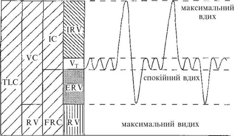
Fig. 16. Volumes and capacities of pulmonary ventilation (labels as in text)
The lungs are perfused by the entire Cardiac Output pumped into the lesser (pulmonary) circulation, which averages 10 L/min (about 5 L/min at rest). Blood flows through the alveoli at a rate of 1 L/s. Under a pressure of 2 cm $H_2O$, an erythrocyte spends about 1 s in a capillary, passing through 2-3 alveoli during this time, with only one-third of this duration remaining for the complete exchange of oxygen and carbon dioxide between the alveolar air and the blood.
The ventilation-perfusion ratio—The ratio of minute ventilation to minute pulmonary perfusion—is 8 L : 10 L = 0.8. This means that for every liter of blood flowing through the pulmonary capillaries per minute, 0.8 L of air passes through the alveoli.
Ventilation-perfusion relationships play a crucial role in respiratory gas exchange. For instance, during airway obstruction, including Atelectasis or impaired patency of the tracheobronchial tree, this ratio decreases, approaching 0 in complete obstruction. Conversely, when perfusion is reduced, the ventilation-perfusion ratio increases, approaching infinity in pulmonary artery embolism:

In emergency medicine, clinicians much more frequently encounter uneven ventilation-perfusion ratios across different Regions of the lungs. This is one of the primary causes of acute respiratory failure (ARF) in clinical scenarios such as Shock, Pneumonia, and others.
Alveolar-capillary gas diffusion depends primarily on the gas tension on both sides of the membrane, as well as on membrane permeability and the diffusion capacity of the gas.
Gas tension is characterized by partial pressure—the pressure that a component of a gas mixture would exert if it alone occupied the volume of the entire mixture. For example, ambient air contains 21% oxygen and 0.03% carbon dioxide. On average, atmospheric pressure at sea level is 760 mm Hg; it decreases with altitude, dropping, for instance, to 230 mm Hg at the world's highest peak, Mount Everest. Consequently, the partial pressure of oxygen ($p_{O_2}$) in the air at sea level is 21% of 760, which equals 159 mm Hg, whereas on Mount Everest it is 21% of 230, or 48 mm Hg. Similarly, we find that the partial pressure of carbon dioxide ($p_{CO_2}$) in the air at sea level is 0.23 mm Hg, and on Mount Everest 0.07 mm Hg, which in clinical and physiological calculations can be assumed to be 0.
Gas diffusion from a mixture into a liquid also depends on partial pressure, because The amount of gas dissolved in a liquid is directly proportional to the partial pressure of the gas above the liquid. Thus, the statement "the partial pressure of oxygen in Blood Plasma is 100 mm Hg ($p_{O_2} = 100$ mm Hg)" means that the plasma contains as much oxygen as it would if it were in direct contact with air in which the partial pressure of oxygen is 100 mm Hg. These considerations apply equally to the fluids of the interstitial and cellular environments. The dynamics of gas partial pressures in various PARTS OF THE human gas transport system are illustrated in Fig. 18.

Fig. 17. The normal breathing process

Fig. 18. Partial pressure of gases in various compartments of the human gas transport system
At an atmospheric pressure of 760 mm Hg, $p_{O_2}$ in the atmosphere is 159 mm Hg. In inhaled air, mixed with 16% Water vapor, $p_{O_2}$ drops to 150 mm Hg, and in the alveolus, due to the admixture of water vapor and carbon dioxide, it further decreases to 105 mm Hg. In venous blood flowing to the alveoli via the pulmonary artery system, $p_{O_2}$ is 40 mm Hg; therefore, oxygen diffuses from the alveolar lumen into the blood, raising plasma $p_{O_2}$ to 100 mm Hg. In tissue capillaries, oxygen diffuses from the blood into the tissues, where $p_{O_2}$ is 40 mm Hg, meeting their oxygen demand, which averages 250 mL/min under resting conditions. At the end of the capillary bed, plasma $p_{O_2}$ drops to 40 mm Hg and remains at this level in the venous blood until it reaches the pulmonary capillaries surrounding the alveoli once again.
Simultaneously, carbon dioxide diffuses in the direction opposite to oxygen diffusion. Produced in the tissues, $CO_2$ diffuses into the tissue capillaries, raising the $p_{CO_2}$ of venous plasma to 47 mm Hg. Venous blood maintains this level until it encounters the alveolar-capillary membrane, where carbon dioxide diffuses into the alveolar lumen, is eliminated into the atmosphere via exhaled air, and lowers arterial $p_{CO_2}$ to 40 mm Hg.
When ventilation-perfusion ratios change, the partial pressure of gases in the lungs alters accordingly (Fig. 19).
However, simple dissolution of gases in the liquid portion of the blood is entirely insufficient for transporting gases in the required volumes. For instance, the total circulating blood volume of 5 L (2 L of erythrocytes and 3 L of plasma) is capable of transporting only 9 mL of dissolved oxygen. If a person breathes pure (100%) oxygen, the amount dissolved in all the blood increases to 68.4 mL, and during respiration in a hyperbaric chamber with an oxygen environment at 3 atm, it reaches 205.2 mL. This clearly highlights the vital role of erythrocyte Hemoglobin in gas transport, particularly for oxygen; 1 g of hemoglobin can bind a maximum of 1.39 mL of oxygen (Hüfner's constant).
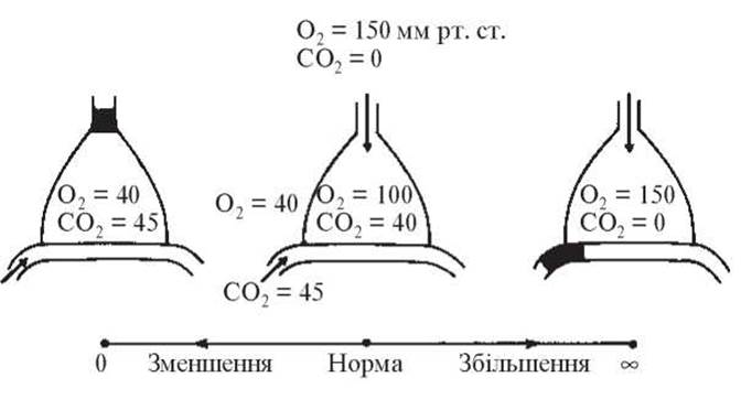
Fig. 19. Alveolar $O_2$ and $P_{CO_2}$ under altered ventilation-perfusion ratios

Fig. 20. Oxyhemoglobin dissociation curve. Dependence on Temperature (a), pH (b), and $P_{CO_2}$ (c)
Consequently, with a blood hemoglobin level of 145-150 g/L, the total blood volume can transport 1000 mL of oxygen. The primary factor determining both the saturation of blood hemoglobin with oxygen and the release of oxygen to the tissues (desaturation) is the difference in oxygen partial pressure across the alveolar-capillary and capillary-tissue membranes.
An equally important role in Oxygen transport is played by Changes in the binding affinity between hemoglobin and oxygen, as illustrated by the oxyhemoglobin dissociation curve (Fig. 20). It demonstrates that at a temperature of 37 °C, pH 7.4, and $p_{CO_2}$ of 40 mm Hg, hemoglobin binds oxygen most intensely when $p_{O_2}$ is 20-70 mm Hg, while complete hemoglobin saturation (99-100%) is achieved at a $p_{O_2}$ of 90-100 mm Hg. However, an increase in temperature and The Development of acidosis shift the dissociation curve to the right, which signifies a decreased affinity of hemoglobin for oxygen: oxyhemoglobin forms with difficulty but readily releases oxygen to the tissues. Conversely, a drop in temperature and the onset of alkalosis shift the curve to the left, indicating an increased affinity of hemoglobin for oxygen: oxyhemoglobin forms rapidly, but releases oxygen with difficulty.
Carbon dioxide is transported by the blood in two different forms: a minor fraction (about 10%) is physically dissolved, while the major fraction is chemically bound to hemoglobin (20%) and carried as sodium bicarbonate (70%). Therefore, it must be emphasized that erythrocytes, equipped with hemoglobin and Carbonic anhydrase, are just as vital for the chemical binding of 90% of transported $CO_2$ as they are for oxygen transport.
Gas Exchange in the lungs and its transport by the blood are carried out solely to support tissue respiration, which is the ultimate goal of gas exchange. The essence of tissue respiration lies in the ability of living tissue, via an enzymatic system, to rapidly and completely (down to water and carbon dioxide) oxidize organic substances that are normally relatively inert to molecular oxygen.
Based on blood gas tension data, acute respiratory failure (ARF) is defined as a condition in which pO2 is below 60 mmHg and pCO2 is above 50 mmHg, or when normal blood gas tension (pO2 60-100 mmHg, pCO2 35-45 mmHg) is maintained only by engaging additional respiratory reserves (compensated ARF).
Causes of ARF classified by the anatomical Location OF THE impairment:
— Brain (Disorders of the respiratory center due to Hypoxia and ischemia resulting from trauma, cerebrovascular accidents, poisoning, etc.);
— Spinal Cord (trauma, poliomyelitis);
— motor nerves (polyneuritis);
— neuromuscular junctions and muscles (water-electrolyte balance disorders, botulism, Muscle relaxants, myasthenia gravis);
— chest wall (bone fractures, kyphoscoliosis, Ankylosing spondylitis, pneumothorax, and hemothorax);
— Pulmonary Circulation (shock-induced shunting, decreased pulmonary artery pressure caused by ganglion blockers and hypovolemia, Pulmonary Embolism, fat embolism);
— lungs and airways (bronchial asthma, acute Bronchitis, pneumonia, Chronic bronchitis, and emphysema).
Most often, ARF results from a combination of factors. Precipitating triggers primarily include infection, surgery, intercurrent illness, and The Use of respiratory-depressant medications.
The pathogenesis of ARF development can vary depending on which link in the complex physiological chain described above is the primary site of respiratory impairment.
Pathological disturbances may primarily affect ventilation (as predominantly seen in impaired respiratory Regulation at the level of the brain, spinal cord, and neuromuscular conduction, tracheobronchial obstruction, or chest wall defects), perfusion (shock, pulmonary hypotension, pulmonary vascular embolism), ventilation-perfusion mismatches (inflammatory lung diseases, atelectasis), or alveolar-capillary diffusion (inflammatory membrane thickening, interstitial pulmonary edema).
However, whichever factor acts as the primary trigger, it is never truly isolated. It may operate in parallel with other factors from the very beginning—for instance, in traumatic injuries, where ventilatory impairment due to central regulation disorders, Tongue drop, and mucus accumulation in the Trachea during unconsciousness combines with perfusion deficits (resembling shock lung) and impaired alveolar-capillary diffusion caused by interstitial edema. Even when the primary factor initially acts in isolation, it inevitably pulls other factors into the pathological process. This occurs, for example, in narcotic analgesic overdose, where hypoventilation leads to impaired tracheobronchial tree drainage with segmental atelectasis, hypoxic pulmonary vasoconstriction with perfusion disorders, and so on.
The consequence of any of these mechanisms is arterial hypoxemia (insufficient oxygen in the blood) and hypercapnia (elevated carbon dioxide concentration in the blood).
The Mechanism of hypoxemia in ARF is associated with:
— inadequate pulmonary ventilation;
— decreased oxygen diffusion across the alveolar-capillary membrane;
— reduced alveolar perfusion (arteriovenous shunting in shock) or ventilation-perfusion imbalances.
In all these cases, hypoxemia leads to an inadequate supply of oxygen to the body's tissues, a condition known as hypoxia. There are numerous types of hypoxia. The form developing As a result of hypoxemia is called Hypoxic hypoxia, also referred to as respiratory hypoxia. Additionally, hypoxia may be linked to a lack of oxygen in the breathing mixture (exogenous hypoxia), a reduction in the oxygen-carrying capacity of the blood due to anemia, hydremia, or hemoglobin blockade (hememic hypoxia), circulatory disturbances (circulatory hypoxia), and the suppression of redox processes in tissues (histotoxic or tissue hypoxia); however, these types of hypoxia are not directly related to ARF.
Hypercapnia in ARF is almost invariably associated with alveolar hypoventilation or an increase in dead space, since carbon dioxide production (200 mL/min) changes very little, and the diffusion of CO2 in fluid media (blood and the alveolar-capillary membrane) significantly exceeds that of oxygen.
Hypoxia increases cellular membrane permeability, resulting in cerebral edema. The sympathoadrenal system is stimulated, leading to catecholaminergic states that cause arteriolar spasm and impair microcirculation. From this point on, the rheological properties of blood begin to deteriorate. Sluggish blood flow promotes cellular aggregation and thrombosis, followed by disseminated intravascular coagulation (DIC), blood sequestration, and a decrease in circulating blood volume (CBV). Hypovolemia reduces cardiac output, which in turn triggers further arteriolar constriction, locking in a vicious cycle.
In the lungs, hypoxia causes vasoconstriction, interstitial edema, decreased surfactant production, reduced compliance of lung tissue, and increased airway obstruction. Pulmonary Hypertension leads to right ventricular failure. When hypercapnia is superimposed on hypoxia, the patient's condition deteriorates even further.
Hypercapnia is one of the key factors stimulating respiration in healthy individuals. However, in ARF, this factor's effect has low efficacy, and once pCO2 reaches 90-100 mmHg or higher, the stimulating effect of CO2 on the Central Nervous system shifts to a depressant effect, known as "carbon dioxide narcosis."
By inducing cerebral vasodilation, hypercapnia contributes to elevated intracranial pressure. Cardiac output increases, and peripheral vessels dilate. The oxyhemoglobin dissociation curve shifts to the right, which means lower oxygen saturation in the blood, but easier release of oxygen to the tissues.
The Clinical presentation of ARF depends on its underlying cause and primary pathophysiological mechanism. It is characterized by a combination of hypoxia and hypercapnia along with their downstream effects, as well as the manifestation of compensatory respiratory mechanisms. Blood gas analysis and acid-base balance (ABB) testing are invaluable tools in diagnosing ARF. The rate of onset significantly influences the clinical manifestations. When developing gradually, the course of ARF is typically divided into 4 severity grades:
— Grade 1 — mild dyspnea (respiratory rate increased by 10–20%), a resting pulse-to-respiration ratio of 3:1 (normally 3:1 to 4:1 depending on age), mild cyanosis of the Lips, and a blood pH of 7.35;
— Grade 2 — more pronounced dyspnea (respiratory rate increased by 20–30%), tachycardia with a 30% increase in Heart rate, a pulse-to-respiration ratio of (2.5–2):1; intermittent acrocyanosis; mild diaphoresis, restlessness, or mental depression may occur; pH drops to 7.3, PaO2 is 80–60 mmHg, and PaCO2 is 45–60 mmHg;
— Grade 3 — shallow, rapid breathing and tachycardia, a pulse-to-respiration ratio of (2–1.5):1; an ashen Skin tone, cyanosis of the lips; excitation gives way to lethargy, pH drops to 7.2, Pa O2 is 60–40 mmHg, PaCO2 is 70 mmHg and higher;
— Grade 4 — development of hypoxic coma; spasmodic respiratory movements; Mouth closed, gasping for air (“fish-mouth breathing”); a pulse-to-respiration ratio of (8–9):1; loss of consciousness; ashen skin tone, with purplish-blue spots appearing on the extremities and trunk; pH down to 7.15.
When ARF develops rapidly, staging is difficult to determine. In cases of severe respiratory impairment, hypoxia can reach a life-threatening degree within minutes, as confirmed by theoretical calculations. For instance, the total oxygen capacity of the body is 1.5 L, and the oxygen requirement is about 250 mL/min. Even accounting for the complete utilization of the body's oxygen reserves—which is physiologically impossible—life cannot be sustained for more than 6 minutes. Furthermore, prior inhalation of 100% oxygen extends this timeframe only twofold.
In cases of ARF constituting a medical emergency, where a delay of even a few minutes in initiating intensive care can be fatal for the patient, symptoms related to the Etiology AND PATHOGENESIS of ARF are indistinct, and the clinical picture is largely confined to signs of hypoxia and hypercapnia. These include tachypnea, mental and physical restlessness, and progressive impairment of consciousness leading up to coma.
Convulsions may develop against the Background of a coma. Involvement of accessory muscles in the respiratory act is observed, particularly when airway patency is compromised and airway resistance increases. The skin and mucous membranes are cyanotic; in extremely severe ARF accompanied by marked microcirculatory disorders, the skin becomes ashen, cold, and covered in cold sweat. In anemic patients (hemoglobin below 50 g/L), the skin is pale without cyanosis. A pink facial complexion is preserved in ARF only in patients with hemic and histotoxic hypoxia (carbon monoxide or cyanide poisoning). A brick-red facial flush with profuse sweating occurs when hypercapnia predominates over hypoxia, although such a presentation is atypical for severe ARF.
Tachycardia giving way to bradycardia, and arterial hypertension transitioning to hypotension, are frequently noted. Diuresis decreases, intestinal paresis develops, and acute stress ulcers of The Stomach and intestines may form.
Patients with severe ARF require intensive nursing care, monitoring of respiration (respiratory rate, tidal and minute volumes, inspired oxygen and expired carbon dioxide concentrations, tissue pO2 and pCO2 via polarography, and oximetry) and hemodynamics. Systematic laboratory control of acid-base balance and blood gases is performed (using Astrup's micromethod).
The Treatment of severe forms of ARF is so urgent that it must precede Diagnostics.
The principles of intensive care for ARF include:
— restoring and maintaining airway patency;
— in cases of significant respiratory depression — mechanical ventilation until the situation is clarified and effective treatment is initiated;
— oxygen inhalation;
— treatment of underlying causes amenable to immediate therapy (bronchodilators for bronchial asthma, nalorphine for morphine poisoning, etc.);
— suctioning of secretions from the airways;
— monitoring the adequacy of ventilation during spontaneous breathing;
— strict adherence to asepsis and antisepsis;
— antimicrobial therapy;
— management of concomitant pathological processes, whether related to the Respiratory system or not (pneumothorax, hemothorax, heart failure, Homeostasis correction);
— if prescribed treatment proves ineffective — mechanical ventilation and other measures within specialized intensive care units and wards.
Methods for restoring and maintaining airway patency include: HEAD tilt, jaw thrust, and mouth opening (the so-called “triple airway maneuver”); insertion of airways, tracheal intubation, conicotomy, tracheostomy, and tracheal suction. The use of anti-inflammatory and anti-edema agents (intravenous administration of corticosteroids, referred to as “intravenous tracheostomy”) and antispasmodics, including aerosol inhalations, is also recommended.
If, upon restoring airway patency, breathing is absent, marked hypoventilation is observed, or breathing is performed with excessive effort leading to progressive respiratory muscle fatigue, urgent transfer to mechanical ventilation by any available method is recommended, starting with mouth-to-mouth resuscitation or portable manual resuscitators.
However, during the course of intensive care—despite airway maintenance, oxygen inhalation, and medical treatment—ARF may progress, reaching Grade 2. Consequently, with a respiratory rate exceeding 30 breaths per minute, pronounced tachycardia over 100 bpm, cyanosis, diaphoresis, restlessness, or mental depression, accompanied by laboratory signs of hypoxemia and decompensated respiratory acidosis (pH<7.3, PaO2 <60 mmHg, PaCO2 >60 mmHg), further escalation of care is warranted.
In such cases, transitioning to mechanical ventilation using specialized equipment is recommended, connected to the patient via oro- or nasotracheal intubation. If prolonged support is anticipated for an unpredictably long period, a tracheostomy should be performed instead. Therefore, the indications for mechanical ventilation include the absence of spontaneous breathing, pathological breathing patterns (including pre-agonal and agonal states), and the aforementioned symptoms of pronounced hypoxia and hypercapnia.
In patients with pneumonia and other pulmonary diseases ("parenchymal ARF"), mechanical ventilation is always merely an auxiliary palliative method. It can be effective only if a reserve inspiratory and expiratory volume is present; otherwise, it may even yield a negative effect. In patients with parenchymal ARF, mechanical ventilation should be initiated with the expectation of continuous application (without frequent attempts to switch to spontaneous breathing) for 2–3 days or longer. This is because, unlike ventilatory ARF (characterized by "conventionally healthy lungs"), rapid morphological and functional changes do not occur in the lungs under this pathology. Selecting the appropriate ventilation mode is challenging, and the adequacy of ventilation should be assessed exclusively under the control of acid-base balance (ABB) and blood gases.
Among the types of mechanical ventilation, a distinction is made between external and internal.
External Methods of mechanical ventilation are outdated and practically unused. They substituted the function of the diaphragm—such as the "pulmonary belt" or "rocking bed"—or artificially created negative extrathoracic pressure during inspiration, as seen in the "cuirass respirator" and "iron lung."
Internal or insufflation methods (derived from insufflatio) of mechanical ventilation are the only ones used today. With these methods, gas is directed into the lungs under positive pressure, which is why they are referred to as intermittent positive pressure ventilation (IPPV). Their advantage lies in The ability to adequately ventilate the lungs despite increased pulmonary resistance or decreased lung compliance, the accessibility of the patient's body for examination and manipulation, and the facilitation of tracheal and bronchial suctioning.
At the same time, during IPPV, the fluctuating positive pressure in the airways impedes the venous return of blood to The Heart and may impair cardiac filling. This drawback can be eliminated by actively creating negative pressure in the airways during expiration (active expiration). This method is termed intermittent positive-negative pressure ventilation (IPNPV); however, the risk of pulmonary edema serves as a contraindication for IPNPV. In such cases, IPPV with positive end-expiratory pressure (PEEP) is recommended, which in this context is referred to as continuous positive airway pressure (CPAP).
In recent years, mechanical ventilation equipment has become highly sophisticated. Utilizing new-generation microprocessors and computer technology, intelligent ventilation systems are designed to adapt to the specific needs of the individual patient—such as controlled ventilation, assisted controlled ventilation, and others. Assisted mechanical ventilation is based on a triggering mechanism that responds to the faintest attempt by the patient to inhale with a full breath. If the pause following a regular breath exceeds the expected duration, the system switches back to standard mechanical ventilation mode.
State-of-the-art devices are capable of high-frequency ventilation (HFV), which is ventilation either without respiratory movements or with movements where the tidal volume is smaller than the dead space volume. Gas exchange is maintained by a significant gas flow driven by turbulence generated at oscillation frequencies of 60–300 cycles per minute. These methods enhance gas diffusion and improve gas distribution within the lungs. They are utilized when maintaining a sealed breathing circuit is impossible, prove effective in treating acute respiratory distress syndrome in both adults and children, and serve as adjuncts to any mode of spontaneous and mechanical ventilation.
Physiologically, membrane oxygenation is closely related to mechanical ventilation. It is performed extracorporeally using an oxygenator connected to the patient's Vascular System.
Additionally, Oxygen therapy—as one of the treatment modalities for ARF accompanied by hypoxia—is employed alongside any form of pulmonary ventilation, most frequently via the inhalation of oxygen-enriched mixtures (oxygen-air or oxygen-helium combinations), including under hyperbaric conditions.
It is important to emphasize the exceptional significance not only of providing emergency care for ARF with subsequent treatment and rehabilitation, but also of preventing respiratory disorders in any critically ill patient through meticulous general therapeutic care. This includes optimal patient positioning in bed, pain management without depressing motor activity, sputum liquefaction and clearance via coughing, nutritional support via any appropriate route (oral, enteral via tube, or parenteral), hygiene of the Oral Cavity and Upper Respiratory Tract, and psychological support. The latter promotes the rational expenditure of respiratory energy, fosters crucial mutual understanding between the physician and the patient, and instills confidence in a successful recovery.
1. Bunyatyan, A. A., Ryabov, G. A., & Manevich, A. Z. (1984). Anesthesiology and Reanimatology. Moscow: Meditsina, pp. 40-63, 366-375.
2. Glumcher, F. S. (1999). Anesthetic Management and intensive care in reconstructive surgery of the lower airways (Author's Abstract of Doctoral dissertation). Kyiv, 32 p.
3. Zilber, A. P. (1989). Respiratory Failure. Moscow: Meditsina, 512 p.
4. Zilber, A. P. (1996). Studies in Critical Care Medicine (Vol. 2: Respiratory Medicine). Petrozavodsk: Petrozavodsk University Press, 488 p.
5. Chuev, P. N., Molchanov, V. I., Vladyka, A. S., et al. (2003). Intensive Care Medicine: A Textbook. Simferopol: Tavriya, pp. 121-144.
6. Koryachkin, V. A., & Strashnov, V. I. (Eds.). (2002). Intensive Care of Life-Threatening Conditions. St. Petersburg: Saint Petersburg Medical Publishing House, 286 p.
7. Kassil, V. L. (1987). Artificial Pulmonary Ventilation in Intensive Care. Moscow: Meditsina, 256 p.
8. Kassil, V. L. (1998). Respiratory Support. Moscow: Meditsina, 320 p.
9. Kurapov, E. P., Khripachenko, I. A., & Vorkhlik, M. I. (1998). Proteolytic Enzymes and methods of their administration in the comprehensive treatment of acute respiratory distress syndrome. Pain, Anesthesia and Intensive Care, (4), 37-41.
10. Milic-Emili, J. (1997). Mechanics of breathing. In Current problems of Anesthesiology and Reanimatology (pp. 203-206). Arkhangelsk; Tromsø.
11. Mitin, Y. V. (1986). Acute Laryngotracheitis in Children. Moscow: Meditsina, 208 p.
12. Navrátil, M., Kadlec, K., & Daum, S. (1966). Pathophysiology of Respiration. Moscow: Meditsina; Prague: State Medical Publishing House, 372 p.
13. Glumcher, F. S., Makarov, A. V., Skubriy, V. M., et al. (2003). Results of using Modern Methods of respiratory support. Pain, Anesthesia and Intensive Care, (2), 8-16.
14. Sykes M. K., McNicol M. W., Campbell E. J. M. Respiratory Failure. — Moscow: Meditsina, 1974. — 344 p.
15. Simbirtsev S. A., Belyakov N. A. Pulmonary Microembolism. — Leningrad: Meditsina, 1986. — 216 p.
16. Titov I. I. Acute Lung Injury Syndrome in Critical States and Methods of Its Correction (Experimental and Clinical Study): Author's Abstract of Diss. ... Dr. of Med. Sci. — Kyiv, 2003. — 36 p.
17. Tracheostomy as an Alternative to Prolonged Intubation / W. Hackl, B. Regal, K. Strasser, M. Watzak-Helmer // Current Problems of Anesthesiology and Intensive Care. — Arkhangelsk; Tromsø, 1997. — P. 221-222.
18. Falx C. Management of Severe ARDS // Ibid. — 1998. — P. 240.
19. Kharitoshina M. V., Molchanov V. I. Optimization of Bronchial Asthma Treatment with "Sucrim" // Achievements and Prospects of Modern Anesthesiology and Intensive Therapy. — Dnepropetrovsk, 2003. — P. 159-162.
20. Shlapak I. P., Pylypenko M. M. Guidelines for Respiratory Support: Textbook for Resident Physicians and Postgraduate Students. — Kyiv: Logos, 2003. — 134 p.
21. Shlapak I. P., Titov I. I. Latest Trends in the Treatment of Acute Lung Injury Syndrome // Proceedings of the 3rd National Congress of Anesthesiologists of Ukraine. — Odesa, 2000. — P. 219.
22. Yakovleva I. I., Timokhov V. S. Pathogenesis and Treatment of Adult Respiratory Distress Syndrome in Patients with Multiple Organ Failure // Anesthesiology and Reanimatology. — 1996. — No. 1. — P. 75-80.
23. Tchuyev P. M., Vladyka A. S. Intensive Care in Emergency. — Odessa: The Odessa State Medical University, 2005. — P. 25-35.
24. Intensive Care Medicine: Textbook / P. N. Chuev, V. I. Molchanov, A. S. Vladyka et al. — Simferopol: Tavria, 2006. — P. 119-201.
25. Irwin R. S., French C. T., Mike R. W. Respiratory Adjunct Therapy // Intensive Care Medicine / Ed. by J. M. Rippe, R. S. Irwin, M. P. Fink, F. B. Cerra. — Boston; New York; Toronto; London: Little, Brown & Co., 1996. — Vol. I. — P. 773-787.
26. Irwin R. S., Pratter M. R. Physiologic Approach to Managing Respiratory Failure // Intensive Care Medicine / Ed. by J. M. Rippe, R. S. Irwin, M. P. Fink, F. B. Cerra. — Boston; New York; Toronto; London: Little, Brown & Co., 1996. — Vol. I. — P. 581-587.
27. Schuster D. P., Kolleff M. H. Acute Respiratory Distress Syndrome // Intensive Care Medicine / Ed. by J. M. Rippe, R. S. Irwin, M. P. Fink, F. B. Cerra. — Boston; New York; Toronto; London: Little, Brown & Co., 1996. — Vol. I. — P. 773-787.
Last update: 11/08/2026
Editorial and Educational Adaptation: This material has been compiled based on the primary/original source text. The project team performed an editorial review, corrected technical inaccuracies, structured sections, and adapted the content for an educational format.
What was processed:
- elimination of formatting defects (OCR errors, structural breaks, corrupted characters);
- editorial organization of content;
- standardization of terminology in accordance with academic sources;
- verification of factual statements against the original source text.
All mentions of the author, publication year, and origin of the primary text have been preserved in accordance with the source.