NEONATAL SURGERY - 1976
1. GENERAL PROVISIONS
Class="center">
1. Anatomical and Physiological Characteristics of the Newborn Infant's Organism
The anatomical and PHYSIOLOGICAL CHARACTERISTICS OF the newborn infant's Organism, which determine the specific nature of its reactions to various environmental influences in normal and pathological situations, are detailed in numerous, now classic, works by domestic and foreign researchers (N. P. Gundobin, 1906; F. I. Walker, 1938; I. A. Arshavsky, 1959; G. Fanconi, A. Wallgren, 1960; D. Vobev, I. Ivanova, 1969; E. Ch. Novikova et al., 1971, etc.).
A new milestone in the understanding of Embryogenesis and the patterns of pre- and Postnatal development in animal and human organisms was The Doctrine of systemogenesis developed by P. K. Anokhin (1948). According to this concept, the selective and asynchronous (heterochronic) development of morphological structures united by a common specific function is a vital prerequisite for providing the organism with timely adaptive reactions aimed at its survival during a given time period. Systemogenesis and the harmonious system of normal development (pormogenesis) of the organism served as the starting point for the development (S. Ya. Doletsky, 1968) of The Theory of relative immaturity and the disparities in the GROWTH AND DEVELOPMENT of Organs and systems in newborns. This theory facilitates the understanding of the specific course of pathological processes in infants, the justification of therapeutic principles for various pathological conditions, and the assessment of long-term prognosis.
Weight, height, and body proportions. In recent years, considerable attention has been paid to The Study of the anatomy and PHYSIOLOGY OF THE growing organism (B. F. Shagan, 1959; A. Andronescu, 1970, etc.). These anatomical and physiological features are more pronounced the younger the child is. Knowledge of the age-related CHARACTERISTICS OF THE newborn is essential for the timely and rational Prevention of potential diseases, the creation of optimal conditions for normal development, and the proper Organization of care. Body length and weight are the Main criteria for assessing maturity and full-term status in newborns.
In recent years, the indicators of physical development in newborns have increased. Average height has increased by 0.8 cm and currently stands at 51–52 cm. The weight of boys reaches 3400–3500 g, and that of girls 3250–3400 g (A. F. Tur, 1971). This is attributed to the predominance of first-born infants among newborns, whose birth weight is known to be lower than that of subsequent children. According to R. B. Kogan, the proportion of first-borns in Moscow in 1964 was 77%. According to most authors, the minimum weight of a full-term newborn is 2500 g, and the maximum is 4000–4500 g. Weight variations depend on the individual characteristics of the parents, their health status, maternal Nutrition during Pregnancy, etc.
The body proportions of a newborn differ significantly from those of an adult. The HEAD accounts for 1/4 of the body length, reaching a circumference of 32–34 cm, which exceeds the chest circumference by 2 cm. A characteristic feature of the newborn is an eightfold predominance of the cerebral cranium over the facial part (compared to 2 times in adults). The extremities are relatively short. There is no difference between the lengths of the upper and lower extremities. Data on the dimensions of the newborn's body parts are presented in Table 1.
Table 1. Main dimensions (in centimeters) of newborns (according to A. F. Tur, 1967)
Parameters |
Boys |
Girls |
Height |
50.8 |
50.0 |
Trunk length |
21.4 |
21.2 |
» Arm length |
21.4 |
21.0 |
» Leg length |
20.5 |
20.3 |
Head circumference |
32.7 |
32.6 |
» Chest circumference |
28.2 |
28.5 |
During the neonatal period, some of the listed parameters change significantly. The first days of life are marked by the so-called physiological weight loss, primarily due to the elimination of intestinal contents, the desiccation of the umbilical cord remnant, and fluid loss through the Kidneys, Lungs, Skin, etc. Physiological weight loss accounts for 6–8% and sometimes up to 10% of the initial body weight. The maximum weight reduction is observed on the 2nd to 3rd day. A weight drop of 500 g or more generally indicates a pathological condition associated either with underfeeding or illness in the newborn. Birth weight is normally regained by the 7th to 10th day, and by the end of the neonatal period, the infant gains 700–800 g.
The body length of the newborn may decrease During the first days of life As a result of the resolution of cranial deformations caused by labor. By the end of the neonatal period, the infant grows by 1 cm.
Thus, throughout the neonatal period, interconnected processes of body mass increase, morphogenesis, and actual development, or differentiation of organs and Tissues, proceed intensively. Quantitative, qualitative, and temporal disparities among these processes determine the Specific features of certain transitional states and diseases characteristic exclusively of infants in their first weeks of life.
Skin, mucous membranes, subcutaneous adipose tissue. The total skin surface area of a newborn is 6 to 8 times smaller than that of an adult, amounting to 0.25 m2. The appearance of the skin is characteristic: it has a pale pink color, is succulent and even somewhat edematous due to its high Water content, and appears delicate and velvety thanks to abundant Blood supply. The newborn's skin is smooth and soft, which is due to the loose Structure OF THE epidermis and dermis, as well as the more delicate architecture of Collagen and elastic fibers. The underdeveloped basement membrane and poorly developed stratum corneum reduce the adhesion between the epidermis and dermis, making the infant's skin easily vulnerable.
At birth, the skin is covered with a grayish-whitish or yellowish cheesy substance called vernix caseosa. It is most prominent on the face, ears, axillae, and inguinal folds. The lubricant consists of soluble fats and modified epithelial Cells, and is rich in Cholesterol and Glycogen. S. Ya. Golosovker, P. Popchristov, and other authors point out the protective properties of this substance, which shields the newborn's skin from infection. After the first bath, the substance is removed, exposing the stratum corneum, as a result of which the skin acquires a pale cyanotic hue. Any potential thermal irritations cause capillary dilation and A change in color to bright red (physiological catarrh of the skin or erythema neonatorum), which disappears by the 3rd day of life. The erythema is replaced by general desquamation, which can last for up to 2 weeks. The shedding of the newborn's skin occurs as a result of postpartum desiccation, physiological parakeratosis, and the weak functioning of the glandular apparatus.
Sweat Glands are underdeveloped, particularly on the elbows, hands, and ankle regions. In these areas, the skin tends to be coarse, dry, and pale. It should be noted that in hemorrhagic disease, cutaneous hemorrhages appear primarily in these areas. Well-functioning Sebaceous Glands sometimes become visible due to the excessive accumulation of secretions in their ducts. They can be seen on the skin of the Nose as yellowish-white dots (milia).
Most of the newborn's skin, especially on the shoulders and back, is covered with soft downy Hair (lanugo), which usually falls out during the 1st week of life. Fingernails and toenails are well developed and, in full-term infants, reach the tips of the digits. A weaker Development of the nail plate is not a sign of immaturity. Congenital absence of Nails (anonychia) is extremely rare.
Red spots of various Sizes and Shapes, resulting from the dilation of intracutaneous capillaries, are frequently observed on the heads of newborns. These spots are located on the forehead, the bridge of the nose, and the eyelids; they resemble flat hemangiomas, but unlike them, they disappear spontaneously.
The mucous membranes are bright and somewhat dry due to the insufficient development of glands. The poorly developed epithelial lining makes them delicate and highly vulnerable.
The subcutaneous adipose tissue in the newborn is well developed throughout the entire body, including the extensor surfaces of the joints, where it is subsequently absent. At birth, the child has 5 times more adipose tissue relative to body weight than an adult. It lacks a differentiated structure, is functionally labile, and can transform into hematopoietic or fat-storing tissue. It is currently proven that adventitial mesenchymal Cells of the newborn's adipose tissue possess hematopoietic function. In certain PARTS OF THE body, the tissue has an entirely embryonic character and is closely associated with branching Blood Vessels (Becker, 1954). The chemical composition of the fat is characterized by an elevated content of saturated acid derivatives—stearic and palmitic acids—which results in a denser subcutaneous adipose tissue and its easier solidification under cooling conditions. The latter explains The Development of subcutaneous fat necrosis, sclerema, and other conditions in certain cases.
These characteristics of the skin, mucous membranes, and subcutaneous adipose tissue must be taken into account in all pathological processes in newborns.
Respiratory organs, chest. By the time of birth, the respiratory organs have not reached maturity. The newborn's nose is short, with a poorly developed nasal bridge and narrow choanae. The mucous membrane of the nasal choanae is delicate, with numerous Blood and Lymphatic vessels; the Paranasal Sinuses are poorly developed. The maxillary sinuses are the most prominent, while the frontal and sphenoid sinuses are practically absent. The Pharynx is narrow and small, with an underdeveloped lymphatic ring.
The Larynx is funnel-shaped with an expanded upper portion and is located
2 to 3 vertebrae higher than in adults. The cartilages forming the larynx are thin, elastic, and pliable. The small lumen of the larynx, along with an Abundance of blood vessels and lymphoid tissue, leads to a more rapid onset of stenosis during inflammation or trauma compared to older children.
In newborns, the Trachea varies in shape from wide and short to narrow and long. Its cartilaginous rings are soft, easily compressible, and prone to displacement. The length of the trachea is 4 — 5 cm; its mucous membrane is rich in blood vessels but somewhat dry due to the scarcity of mucous glands. At the level of the III — IV thoracic vertebrae, the trachea bifurcates into the two main Bronchi. The right main bronchus diverges at an angle of 15 — 20° and appears as a direct continuation of the trachea. The left main bronchus is longer and diverges at an angle of 20 — 40°. The lumens of the segmental bronchi are narrow, and their walls contain few elastic fibers. The mucous membrane of the bronchial tree is loose, highly vascularized, and prone to Swelling, which easily causes bronchial obstruction.
During the first hours after birth, the lungs undergo changes in shape, weight, and position. With the first breath, their expansion begins and continues for several days. Certain alveoli, predominantly in the posterior-inferior Regions of the lungs, may remain unexpanded throughout the entire neonatal period, as diaphragmatic respiratory movements initially occur predominantly in the anteroposterior direction. With the initial breaths, the greater portion of air enters the lower regions of the left lung; on the right side, the relatively large Liver restricts diaphragmatic movement. However, the right lung subsequently expands faster and more effectively because it is ventilated via a wider and shorter bronchus. The weight of the lungs reaches 50 g; the right lung is somewhat larger than the left. The newborn's lungs are plethoric due to wide capillaries and lymphatic clefts. The interstitial tissue is well developed, but elastic fibers are insufficient, which reduces lung aeration, predisposes to Atelectasis, and facilitates the development of inflammatory processes.
The aforementioned Anatomical Features determine the physiological changes in respiration. In the fetus, gas exchange occurs via placental Circulation. With the birth of the child and the first breath, pulmonary respiration is established. It is generally accepted that the primary stimulus for the first breath is an elevated carbon dioxide concentration in the blood, which excites the respiratory center (Yu. F. Dombrovskaya, 1957; A. F. Tur, 1967, et al.). Owing to the insufficient Differentiation of the newborn's Nervous system, its regulatory influence on the respiratory center is inadequate, which explains the lability and specific Characteristics of Respiratory parameters.
The respiratory rate is 40 — 60 breaths per minute. Such tachypnea is not pathological and is associated with shallow breathing. Rapid and superficial breathing is caused by the newborn's increased oxygen demand. The minute respiratory volume is 800 — 900 ml. This parameter depends on the basal metabolic rate and changes not only in respiratory disorders but also in circulatory disturbances. The vital capacity of the lungs is approximately 140 ml.
The anatomical and functional immaturity of the newborn's Respiratory system predisposes them to a higher incidence of generalized pulmonary diseases accompanied by severe respiratory failure.
Cardiovascular system. By the end of the 2nd month of intrauterine life, placental circulation develops, providing the fetus with everything necessary for development. There is no direct communication between the fetal and maternal blood. The transfer of oxygen and nutrients is accomplished through the villous epithelium and the capillary endothelium located within the villi.
Arterial blood enters the fetal body via the umbilical vein and divides into two streams. A smaller portion of the oxygen-rich blood goes to the liver, while the larger portion flows through the ductus venosus into the INFERIOR VENA CAVA, where it first mixes with the venous blood from the lower half of the body. Blood from the inferior vena cava enters the right atrium, where it merges with the venous blood of the SUPERIOR VENA CAVA. Subsequently, the blood in the right atrium divides into two streams. One stream enters the right ventricle, from which the bulk of the blood is shunted through the ductus arteriosus into the descending aorta, while a smaller portion passes through the lungs and enters the left atrium. The second stream passes directly from the right atrium through the foramen ovale into the left atrium, mixes with the blood coming from the non-functioning lungs, and is directed into the left ventricle. The greater part of the blood from the descending aorta returns via the umbilical Arteries to the Placenta (Fig. 1, a).
Fig. 1. Diagram of fetal (a) and neonatal (b) circulation.

Thus, multiple instances of arterial and venous blood mixing occur during embryonic circulation. At the same time, the organs performing the most vital Functions for the fetus receive the blood richest in oxygen. These include the liver, Brain, and myocardium. The pelvic organs and lower extremities receive blood that is relatively poor in oxygen. According to S. Ya. Doletsky (1968), among the causes of the prevalence of certain developmental anomalies in the zone of relative Hypoxia, the latter may play a certain role in teratogenesis in general, as well as in the selective vulnerability to conditions such as calcinosis.
From the moment of birth, a dramatic reorganization of the circulation occurs: placental circulation ceases, and the pulmonary (lesser) circulation begins to function (Fig. 1, b). This leads to an increase in pressure in the left atrium (due to increased blood inflow from the functioning lungs) and a decrease in pressure in the right atrium (due to the cessation of BLOOD FLOW IN the umbilical vein). This pressure shift in the atria halts the shunt of blood through the foramen ovale and leads to its gradual closure. At the age of 6 — 8 months, it closes completely, and from that moment, the SYSTEMIC AND PULMONARY circulations begin to function independently. The expansion of the lungs and the associated change in Heart position impede Blood flow through the ductus arteriosus. The mixing of arterial and venous blood ceases; the ductus arteriosus becomes non-functional and subsequently obliterates, transforming into the ligamentum arteriosum magnum.
The newborn's heart exhibits distinctive features. Relative to body weight, it is larger than in an adult, accounting for 0.8% of the child's body weight (20 — 24 g). According to A. B. Volovik (1952), the length of The Heart is 3.1 cm, its width is 4 cm, and its thickness reaches 1.85 cm. The thickness of the right and left ventricular walls is almost equal — about 5 mm. The volume of the heart chambers reaches 20 cm3.
It is believed that in the newborn, the right ventricle predominates over the left (F. I. Valker, 1938). With age, the Muscle of the left ventricle increases significantly, unlike the right ventricle, which remains almost unchanged. The atria and arterial vessels are relatively large compared to the ventricles.
During the 1st month of life, the heart lags behind other organs in weight, but subsequently grows intensively. Heart growth is accompanied by changes in its topography. A transverse position of the heart is characteristic of the newborn. As the Ribs and Diaphragm descend, the heart assumes an oblique position. The apex of the heart is typically formed by both ventricles. Less frequently, the apex is formed by a single ventricle, most often the left.
Histologically, the myocardium has a delicate structure. Muscle fibers are thin and short, and are arranged more compactly. Elastic fibers are poorly developed. The network of blood vessels is well pronounced and features numerous anastomoses.
Cardiac innervation is embryonic in nature. By the time of birth, the centers of the sympathetic and vagus nerves appear morphologically developed. The peripheral endings of both nerves are well formed, but since the sympathetic nerve appears earlier than the vagus during embryogenesis, sympathetic predominance persists after birth. This accounts for the infant's rapid and labile pulse. The heart rate is 120 — 160 beats per minute. The ARTERIAL BLOOD PRESSURE of a child on the 1st day of life is 67.7 ± 0.49 — 36.5 ± 0.43 mm Hg. By the 10th day, it correspondingly rises to 77.2 ± 0.75 mm Hg (V. P. Buyko, 1967; A. A. Makarov, 1973). Venous pressure is 70 — 90 mm H2O. The circulating blood volume does not exceed 85 ml/kg, which must be taken into account when determining the extent of blood loss in various diseases and during surgery.
The specific Features of the newborn's myocardium determine a characteristic radiological picture, which is distinguished by poor differentiation of the cardiac borders. Along the right border, the cardio-vascular angle is not always present; the lower border appears more convex and is formed by the right atrium. The left ventricle and great vessels (aorta and pulmonary artery) form the lower and upper borders, respectively, along the left edge of the cardiac shadow (O. L. Tsymbal, 1968). The ROOT vascular pattern is barely discernible, as the lung roots are obscured by the cardiac shadow. In most cases, heart size is determined by the width of its chambers rather than myocardial hypertrophy.
Blood. The primary hematopoietic organ in newborns, as well as in older children, is the Bone Marrow of flat and tubular bones. The presence of accessory extramedullary hematopoiesis sites in the liver, Spleen, and reticuloendothelial system is characteristic. Newborn blood is denser and more viscous, and possesses a high specific gravity and increased osmotic resistance of erythrocytes (Table 2).
Table 2. Physical Properties of neonatal blood (after A. F. Tur, 1963)
Blood properties |
Values |
Specific gravity |
1.060 — 1.080 |
Clotting time |
4.5 — 6.5 s |
Bleeding time |
2 — 4 min |
ESR |
2 mm/h |
Viscosity |
14.8 — 10.0 |
Erythrocyte osmotic resistance |
0.48 — 0.52 — 0.24 — 0.28 |
The morphological COMPOSITION OF THE blood is characterized by a high count of erythrocytes, leukocytes, and other formed elements. The erythrocyte count ranges from 5,000,000 to 7,000,000. There is no correlation between the erythrocyte count and the child's weight. Marked anisocytosis is present, which
persists for 5 — 7 days. During the first 24 hours of life, the erythrocyte count increases and then decreases. A characteristically high Hemoglobin content — 20 — 24 g% — is observed. Two Types of hemoglobin are distinguished: adult type (HbA) and fetal (HbF). The latter is more stable and has a higher affinity for oxygen. By birth, the infant possesses both types of hemoglobin, with HbF predominating (80%).
The leukocyte count is elevated to 20,000 — 30,000. The leukocyte differential is distinctive, with neutrophils predominating up to the 5th — 6th day. Subsequently, their number decreases, while the lymphocyte count increases. Changes in the leukocyte differential are presented in Table 3.
Table 3. Leukocyte differential of neonatal blood (after A. F. Tur, 1963)
Age |
Neutrophils |
Eosinophils |
Basophils |
Monocytes |
Lymphocytes |
0 — 6 h |
65.5 |
3.0 |
0.75 |
8.0 |
22.5 |
6 — 12 » |
68.0 |
1.5 |
0.5 |
9.5 |
20.0 |
12 — 24 » |
64.0 |
2.0 |
0.25 |
9.5 |
24.0 |
2 days |
62.0 |
3.0 |
— |
10.5 |
24.0 |
3 » |
55.0 |
3.0 |
— |
11.0 |
30.5 |
4 » |
48.5 |
3.5 |
— |
11.0 |
36.5 |
5 days |
44.5 |
3.0 |
— |
11.0 |
40.5 |
6 » |
37.0 |
3.0 |
0.25 |
11.0 |
48.0 |
7 » |
35.5 |
3.5 |
0.5 |
11.0 |
44.0 |
8 » |
34.0 |
4.0 |
0.5 |
11.0 |
50.25 |
9 » |
35.0 |
3.5 |
0.5 |
11.0 |
49.25 |
10 » |
33.5 |
3.0 |
0.5 |
12.0 |
51.0 |
11 » |
34.0 |
2.5 |
0.5 |
12.5 |
50.0 |
12 » |
29.5 |
3.0 |
0.5 |
11.5 |
55.0 |
The morphological composition of a newborn's blood is characterized not only by an elevated content of formed elements but also by a high number of young cells, which is due to the lability and immaturity of the Hematopoietic organs.
Abdominal organs. The position and dimensions of the abdominal organs in newborns exhibit considerable individual variations. This can be seen in the length of various segments of the intestine (Table 4). Peritoneal recesses and fossae are sufficiently well-defined by birth and deepen as the child grows. The volume of the Abdominal cavity varies, depending on body weight, gestational age at birth, and the height of the diaphragm.
Table 4. Length of various segments of the newborn intestine (According to the Department of Operative Surgery and Topographic Anatomy, Leningrad Pediatric Medical Institute, 1970)
Intestinal segments |
Length (cm) |
|
greater curvature |
5,6 — 12,8 |
lesser » |
3,3 — 6,5 |
150,0 — 300,0 |
|
Large » |
35,0 — 66,0 |
vermiform Appendix |
1,5 — 6,5 |
cecum |
1,0 — 3,0 |
ascending colon |
2,0 — 9,0 |
transverse colon |
11,5 — 27,0 |
descending colon |
3,0 — 12,0 |
sigmoid colon |
12,0 — 29,0 |
The Stomach can vary in shape, appearing sac-like, hook-shaped, or stocking-like (V. K. Sobolev, 1970). The most pronounced differences in the stomach's outline appear in the fundus region, where single or double diverticula-like outpouchings may occur. The pyloric part is long, cylindrical in shape, and projects anteriorly to the porta hepatis. The physiological capacity of the stomach during the first 24 hours of life is 7 — 10 cm3, reaching 90 cm3 by the 10th day (A. A. Deshin, 1929). The wall is thin, pliable, and easily traumatized during various manipulations.
The mucous membrane is thicker than that of an adult, with poorly defined folds. The number of folds increases during the first week of life. The growth rate of the mucous membrane is accelerated during the neonatal period, which, combined with its loose fixation to the underlying layer, can lead to mucosal prolapse into the pyloric lumen and partial obstruction.
The muscular layer is poorly developed; the superficial layer is very thin and may sometimes be absent; the middle layer is well-defined and forms a strong sphincter at the level of the pylorus; the deep layer is poorly developed by birth. The cardiac sphincter is underdeveloped and gaping, which causes frequent regurgitation.
Duodenum. Data from X-ray anatomical studies of the duodenum in newborns (L. K. Zholobov, G. V. Petkevich, 1970) allow it to be classified by shape into: annular (34%), II-shaped (27%), U-shaped (17%), horseshoe-shaped (14%), and V-shaped. In older children and adults, the II-shaped form is more common (41%). The junction of the duodenum and the jejunum is located at the level of the I — II lumbar vertebrae. Due to the lack of adipose tissue in the retroperitoneal space and weak attachment to other organs, the newborn's duodenum is highly mobile. The mucous membrane features prominent circular folds that easily flatten out in the first weeks of life due to the underdevelopment of the muscular layer and Connective Tissue. The Glands of the mucous membrane are better developed than in the rest of the intestine.
The small intestine in a newborn begins at the level of the II lumbar vertebra in 54% of cases, and at the level of the I in 41%, which is significantly higher than in adults. In the upper parts of the abdominal cavity, the loops of the small intestine lie beneath the liver, while in the lower parts, they lie against the anterior abdominal wall. The Blood supply to the small intestine is characterized by an uneven distribution of vessels along its length (E. M. Margorin, 1970). In terms of arterial and venous density, the middle third of the small intestine holds the leading position. The largest vessel diameter also corresponds to the middle section, while the smallest corresponds to the initial and terminal sections of the intestine.
The mucous membrane is highly permeable. Circular folds are predominantly found in the initial segment of the jejunum. The muscular layer is underdeveloped. A characteristic feature is the uniform thickness of all layers of the newborn's intestinal wall, whereas in adults, the muscular layer accounts for half of its thickness.
The Large Intestine varies in shape, size, and position during the first weeks of life. As a rule, by birth, the left half of the large intestine is more developed than the right. In newborns, the features distinguishing the small intestine from the large one are often absent or poorly defined—epiploic appendages are barely outlined, and the taeniae and haustrae are poorly defined and almost indiscernible on a distended intestine.
Comparative Characteristics of Individual topo-anatomical parameters in newborns and adults

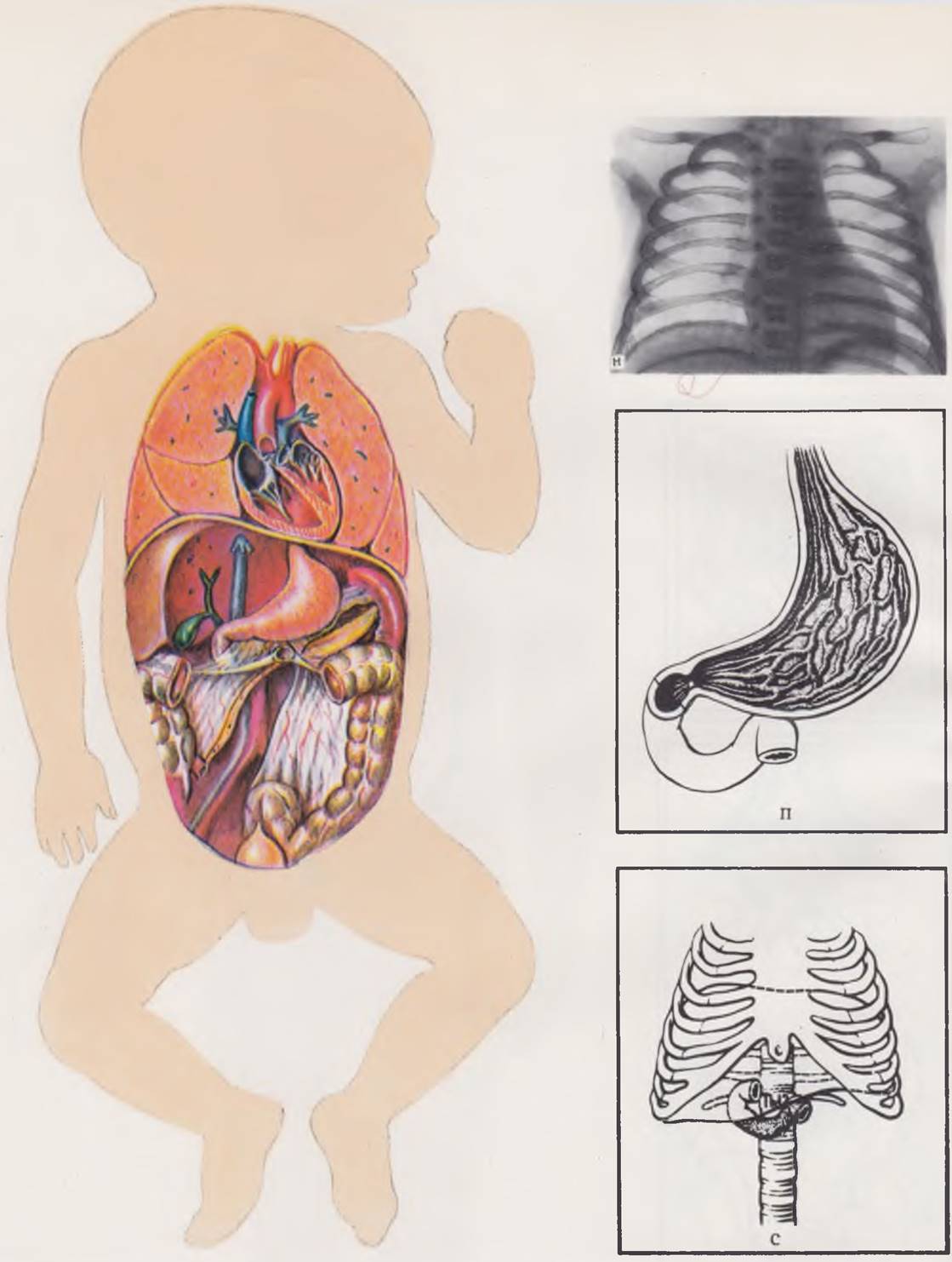
The body of a newborn is characterized by different Proportions of the head and trunk relative to the limbs compared to an adult. Figures (a) and (b) illustrate the differences in the ratios of body segments between a newborn and an adult (after Startz).
Newborns more frequently exhibit a pyramidal shape of the chest (c). In contrast to The structure of an adult chest (d), the ribs are positioned almost horizontally, and the configuration of the superior thoracic aperture resembles an oval elongated forward. The wide epigastric angle significantly facilitates access to the diaphragm and thoracic organs from the abdominal cavity.
The Muscles and aponeurotic formations of the anterior abdominal wall in newborns are poorly developed. Unlike the structure of the adult abdominal wall, newborns feature broad aponeurotic bands extending from the costal margin to the inguinal ligament between the semilunar line (Spigelian line) and the lateral edge of the rectus abdominis muscle. The linea alba is characterized by considerable width and minimal thickness. These zones, devoid of a muscle layer, represent the weakest areas of the anterior abdominal wall. Laparotomy performed through incisions in these aforementioned zones is most frequently complicated by eventration.
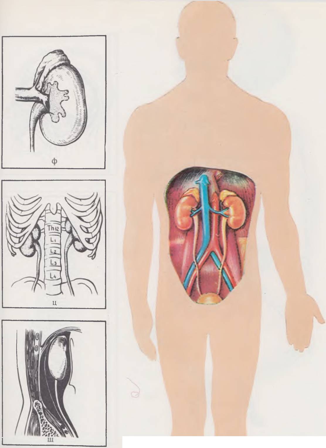
There are substantial differences in the Anatomy of the inguinal region between a newborn (e) and an adult (f). In newborns, the crura of the aponeurosis and fibrae intercrura les are poorly developed. The inguinal triangle (interfoveolar space) is filled by the m. cremaster. The Inguinal Canal is short and wide, and its direction is nearly straight. The superficial inguinal ring is located at the level of the suprapublic arched fold.


General differences in the size and topography of Internal Organs between newborns and adults. A large Thymus gland and a transversely positioned heart make the anterior Mediastinum of a newborn relatively wide.
At birth, the liver occupies more than half of the abdominal cavity. Its right and left lobes are of almost equal size. The left lobe occupies a large portion of the left dome of the diaphragm, separating the spleen from it over a considerable distance (g, h). Upon opening the abdominal cavity of a newborn, only a small part of the stomach is visible; its main bulk is hidden beneath the left lobe of the liver. The greater omentum is poorly developed, short, and covers only a portion of the intestines in the left half of the abdomen.

The cecum of a newborn (i) has a funnel-shaped form and, unlike in adults (j), is located at the level of the iliac crest.
Due to the underdevelopment of the lesser pelvis, the Urinary Bladder of a newborn (l) projects high into the abdominal cavity. In contrast to its spherical shape in adults (m), it has a fusiform or pear-shaped configuration. The Peritoneum covers only the posterior surface of the organ, which allows for extraperitoneal approaches to the urinary bladder.

The heart of a newborn has an oval shape, which is associated with the relatively large size of the atria and the underdevelopment of the ventricles. When compared with the structure of an adult heart, one's attention is also drawn to the relatively narrow lumen of the venous trunks. The features of the skeletotopy and shape of the heart are clearly visible when comparing Overview X-rays: h — overview chest radiograph of a newborn, o — overview chest radiograph of an adult.
The stomach of a newborn is positioned more vertically than in adults. The cardiac region and fundus are indistinctly defined (p). The pyloric region has a cylindrical shape and is projected anterior to the porta hepatis. The muscular ring of the cardiac part is almost absent. The mucosal folds are poorly developed.
The duodenum in newborns is most commonly annular in shape. The superior horizontal part of the duodenum in newborns (c) is located higher than in adults (t). Unlike in adults, the liver anteriorly covers not only the superior horizontal branch, but also the descending segment of the duodenum.

The kidneys of a newborn are relatively large and have a lobular structure. Superiorly and anteriorly, they are covered to a large extent by large Adrenal Glands (y). The renal pelves are most often located intrarenally. The Ureters have a relatively wider lumen and a more tortuous course. In newborns, the kidneys are located lower (x) than in adults (c). A characteristic feature of neonatal anatomy is the extremely poor development of retroperitoneal adipose tissue (ch), which determines the high mobility of organs and The complexity of performing lumbar novocaine blocks and diagnostic Procedures.

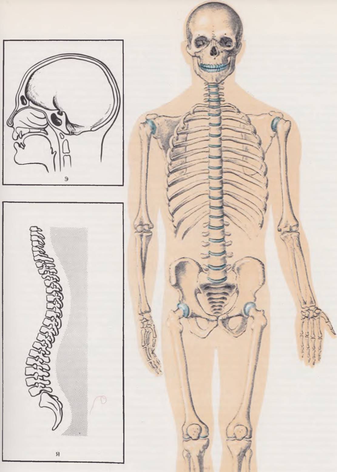
The Skeleton of a newborn is characterized by the underdevelopment of Bone tissue. The epiphyses of the bones are represented by Cartilage, in which ossification centers appear at various times after birth.
The Skull OF A newborn has significant distinctive features. Due to the rapid development of the brain and Sense Organs, the difference in size between the cranial and facial parts of the skull in a newborn (sh) is even more pronounced than in an adult (e). The BONES OF THE skull are thin, elastic, and mobile, as they are connected by connective tissue layers. The frontal and sphenoid sinuses are practically absent.

The movable part of THE Vertebral Column in a newborn (yu) lacks the curvatures observed in adult humans (ya), which serve as an adaptation of the human spine to an upright body posture. The almost straight axis of the spine causes the kidneys to protrude significantly into the abdominal cavity, making them easy to palpate.

Cecum. High and low positions of the cecum relative to the anterior superior iliac spine are distinguished. As a rule, the cecum is located 2 — 3 cm away from it and occupies an intermediate position. The apex of the cecum may transition into the appendix without clear boundaries. Its lumen is relatively larger than in adults. The appendix communicates with the cecum via a large opening, which facilitates the efficient evacuation of the appendix contents and explains why acute appendicitis is rare in newborns.
The ileocecal valve is represented by a delicate mucosal fold, is positioned transversely, and has a well-developed anterior lip.
The ascending colon is short and occupies either an extremely lateral position or approaches the midline.
The transverse colon is the longest part of the large intestine. Its position in the infant's abdominal cavity is determined by its relationship with the liver: the colon may lie beneath it only along its right edge, along both the right and left edges simultaneously with the middle section sagging into the abdominal cavity, or it may be completely covered by the liver (E. A. Alkhimovich, 1970). A low position of the transverse colon is also distinguished, when it lies 1 cm above the Pubic Symphysis, and a high position — up to 8.5 cm.
The descending colon and its splenic flexure may occupy a lower or higher position.
The sigmoid colon is the most developed and variable segment of the intestine in a newborn. It is characterized by a relatively greater length and tortuosity. It is located high in the abdominal cavity, has a long mesentery, and can easily shift as far as the right iliac fossa.
The greater omentum of a newborn contains all structural elements and is distinguished by pronounced plastic properties, which is explained by the high content of histiocyte-like cells in the omental tissue and the presence of a well-developed lymphatic network by the time of birth. The dimensions of the omentum are 2.5 — 8.5 cm in length and 4 — 12.5 cm in width. This accounts for the wide variations in its surface area — from 28 to 115 cm2 (E. M. Margorin, 1970). The omentum consists of individual segments. A single-lobe shape of the omentum is most common, although two-lobe and multi-lobe configurations are possible. The number of segments depends on the distribution of intra-organ arteries. The anterior and posterior duplicatures in early childhood are not fused and have their own developed arterial vessels (V. I. Shifrin,
1970), which makes it possible to elongate the omentum and use it in plastic surgeries.
The liver is the largest internal organ. The weight of the organ is about 5% of the child's total body weight (F. I. Valker, 1938). The lower edge of the liver protrudes 2 — 3 cm below the costal margin. The infant's liver is easily displaced due to the underdevelopment of the fixing apparatus and the mobility of the diaphragm.
The elements of the hepatoduodenal ligament are arranged in the same order as in adults; from left to right: the hepatic artery, portal vein, and common Bile duct. On the right, instead of the common bile duct, the portal vein, cystic artery, or hepatic artery may be located. A powerful hepatic nerve plexus surrounds the vessels and ducts. The total length of the ligament reaches 1 — 2 cm (G. A. Bairov, A. G. Pugachev, A. N. Shapkina, 1970).
The Gallbladder most frequently has a cylindrical shape. Pear-shaped, spindle-shaped, and S-shaped forms also occur. The latter two are characterized by a saccular protrusion of the wall in the neck region, which predisposes to bile stasis. The most typical position of the gallbladder in newborns is "hidden" beneath the liver. The length of the gallbladder is 1.5 — 5 cm, and its width is 0.5 — 1.5 cm.
The Pancreas has an elongated shape, measuring 4.5 — 7 cm in length. It may be curved or form a ring that encircles the wall of the duodenum (annular pancreas). The gland lies horizontally at the level of the XII thoracic or I lumbar vertebra. In cross-section, the organ can be oval or triangular. In the former case, two surfaces are distinguished—anterior and posterior; in the latter, anterior, posterior, and inferior.
In newborns and infants during the first months of life, the spleen has a lobular structure that smooths out with age. At birth, the spleen is a mobile, rounded organ with a diameter of about 5 cm and a weight of 7 — 10 g. It is located at the level of the IX — XI ribs and is separated from the diaphragm by the left lobe of the liver. Anteriorly, it is covered by the transverse colon and the Cytology/practical/108.html">Fundus of the stomach.
Urogenital System. The kidneys of a newborn exhibit pronounced Structural and functional characteristics that define them as an immature organ. Their relative weight and volume are greater than those of an adult. A newborn's kidneys weigh about 11,5 g, accounting for approximately 0.76% of total body weight. The substantial development of the cranial portion causes The Kidneys in the neonatal period to be triangular rather than bean-shaped, as in adults. Situated on either side of the spine, they protrude into the abdominal cavity more than in older children. This makes the newborn's kidneys easily palpable, especially when the organ is enlarged. The longitudinal axes of the kidneys run almost parallel to the spine.
The newborn's Kidney has a lobular structure. On average, each kidney has 14 lobules separated from one another by grooves of varying size and depth. The surface of a lobule corresponds to the renal pyramids, while the grooves correspond to the renal columns. The superficial lobular division of the kidney disappears by the age of three (A. Andronescu, 1970). The kidneys are covered by a fibrous capsule, around which lies a thin layer of adipose tissue—the future adipose capsule.
The renal hilum projects at the level of the II lumbar vertebra, and the renal vessels often follow an oblique course. Their relative length is greater than in adults. The difference in diameter between the afferent and efferent vessels is pronounced—the renal vein has a smaller caliber compared to the renal artery. The renal artery divides into 2 — 3 main branches, which supply blood to strictly defined segments. In most cases, a single segmental artery supplies a segment, and very rarely two. Within the organ, the arteries divide according to three well-known branching patterns: main, ramified, and mixed. With age, the diameter and length of the segmental vessels increase.
In the kidneys of newborns, as in adults, 4 — 5 segments can be identified (M. Kazartsev, 1969). It is important to note that the intersegmental boundaries On the surface of the organ do not correspond to the interlobular grooves.
The renal pelvis in newborns has an ampullary shape and is relatively wider than in subsequent growth periods.
The cortical layer of the kidney is narrow (2 mm). The medullary layer is well developed. The cortex-to-medulla ratio is 1:4 (compared to 1:2 in adults). Malpighian corpuscles are located directly against the fibrous capsule. The loops of Henle are short and do not extend beyond the cortical layer. Convoluted tubules are poorly developed, and their diameter is half that of adults. The renal calyces are thin. The epithelium of the glomerular capsule is cuboidal.
THE POSITION OF the kidneys relative to other organs differs from that in adults. Frequently, the cecum and appendix lie adjacent to the lower pole of the right kidney. The left kidney may briefly contact the spleen, and the tail of the pancreas approaches its upper pole.
The Ureter is long and frequently tortuous. Its lumen is relatively wider due to the poor development of the muscular layer and elastic fibers. Ureteral kinks are pronounced where it crosses the iliac vessels and enters the urinary bladder wall. With age, the ureters acquire a straighter course. Their length in newborn boys is about 6.5 cm, and in girls, 5.5 cm. Upon entering the urinary bladder, an infant's ureters adjoin the bladder wall over a greater distance than in older children.
Urinary bladder. Connective and adipose tissue surrounding the urinary bladder is poorly developed, making it highly mobile. In newborns, the fundus of the urinary bladder is practically absent, as the trigone lies vertically and appears as a direct continuation of the posterior wall. The bladder capacity is 50 — 80 ml.
The bladder wall is thicker than in adults, while its density is the same. The mucous membrane is well developed and rich in folds, predominantly vertical in direction. The connective tissue component of the mucosa is significantly developed. The muscular layer is weak, and elastic tissue is practically absent.
The Urethra is relatively larger in size during the neonatal period. In male infants, it reaches 5 — 6 cm in length. The urethra in girls is proportionally wider than in adults, runs obliquely, and reaches 1 cm in length. The internal urethral orifice is round with smooth walls. Later, it becomes slit-like and acquires distinct folds. The external orifice is gaping. Like in adults, the urethra features narrow segments—the external orifice and the transition point to the membranous part. The urethral tunics are poorly developed. The mucous membrane appears smooth, devoid of folds and transverse wrinkles, and its glandular apparatus is not fully formed.
Functional Characteristics of the newborn kidney. The placenta is the primary excretory organ during the intrauterine period. The transition to extrauterine life is accompanied by an increased load on the newborn's kidneys, as placental excretion ceases. Clinically, this may manifest as transient renal failure in the form of so-called physiological azotemia. This manifestation is likely caused by dehydration and Protein Catabolism (Yu. E. Veltishchev, 1967).
Glomerular Filtration in full-term newborns (calculated relative to body surface area) averages 30 — 50% of adult values (Winberg, 1959). After the first week of life, filtration capacity increases and approaches adult levels relative to total body water content. The lower performance of the glomerular apparatus in the neonatal period is explained by morphological features. The visceral layer of the capsule is formed by tall epithelium, which hinders filtration processes. The diameter of the glomeruli, especially in the outer cortical regions, is so small that up to 50 glomeruli can be seen in a single microscopic field, whereas in adults there are only 5 — 6 (E. P. Semyonova, 1950).
Due to these characteristics, the total surface area of the filtering apparatus per unit of organ weight is significantly smaller in a newborn than in an older child.
The concentrating capacity of the kidneys in the neonatal period is characterized by a low capacity for osmotic concentration. The kidneys of infants in their first weeks of life excrete urine that is hypotonic relative to Blood Plasma, which protects the infant's tissues from fluid overload. With age, as the osmoregulatory function matures, the urine becomes more hypertonic. The instability of osmoregulation in newborns is caused by a wide range of fluctuations in BLOOD OSMOTIC PRESSURE, which depends on food intake and varying dietary protein content, whereas in adults this value remains constant (E. A. Zaryanova, 1951; Keitel, 1958). The causes of the limited concentrating ability in newborn kidneys have not yet received a satisfactory explanation. A. G. Ginetsinsky (1952) believes that renal tubules in the neonatal period are insensitive to the antidiuretic hormone, even though by birth the child has sufficient hormone amounts to produce concentrated urine, and the water-salt balance regulatory system is close to functional maturity. Edelmann and Barnett (1960) attribute an important role in the reduced concentrating capacity of infant kidneys to a low glomerular filtration rate and insufficient delivery of osmotically active substances—sodium chloride and urea—into the renal tubules, which determine the Osmotic Pressure of urine.
The physiological characteristics of the kidneys also explain certain Properties of the urine: the initial portions are light-colored with a low specific gravity (1008 — 1013); during the period of maximum weight loss, the urine darkens and its specific gravity increases; the urine of infants in their first weeks of life is distinguished by an elevated content of urate salts, which rapidly precipitate, and the constant presence of protein. The latter is termed "physiological albuminuria." It is caused by increased permeability of the glomerular and tubular epithelium, which becomes exacerbated during starvation and dehydration.
The daily urine output after birth is negligible, reaching 25% of the volume of milk ingested. This accounts for the low frequency of urination (4 — 5 times per day). By the 7th — 8th day of life, the volume of excreted urine doubles, and the frequency of urination reaches 15 — 25 times.
Central and Peripheral Nervous System. The transition from the stable conditions of intrauterine development to the constantly changing environment places high demands on the newborn's nervous system.
Under normal intrauterine development and normal delivery, a child is born with a sufficiently developed yet undifferentiated nervous system. Its further development continues into the extrauterine period. Abnormal labor and asphyxia can delay the development of The Nervous System due to irreversible changes or the death of Nerve Cells (B. N. Klosovsky, 1949).
The newborn's brain is immature, and its differentiation and myelination are incomplete. Only the main sulci are distinguishable on the surface of the cortex, and they are less pronounced and shallower than in older children. Minor, secondary sulci appear only after birth. The weight of a newborn's brain is about 350 g, which is a quarter of an adult's brain weight (S. I. Nersesyants, 1968). Subsequently, a gradual weight increase is observed, though not through the multiplication of cellular elements in the brain tissue, the total number of which reaches about 16 billion by birth and remains unchanged thereafter (D. S. Futer, 1965). The newborn's brain is rich in water. With age, the fluid volume decreases in proportion to the increase in solid cortical constituents. The cerebral hemispheres consist almost entirely of Gray matter; White matter begins to appear after birth. The brain does not completely fill the cranial cavity, and the spaces filled with CEREBROSPINAL FLUID are wide.
The Cerebellum is relatively less developed and lies almost entirely beneath the cerebral hemispheres, which cover it.
In the Early stages of embryonic development, the Spinal Cord fills the entire vertebral canal. Beginning in the 3rd month of intrauterine life, the longitudinal growth rate of the spine accelerates, leaving a portion of the spinal canal unoccupied. The length of the spinal cord is 14 cm, and its lower edge reaches
the II lumbar vertebra or the upper edge of the III lumbar vertebra. This is clinically significant when performing a spinal puncture in a newborn. As the lower end of the spinal cord shifts upward, the downward angle of the nerve roots increases, forming the cauda equina. The newborn's spinal cord features well-defined cervical and lumbar enlargements. Cerebrospinal fluid is relatively sparse in infants and is under lower pressure.
By birth, peripheral nerves are anatomically developed and capable of supporting the function of their innervated organs. Nerve fibers exhibit numerous expansions caused by traversing blood vessels and the accumulation of interstitial tissue.
An important morphological feature of the newborn nervous system is the absence or insufficiency of nerve fiber myelination. Lacking a myelin sheath, nerve impulses travel to the Cerebral Cortex significantly slower and may even spread to adjacent fibers, preventing The formation of localized excitation foci in the cortex. Consequently, general reactions predominate over local ones in newborns.
According to the theory developed by P. K. Anokhin et al. (1933, 1948, 1966), a child's growth involves the sequential maturation of specific structural relationships within the Central Nervous System—a phenomenon known as heterochrony. This sequence of maturation determines the chronological order in which Various Forms of adaptive behavior emerge. This concept is supported by the neurophysiological analysis of specific neonatal reactions. Infants exhibit only primitive spinal Reflexes, made possible by the early myelination of the corresponding reflex systems. This is particularly evident in the dynamics of Facial Nerve myelination (E. L. Golubeva, 1961). Its branches innervating the lip area are myelinated between 21 and 24 weeks of intrauterine life, whereas the other branches acquire their myelin sheath much later. This fact indicates the early Formation of the morphological basis for the sucking reflex, which is well-developed by birth. Older phylogenetic systems—such as the spinal cord, Medulla Oblongata, and others—are more developed and have a higher myelin content by the onset of extrauterine life than younger CNS structures like the cortex and corpus striatum.
The newborn nervous system's initial environmental contacts act as a tangible stimulus triggering intensive myelination, which is already clearly evident by two weeks of age. Subsequent myelination occurs in a staged, heterochronic manner following a specific sequence: cutaneous receptors, organs of equilibrium, olfactory and auditory analyzers, and visual receptors.
In parallel with the development and functional maturation of the nervous system, the neuroreflex activity of the growing organism emerges, develops, and grows more complex. In newborns, the Influence of the subcortical region on vital processes is prominently manifested. An infant's movements are regulated primarily by subcortical centers (A. F. Tour, 1967). Continuous, uncoordinated movements gradually acquire a definite direction. The most specific reactions in newborns include the Moro reflex, grasping reflex, primitive swimming reflex, postural righting reflexes, as well as feeding-related reflexes such as sucking and palatal reflexes (D. S. Futer, 1965). Conditioned reflexes can be formed in newborns; however, they are characterized by lability and low intensity, especially during the first 10 days of life (O. P. Zykova, 1967).
The Autonomic nervous system functions from birth. It was previously hypothesized that the tone of the Sympathetic division of the autonomic nervous system predominates in infants during their first weeks of life (A. L. Epstein, 1925). It has now been proven that during the neonatal period, some organs are influenced by the parasympathetic nervous system, while others are governed by the sympathetic system (A. Kh. Khamidullina, 1966; A. F. Tour, 1967).
METABOLISM. The metabolism of a newborn infant is characterized by high intensity and lability. The neonatal period features intense plastic processes with an elevated synthesis of Proteins, fats, and CARBOHYDRATES.
The metabolic characteristics in the early stages of a child's development are tied to their living conditions. Prolonged Sleep, minimal physical activity, extensive vascularization of organs, and increased carbon dioxide elimination during respiration promote the predominance of accumulation processes over expenditure. Simultaneously, the age-related inadequacy of reflex and humoral regulatory mechanisms makes metabolic processes highly dependent on the external environment, primarily nutrition.
Protein metabolism. During the intrauterine period, the mother's body supplies protein to the fetus. In the final months of pregnancy, the fetus intensively stores proteins in the liver. However, the total protein content in a newborn remains lower than that in older children.
The protein composition of plasma depends on the gestational age at birth and is subject to significant fluctuations, as shown in Table 5. The liver plays the primary role in regulating plasma protein levels in the infant. Under normal nutritional conditions, Plasma Proteins are synthesized in the liver from dietary Amino Acids. During temporary dietary protein shortages, plasma protein levels are maintained through the direct transfer of proteins from the liver into the bloodstream. In cases of prolonged hypoproteinemia, the oxidative function of liver cells rapidly declines, leading to the depletion of AMINO ACID DEAMINATION processes and drastically impairing the liver's detoxifying function.
Table 5. Age-related characteristics of normal serum protein values (according to I. Todorov, 1963)
Protein fractions (%) |
||||||||
Blood |
Total protein (g %) |
albumins |
α1-globulin |
α2-globulin |
β-globulin |
y-globulin |
||
Umbilical |
6.0 (5 ÷ 7) |
58 (47 ÷ 49) |
4 (2 ÷ 5) |
8 (3 ÷ 11) |
10 (6 ÷ 14) |
20 (14 ÷ 26) |
||
Full-term newborn |
5.6 (4.7 ÷ 6.5) |
60 (49 ÷ 71) |
4 (2 ÷ 5) |
8 (5 ÷ 11) |
9 (5 ÷ 13) |
19 (13 ÷ 25) |
||
Premature |
5.1 (4.4 ÷ 5.8) |
|||||||
On the 30th day of life |
4.8 (4.1 ÷ 5.5) |
60 (50 ÷ 70) |
5 (3-6) |
9 (6 ÷ 12) |
10 (4 ÷ 14) |
16 (10 ÷ 22) |
||
The nitrogen balance undergoes several changes during the neonatal period. Initially, There is a distinct nitrogen retention in the body (up to 78%), while its urinary content reaches its lowest level. This is due to the Protein deficiency in the small amount of food the infant receives during the first days of life. Subsequently, the nitrogen balance becomes positive (N. F. Tolkachevskaya, 1947).
Neonatal fat metabolism is closely linked to Carbohydrate Metabolism and is characterized by instability. The fetus synthesizes the bulk of its fat independently. The sources of fat formation during the intrauterine period are maternal carbohydrates and their metabolic products capable of crossing the placental barrier. In recent years, studies have emerged indicating that minor amounts of lipoids can also penetrate the placenta (Dancis, 1962).
A special role during the neonatal period is played by the so-called brown adipose tissue (Fig. 2). It differs from White adipose tissue by its high content of Cell/35.html">Mitochondria, Coenzymes, and Cytochromes, and is characterized by a High Metabolic Rate. Its weight in a newborn reaches up to 30 g. Studies by E. Ch. Novikova et al. (1972) have proven that a newborn's response to cooling is invariably associated with the activation of heat production in brown adipose tissue.
Fig. 2. Localization of brown adipose tissue in a newborn. a — interscapular region; b — external cervical triangle; c — axillary fossa; d — perivertebral adipose tissue; e — perirenal adipose tissue; f — anterior mediastinal adipose tissue; g — adipose tissue surrounding large blood vessels.

From the first day of life—that is, from the onset of breastfeeding—exogenous fat begins to enter the newborn's body intensively, leading to an increase in blood cholesterol, phospholipid, and fatty acid ester concentrations. Lipid levels reach 531 ± 102.5 mg % during the first week of life. In the gastrointestinal tract, fats are broken down by lipolytic Enzymes into glycerol and Fatty acids, which are absorbed through the intestinal wall. In infants suffering from hypotrophy, a significant portion of fat may be absorbed in an unhydrolyzed form. Fat is transported primarily via the Lymph and is stored in the liver, subcutaneous tissue, and lungs (Koldovsky, 1963).
Lipid Metabolism IN newborns is characterized by the rapid depletion of fat reserves. Consequently, infants with functional Disorders of the central nervous system and liver may experience periodic acetonemia and acetonuria accompanied by vomiting.
Carbohydrate metabolism. Sugar breakdown processes in the organs and tissues of a newborn proceed more intensely than in older children and are highly dependent on environmental conditions. In the final 1–2 months of pregnancy, specific glycogen reserves are built up in the fetus's liver, muscles, and even kidneys, exceeding adult reserves by 2–3 times prior to birth. Such an increase in carbohydrates is necessary to maintain Energy balance during the first days of life. The infant enters the world with a fairly high blood sugar level (up to 100 mg%). In cases of asphyxia, prolonged fasting, hypothermia, or increased respiratory activity, glycogen reserves are rapidly depleted.
Thus, during the first days of life, infants exhibit significant instability in blood sugar levels, caused by limited hormonal regulation and the predominance of Glycolysis over glycogenogenesis. The subsequent establishment of Carbohydrate Metabolism During the neonatal period is determined by gestational age at birth, the course of labor, oxygen supply, ambient Temperature, and the timing of the first feeding.
The REGULATION OF CARBOHYDRATE and lipid metabolism in newborns is heavily influenced by elevated Growth Hormone and catecholamine levels, as well as shifts in corticosteroid concentrations (Yu. A. Baryshkov, 1970).
Energy Metabolism. In newborns, the basal metabolic rate is low, proportional to body weight, and amounts to 50–54 cal per 1 kg (A. F. Tour, 1967). The primary Energy Expenditure goes toward growth and cytoplastic processes.
The birth process and the first days of life demand considerable energy expenditure from the newborn. This is associated with birth stress, the onset of independent respiration, temperature changes, and increased muscular and cerebral activity. Postnatal fasting and increased tissue glucose consumption lead to the utilization of the infant's own energy reserves, triggering glycogen breakdown and fat mobilization.
Carbohydrates are utilized primarily during the first postnatal day. Subsequently, glycemia is maintained at a level sufficient to supply the brain tissue with glucose. During this period, The breakdown of brown Adipose tissue is activated, and non-esterified Fatty acids are mobilized; their levels in blood serum during the first week of life range from 1.5 to 1.8 mEq/L, decreasing by the 7th–10th day of life (Yu. A. Baryshkov, 1966).
Water and Electrolyte metabolism. Total body water in newborns ranges from 75 to 80% of body weight, depending on the infant's gestational maturity. In premature infants weighing 1,500 – 2,500 g, total body water reaches 81 – 85%. Body water in newborns is distributed unevenly across tissues, with the majority located in the intracellular fluid, the volume of which per kilogram of body weight is twice that of adults.
Infants are born with physiological hyperhydration and lose excess water during the first days of life. This process underlies physiological weight loss. Because this loss primarily involves extracellular fluid (urine, meconium, etc.), clinical signs of dehydration are absent. Physiological weight loss is driven by a negative water balance in the early postnatal days.
Water is excreted mainly through the lungs and skin (52 – 72%). This is due to renal functional immaturity, a relatively large body surface area, and a high respiratory rate. In older children, 50% of the fluid intake is excreted by the kidneys, while the remainder is eliminated via the skin, lungs, and feces.
Water Metabolism in newborns is closely linked to electrolyte balance. Differences in electrolyte concentrations help distinguish intracellular fluid from extracellular fluid (Figs. 3, 4). The primary cations of the extracellular fluid are sodium, potassium, calcium, and magnesium, which are balanced by chloride, bicarbonate, orthophosphate, and sulfate anions. Some cations in the extracellular fluid of children are bound to organic acids and proteins (Yu. E. Veltishchev, 1967). Sodium and chloride levels remain relatively constant throughout all periods of life. The concentrations of other electrolytes represent strict physiological constants, and any alteration is poorly tolerated by the body.
Fig. 3. Ionogram of the body's extracellular fluid (after Fanconi).

Fig. 4. Ionogram of the body's intracellular fluid (after Fanconi).

The newborn's extracellular fluid is characterized by two distinct features. The first is an elevated chloride concentration (up to 110 mEq/L). This must be taken into account in clinical practice, as administering large volumes of chloride-containing solutions can induce hyperchloremic intoxication, whereas significant chloride loss leads to a drop in blood osmotic pressure, resulting in exicosis due to the shift of intracellular fluid into the plasma. The second feature is that the infant's intracellular fluid exhibits weak buffering capacity owing to low protein and bicarbonate levels. This explains the rapid onset of acidosis in newborns in the presence of various diseases or when large amounts of protein are administered.
Mechanisms regulating electrolyte balance in the neonatal period are imperfect, as they are not fully mature at birth. In particular, due to renal tubular immaturity, Ammonia Production is limited in infants during their first weeks of life, meaning that this crucial base-sparing mechanism is practically non-functional (Yu. E. Veltishchev, 1967).
Anabolic processes predominate in the newborn body; driven by active glycolysis, the acid-base balance of the blood manifests as metabolic acidosis (pH 7.3; BE = -7.15; SB = 18.8).
Thus, metabolism in the newborn infant is characterized by unstable regulation, pronounced lability, and high dependence on environmental conditions, factors that must be carefully considered during preoperative preparation, the surgical Procedure itself, and the postoperative period.
The anatomical and physiological characteristics of the newborn must be taken into account primarily when assessing the infant's baseline condition. To this end, clinical practice routinely employs the Apgar score (Table 6), which reflects the status of the respiratory, cardiovascular, and central nervous systems. Scoring is performed on a scale of 0 to 10 at 1 and 5 minutes after birth. In normal infants, the score should range from 8 to 10; lower scores indicate a compromised condition of the newborn.
Table 6. Apgar score
Score |
|||
Sign |
0 |
1 |
2 |
Heart rate |
Absent |
Less than 100 beats per minute |
Over 100 beats per minute |
Respiration |
Absent |
Slow, irregular |
Good, crying |
Muscle tone |
Limp |
Decreased, mild flexion of extremities |
Active motion |
Reflexes (tested by inserting a catheter into the nose) |
No response |
Grimace or feeble motion |
Movement, cry, cough, or sneeze |
Skin color |
Blue or pale |
Body pink, extremities blue |
Completely pink |
Under pathological conditions, the structural and functional features of the newborn determine the specific clinical manifestations of various diseases and Congenital Malformations, serve as the starting point for justifying Pathogenetic Therapy, and guide the evaluation of immediate and long-term outcomes. Fundamental differences in the anatomy of organs and systems between newborns and adults, which dictate the choice of Surgical Treatment, are illustrated in a series of figures (see atlas).
Last update: 10/08/2026
Editorial and Educational Adaptation: This material has been compiled based on the primary/original source text. The project team performed an editorial review, corrected technical inaccuracies, structured sections, and adapted the content for an educational format.
What was processed:
- elimination of formatting defects (OCR errors, structural breaks, corrupted characters);
- editorial organization of content;
- standardization of terminology in accordance with academic sources;
- verification of factual statements against the original source text.
All mentions of the author, publication year, and origin of the primary text have been preserved in accordance with the source.