BIOCHEMISTRY AND MOLECULAR BIOLOGY - W. ELLIOTT - 2002
CHAPTER 12. REGULATION OF CARBOHYDRATE AND LIPID METABOLISM
This chapter serves as a logical culmination of everything discussed previously regarding METABOLISM, demonstrating how individual metabolic pathways intersect and function together. Up to this point, we have primarily examined each of them separately. Initial consideration of individual metabolic pathways is, in our view, justified because: 1) each pathway is sufficiently complex without the added burden of regulatory mechanisms; 2) it is far more effective to study regulatory issues in relation to the entire ensemble of metabolic processes; 3) one can always return to previously read and studied chapters.
Although all links in metabolism require regulation, the integration and control of carbohydrate and lipid transformations are of particular importance, as these pathways carry exceptionally massive fluxes of substances whose intensity and direction frequently change. This is due to the fact that in animals, periods of feeding alternate with fasting, and periods of rest with Various Forms of activity. All of this requires altering the direction of metabolic fluxes. At the same time, the metabolism of Introduction/36.html">CARBOHYDRATES and Lipids serves as an excellent model to examine all the fundamental principles of regulation.
Since all biochemical reactions are catalyzed by Enzymes, Metabolic Regulation is ultimately the REGULATION OF ENZYMATIC Activity. Therefore, primary attention is focused on the mechanisms regulating enzyme activity. Following this, we will show how regulatory enzymes, whose activity depends on the levels of various metabolites, maintain a balance among metabolic transformations. This is by no means all aspects of regulation, as the METABOLIC ACTIVITY OF individual Cells depends on hormonal and neural signals arriving in accordance with the needs of the whole Organism. In Conclusion, we will analyze how extracellular signals regulate metabolism.
Why is regulation necessary?
It is perfectly clear that all the metabolic processes we have encountered—Glycogen Synthesis and Breakdown, Glycolysis and Gluconeogenesis, fat Synthesis and degradation, The Citric Acid Cycle, electron transport, and so forth—cannot simultaneously proceed at the maximum possible rate for each of them. If metabolic transformations proceed in one direction, the reverse transformations must not take place. The reaction rate of each metabolic pathway must vary over a wide range depending on the organism's current energy demands. For instance, The rate of key metabolic reactions in a person playing tennis increases sixfold, and during even more strenuous physical exertion, fifteenfold.
Let us point out two most important aspects related to the REGULATION OF ENERGY-producing reactions:
✵ energy production must be coordinated with constantly changing energy demands;
✵ the rate and direction of transformations must obey the needs and vital rhythm of the whole organism, which periodically feeds and fasts, occasionally nurses offspring, and sometimes falls ill.
Another, less obvious necessity for regulation is the potential danger of "futile" (or substrate) cycles arising.
The potential danger of "futile" cycles in metabolism
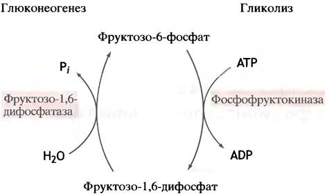
We will discuss this problem using gluconeogenesis and glycolysis as an example. During glycolysis, fructose-6-phosphate is phosphorylated by Phosphofructokinase into fructose-1,6-diphosphate, whereas during gluconeogenesis, diphosphatase hydrolyzes fructose-1,6-diphosphate back to the initial fructose-6-phosphate (Fig. 12.1). Such an uncontrolled sequence of events leads to nothing other than the senseless conversion of ATP energy into heat, much like an electrical short circuit (a technique bumblebees use to warm up their flight Muscles on cold mornings before takeoff).
Class="center">Fig. 12.1. "Futile" cycle at the level of glycolytic phosphofructokinase, which may occur in the absence of regulatory mechanisms
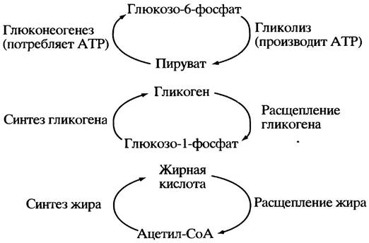
A similar giant "futile" cycle dedicated to the destruction of ATP could link all of glycolysis and gluconeogenesis together in a closed loop (Fig. 12.2). Reactions associated with glycogen and fat synthesis and breakdown could potentially combine into similar cycles, i.e., any paired combinations of synthesis and degradation processes for individual metabolites.
Fig. 12.2. Large-scale "futile" cycles potentially possible in the absence of regulation
Clearly, the breakdown and synthesis of metabolites must be regulated in opposite ways: when one of these processes is activated, the other must be inhibited. Such regulation is only possible if synthesis and breakdown proceed via different metabolic pathways. As we have already noted, for this to happen, both pathways must include at least one irreversible step catalyzed by different enzymes and, consequently, subject to differential regulation. In the case of reversible processes, the forward and reverse reactions are catalyzed by the same enzymes, making their separate regulation impossible.
"Futile" cycles are also called Substrate Cycles, which reflects their essence: the "coupling of reactions" for substrate synthesis and breakdown. Although The Emergence of such large cycles is fundamentally wasteful, they can nevertheless function as elements of regulatory systems. Suppose, for example, that substance A is converted into substance C via an intermediate substance B, with a reversible reaction B <-> A being possible.

The rate of C formation can be reduced either by inhibiting enzyme 1 or by activating enzyme 2. The greatest effect will be achieved if both occur simultaneously. In this case, significantly less inhibitor of enzyme 1 will be needed to completely halt the reaction B —> C. As will be shown below, this exact type of regulation is characteristic of the fructose-6-phosphate to fructose-1,6-diphosphate conversion.
How is enzyme activity regulated?
Metabolic regulation is nothing other than the Regulation of the rate of at least one successive catalytic reaction that together constitute a metabolic pathway. Furthermore, the reaction does not necessarily have to be chemical; for instance, transport Proteins catalyze the translocation of metabolites across membranes. There are two principal ways to reversibly influence the Rate of Enzymatic processes in a Cell.
1. Changing The amount of enzyme.
2. Altering its catalytic activity. Naturally, Methods for irreversible enzyme activation do exist. Examples include the proteolytic conversion of trypsinogen into active Trypsin (see p. 75). However, this chapter focuses exclusively on fully reversible regulatory mechanisms, as only these play a significant role in effective Metabolic control.
Metabolic Control via Changes in Enzyme Abundance
The cellular concentration of a protein can be modulated by altering either the rate of its synthesis or the rate of its degradation. Proteins are short-lived cellular components; the half-lives of Liver enzymes typically range from an hour to several days.
In animals, changes in enzyme abundance make a substantial contribution to metabolic regulation. Such long-term regulation operates on a timescale of hours and days rather than seconds, and is employed to adapt to changing physiological demands. Numerous examples can be cited. Consider lipoprotein lipase in Blood capillaries (see p. 93): its levels correlate with the tissue's demand for lipids, increasing significantly in the Mammary Glands during Lactation, for instance. In response to dietary shifts, liver enzyme levels change within hours to handle unfamiliar, high-fat, or carbohydrate-rich foods. A high-calorie diet increases the abundance of enzymes involved in fat synthesis in the liver, whereas starvation reverses this process within just a few hours. When foreign chemical substances, such as drugs, enter the body, the level of enzymes responsible for their oxidation rises rapidly in the liver (see p. 210). To reiterate, in animals, cellular enzyme levels fluctuate over hours or even days, though this timeframe can be shorter for short-lived enzymes. In general, this mode of regulation (altering enzyme abundance) is utilized by animals only when the system's potential response time to external stimuli can be relatively slow.
In Bacteria, enzyme levels change much more rapidly. When E. coli cells are placed in a medium where lactose serves as the sole carbon source, the synthesis of β-galactosidase—the enzyme that hydrolyzes this sugar—begins immediately. Its concentration becomes detectable within minutes and increases a thousandfold over the course of a few hours. Nevertheless, this is still not the fastest response.
Once the demand for an enzyme ceases, its concentration within The Cell declines. The response time of the system is then determined by the Rate of protein degradation (and, in bacteria, by dilution during Cell Division), correlating with the protein's half-Life in the organism. For rapid, automated metabolic regulation, changing enzyme concentrations is an unsuitable mechanism, as this type of regulation deals only with enzymes already present in the cell at a given moment.
Metabolic Control via Changes in Enzyme Activity
When regulating enzyme activity, it is not the amount of the enzyme that changes, but rather the rate at which it works. The primary advantage of this control mechanism is its speed. To understand the fundamental principles of such regulation, one must examine the Factors influencing the catalytic Properties of the enzyme.
Fundamentals of Enzyme Kinetics
Hyperbolic Kinetics of the "Classical" Enzyme
During Enzymatic Catalysis, a substrate (S) binds reversibly to the Active Site of an enzyme (E) to form an enzyme-substrate complex (ES). Once a chemical reaction has taken place within this complex, the resulting product(s) (P) are released from the active site, leaving the enzyme free for a new interaction and reaction cycle. This conceptual model of enzyme action forms The basis of the Michaelis-Menten formulation for describing the Kinetics of Enzymatic catalysis:
Е + S <-> ЕS —> Е + Р.
At low substrate concentrations [S], the rate of an enzymatic reaction is determined solely by the frequency of collisions between substrate molecules (S) and free enzyme molecules (E). Under these conditions, the concentration of enzyme-substrate complexes [ES] and the reaction rate are both low. However, as [S] increases, the concentration of the enzyme-substrate complex rises, leading to a corresponding increase in the reaction rate. Eventually, a point is reached where a further increase in Substrate Concentration results in virtually all enzyme molecules becoming saturated with substrate and converted into ES complexes, after which any further increase in [S] no longer affects the reaction rate. This state is referred to as enzyme saturation, and the maximum reaction rate is designated as Vmax. Graphically, this relationship is described by a hyperbolic curve (Fig. 12.3). Kinetics of this type are known as Michaelis-Menten kinetics, and enzymes that obey this model are termed Michaelis-Menten enzymes. For a long time, these were the only enzymes known to science and were therefore designated as classical enzymes.
Thus far, we have used the term low substrate concentration, which is not entirely precise, since at the exact same value of [S], one enzyme may be saturated with substrate while another is not. This depends on the tightness of substrate binding by the enzyme or, in other words, the affinity of the enzyme for the substrate. Affinity can be quantified by the dissociation constant of the enzyme-substrate complex or by The change in Free energy during its formation, and it depends on The Nature of the reacting species as well as the number of weak bonds formed between them.
In most cases, the relative affinity of an enzyme for its substrate can be estimated by knowing the Michaelis constant, KM. This parameter is defined as the substrate concentration at which the enzymatic reaction rate is half of the maximum possible rate (see Fig. 12.3) and, as is easy to see, is independent of the Enzyme Concentration. (Note, however, that the true value of KM can only be determined when the rate of dissociation of the ES complex into E + S is significantly greater than the rate of breakdown of ES into E + P.) While numerous graphical methods exist for transforming the Michaelis-Menten model to estimate KM simply and accurately, we will restrict ourselves here to a streamlined Overview of the fundamental Principles of Enzyme kinetics required to understand regulatory mechanisms.
Fig. 12.3. Effect of Substrate concentration on the rate (V) of an enzyme-catalyzed reaction obeying Michaelis-Menten kinetics
KM is the Michaelis constant. The dashed line illustrates The properties of the uncatalyzed reaction; attention should be paid only to the shape of this curve rather than the absolute rate values. The rates of reactions occurring within the cell would be negligibly small were they not catalyzed by enzymes

The larger the value of KM, the lower the affinity of the enzyme for its substrate. Consequently, KM values can be used to compare the affinities of different enzymes for their respective substrates. KM values can range from nanomolar to millimolar concentrations, but generally fall within the 10-6 to 10-3 M range. The intracellular substrate concentration is typically of the same order of magnitude as KM, meaning that the enzyme is not fully saturated with substrate within the cell.
Which enzymes of a metabolic pathway need to be regulated?
The straightforward answer is: those enzymes to which evolution has imparted this property. Every metabolic pathway includes Reactions Catalyzed by specific regulated enzymes. Typically, these are situated at strategic locations, such as at an irreversible step within an otherwise reversible process.
As a rule, the primary strategic control point is the first enzyme of a metabolic pathway. Consider the reaction sequence A —> B —> C —> D —> E, where the pathway culminates in The production of substance E, which is required by the cell. It is crucial that E is not produced in quantities exceeding current cellular demands. The simplest Scheme for the automated regulation of this reaction cascade involves the inhibition of the first enzyme—which catalyzes the conversion A —> B—by the end product (E):
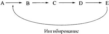
This effect is achieved in two ways: through Enzyme Inhibition and/or a reduction in enzyme abundance. Inhibiting the very first step of the pathway prevents the accumulation of intermediate metabolites B, C, and D generated in subsequent steps. When metabolite E is depleted or its concentration drops, the inhibition ceases, causing the synthesis of E to resume vigorously. In biochemistry, this type of regulation is termed feedback regulation, or end-product inhibition. It is widespread in bacteria (for instance, in Amino acid Biosynthesis) and operates with exceptional precision in these organisms. However, this regulatory type is too primitive for complex processes such as lipid or Carbohydrate Metabolism, where identifying a single end product is far from straightforward. Nevertheless, it enables The regulation of metabolically disparate enzymatic reactions by Key Intermediates (products of intermediate reactions).
In some cases, regulation is exerted not by the end product (via negative feedback), but by the initial reactant (positive feedforward control), which activates enzymes participating in its subsequent conversion along the metabolic pathway.
Nature of Regulatory Enzymes
There are Two main mechanisms for regulating the catalytic activity of enzymes (changes in enzyme quantity are not considered here).
2. Covalent protein modification — typically phosphorylation and dephosphorylation.
Let us examine both of these mechanisms.
Allosteric Regulation of Enzymes
Allosteric regulation plays a crucial role in the control of metabolism. The prefix *allo-* means "other." It implies that, In addition to the substrate-binding site, an enzyme possesses at least one other binding site (and often several) for another compound (or compounds). Low-molecular-weight compounds bound by proteins are generally referred to as ligands; this term is also frequently extended to small proteins. Ligands that bind to allosteric sites are called allosteric effectors or Allosteric regulators. Typically, they bear no structural resemblance to the substrate. The action of effectors is usually observed at a specific substrate concentration. Positive effectors increase enzyme activity upon binding to the allosteric site, whereas negative effectors decrease it. In other words, allosteric effectors can be classified into activators and inhibitors.
In some cases, allosteric inhibitors decrease the rate of an enzymatic reaction by lowering Vmах. More frequently, however, their effect manifests as a reduction in the enzyme's affinity for the substrate. As noted earlier, intracellular enzymes typically operate at substrate concentrations below saturation. Consequently, a decrease in substrate affinity leads to a reduction in enzyme activity, while an increase in affinity results in activation. At saturating substrate concentrations, allosteric effectors do not alter Vmах, even if the enzyme's affinity for the substrate changes. However, under physiological conditions,
where intracellular substrate concentrations are largely far from saturation, effectors can significantly influence the reaction velocity.
Mechanism of Allosteric Enzyme Regulation
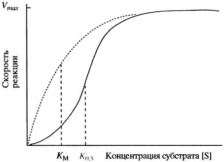
We will now discuss the nature of allosteric enzymes, in which effector binding affects the enzyme's affinity for the substrate (this is the primary type of allosteric regulation). Such allosteric enzymes consist of multiple catalytically active proteins assembled into a single enzyme complex via non-covalent bonds. The components of this complex are called protein subunits, protomers, or monomers. Although each subunit possesses catalytic activity, the overall activity of the enzyme depends on their mutual interaction. Typical curves showing the Dependence of enzymatic reaction velocity on substrate concentration are presented in Fig. 12.4. An allosteric enzyme differs from a "classical" enzyme in that its curve is sigmoidal (also referred to as S-shaped) rather than hyperbolic. What are the implications of this, and why does it occur?
Fig. 12.4. Effect of substrate concentration on the reaction velocity (1) catalyzed by a typical allosteric enzyme. The dashed curve is shown for comparison and represents a "classical" enzyme obeying Michaelis-Menten kinetics. Since THE CONCEPT OF Km lacks physical meaning outside the Michaelis-Menten model, the designation K0.5 is used here
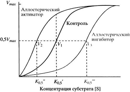
To answer the first question: when an allosteric activator binds to the enzyme, the sigmoidal curve shifts to the left, whereas an allosteric inhibitor shifts it to the right (Fig. 12.5). A leftward shift indicates an increase in the enzyme's affinity for the substrate, while a rightward shift indicates a decrease. The sigmoidal shape of the velocity-versus-substrate-concentration curve indicates that the reaction rate is more sensitive to changes in substrate concentration than in the case of hyperbolic kinetics, especially in the velocity range close to 0.5 Vmах.
Fig. 12.5. Effect of substrate concentration on the reaction velocity (V) catalyzed by a typical allosteric enzyme. V1, V2, and V3 represent the reaction velocities in the absence of a Ligand, in the presence of an allosteric activator, and in the presence of an allosteric inhibitor at a fixed substrate concentration S (K0.5)
Why does the dependence of reaction velocity on substrate concentration assume a sigmoidal shape? The interaction of one subunit of an allosteric enzyme with a substrate facilitates the binding of substrate molecules by other subunits. This phenomenon is termed homotropic cooperative substrate binding. The term homotropic reflects the fact that all subunits bind the same ligand—the substrate. Because the sigmoidal shape of the curve in this case arises solely from substrate binding, interpreting this regulation strictly as the result of an allosteric conformational change (which implies effector interaction at a site distinct from the catalytic site) is not entirely accurate. Nevertheless, cooperativity is frequently classified as an allosteric effect, even though the ligand- and substrate-binding sites are identical. This cooperative interaction can arguably be considered allosteric, as the binding of substrate to each subunit depends on the presence of a ligand at the adjacent subunit.
What is the mechanism underlying the mutual influence between the binding sites of different subunits? Two theoretical models have been proposed, according to which the substrate-binding site can exist in two states differing in substrate affinity. Let us call them the high-affinity and low-affinity states. It is postulated that substrate binding increases the proportion of subunits in the high-affinity state. Both models differ in the specific details of how this mutual influence propagates across the unified protein complex.
Within the framework of the concerted model proposed by Monod, Wyman, and Changeux (Fig. 12.6), all subunits can simultaneously exist in either the high-affinity or the low-affinity state. Both forms of the protein complex exist in a dynamic equilibrium that is skewed toward the low-affinity conformation. The binding of a substrate molecule to any subunit shifts this equilibrium in the opposite direction—toward The formation of the high-affinity protein. Thus, as the substrate concentration increases, a progressively larger number of enzyme molecules transitions into the high-affinity state.
Fig. 12.6. The concerted model of cooperative substrate binding. According to this model, the enzyme exists in two states, T and R, which are in equilibrium; however, in the absence of the substrate, this equilibrium heavily favors the T form. Upon binding of substrate molecules to the R form, the equilibrium shifts to the right, increasing the substrate affinity of all subunits within that molecule. The designations H (High) and L (Low) denote the enzyme's affinity for the substrate

It should be noted that all of the above pertains to cooperative substrate binding rather than to allosteric modifiers or effectors that regulate the enzyme, which is a separate issue. According to this model, allosteric regulators alter THE POSITION OF equilibrium between the low-affinity state of the molecule (designated as T, from the English "tense" state) and the high-affinity state (designated as R, from "relaxed" state). If an allosteric effector binds more tightly to the enzyme in the R state, it will shift the T <-> R equilibrium to the right. This results in enzyme activation, since a greater number of its molecules will adopt the R form at a given substrate concentration. As the concentration of a positive allosteric regulator increases, more enzyme molecules transition into the high-affinity R state, and the velocity-versus-substrate-concentration curve becomes hyperbolic. The reverse situation occurs when a negative regulator binds more tightly to enzyme molecules in the T state, driving other molecules into that same state and thereby reducing the overall rate of the enzymatic reaction.
The sequential model was developed by Koshland, Némethy, and Filmer. They postulated that in the absence of substrate, all enzyme molecules reside in the low-affinity T state, and no pre-existing T-to-R equilibrium exists (since the R state is not independently populated). The binding of a substrate molecule to one subunit induces a conformational change in that subunit (from T to R) as well as in the neighboring subunit, enhancing the latter's affinity for a substrate molecule, which can now bind more readily. The binding of a second substrate molecule facilitates the transition of a third subunit into the R state, and this process repeats sequentially until all enzyme subunits have converted into the high-affinity R state (Fig. 12.7).
Fig. 12.7. The sequential model of cooperative substrate binding by an allosteric enzyme. The binding of a single substrate molecule to a subunit induces a conformational change in that subunit. This facilitates the conformational transition of the second subunit upon interaction with the next substrate molecule, and so on. The cumulative effect is an alteration in the overall conformation of the protein

Both models explain the observed effects satisfactorily and cannot be considered mutually exclusive. The true mechanism of allosteric regulation may well turn out to be intermediate.
Some enzymes, such as glyceraldehyde-3-phosphate dehydrogenase (see p. 112), exhibit cooperativity in substrate binding yet lack known allosteric regulators. Such enzymes are more sensitive to changes in substrate concentration than classical ones, which is likely the cause of their cooperative binding. Later on, we will encounter a more complex variant of allosteric regulation in which the binding site
for the regulator is located on a separate regulatory subunit devoid of catalytic properties. The binding of the allosteric regulator to this site causes the regulatory subunit to dissociate from the complex, whereupon the catalytic subunits alter their affinity for the substrate. While this method of controlling an enzymatic reaction may seem excessively cumbersome, evolution prioritizes system reliability and functionality above all else.
Reversibility of Allosteric Regulation
The most crucial feature of allosteric regulation is its instantaneous action. The allosteric ligand binds to the regulatory site almost instantaneously via non-covalent interactions. The regulator dissociates from the enzyme complex when its concentration in the medium drops, returning the enzyme to its initial state.
Allosteric Regulation: A Metabolic Concept of Enormous Power
A vital aspect of allosteric regulation is that The Structure of the effector may bear no resemblance whatsoever to either the substrate of the given enzyme or any other substance produced during metabolic transformations. This means that any metabolic pathway can be regulatory linked to any other. Metabolite(s) from one cascade of biochemical transformations can act as regulator(s) of another. Furthermore, an enzyme can be controlled not by a single regulator but by multiple ones, each assigned to a specific binding site, thereby receiving regulatory signals from several metabolic pathways and increasing regulatory flexibility.
We have already seen how complex and interdependent the metabolic pathways related to energy production and storage are. In such an intricate system, every compartment must "know" how its "neighbors" are doing by assessing the levels of key metabolites. Is there enough ATP or not? Is The Citric Acid cycle supplied with the right amount of acetyl-CoA? Is glycolysis proceeding too quickly or unacceptably slowly? Imagine the chemical chaos that would ensue if every metabolic pathway failed to continuously adapt to the current situation based on information coming from other metabolic pathways or different parts of its own route! THE PRINCIPLE OF allosteric regulation provided evolution with the opportunity to create numerous communication channels that enable enzymes to receive signals from any point on the metabolic map. As Monod, one of the co-discoverers of allosteric regulation, remarked, without it the existence of a system as complex as a cell would simply be impossible. He called allosteric regulation "the second secret of life" (the first being DNA).
The Role of Phosphorylation in the Regulation of Enzymatic Activity
Now let us turn to the second method of enzyme regulation: phosphorylation. Strictly speaking, this section should be devoted to the broader problem of regulation via Covalent Modification of enzymes. However, phosphorylation is of such paramount importance that discussing this process separately is fully justified.
The principle is extremely simple. There are enzymes called protein Kinases that transfer a phosphate residue from ATP to a target protein. This alters the conformation of the phosphorylated protein and, consequently, its catalytic properties. The target for phosphorylation can be not only an enzyme but also a regulatory protein—an enzyme inhibitor or activator. In this case, phosphorylation affects its regulatory properties. It is hardly surprising that the Introduction of a strongly charged group impacts the STRUCTURE OF THE protein molecule. On the other hand, phosphate groups are easily cleaved by phosphoprotein Phosphatases (Fig. 12.8).
Fig. 12.8. Regulation of Enzyme Activity through phosphorylation. In various cases, the phosphorylated enzyme may be either more or less active than the unphosphorylated one. Regulation is also possible via an inhibitory protein whose activity changes upon phosphorylation
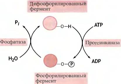
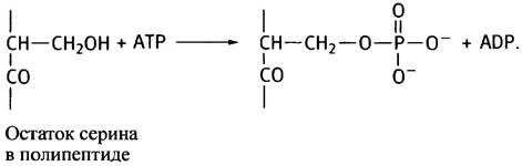
Phosphorylation targets the hydroxyl groups of specific Serine and Threonine residues within the polypeptide chain of the enzyme, which the kinase recognizes by their amino acid environment (Tyrosine phosphorylation is described in Chapter 26 and will not be covered in this section).
The scheme below shows the serine hydroxyl group undergoing phosphorylation:
Thus, we have examined two major mechanisms regulating enzyme activity: allosteric regulation and reversible phosphorylation. We must now consider how these mechanisms are utilized to regulate metabolism.
Regulation of Individual Metabolic Pathways
Two Types of Control: Internal and External (Extracellular)
Allosteric regulation of enzymes occurs with the direct participation of the enzyme and substrate, whereas in regulation via phosphorylation, The activity of the target enzyme depends on The ratio of kinases to phosphatases. What determines this ratio? In most cases (though not always), it is determined by a hormonal signal arriving from the outside of the cell.
Metabolic control is an intricate, complex process. To make its Discussion more accessible, we will divide the material into two broad sections. First, we will discuss intracellular regulation, which coordinates the interaction of metabolic pathways and their individual segments within a single cell to prevent deficits or overproduction of essential substances. At this level, metabolites themselves serve as regulatory signals, and control is typically (though far from always) exerted through allosteric regulation. In some cases, this utilizes feedback principles, such as preventing glycolysis from producing more Pyruvate than the citric acid cycle can process. In other cases, by contrast, positive feedback regulation allows metabolic steps located further down the chain to cope with an increasing influx of substrates.
A completely isolated cell could make do with internal control of metabolic processes. However, in Multicellular Organisms, systems of external life-cycle control operate (external, of course, with respect to the cells, not the organism). Signals reach the cells from Hormones or Neurotransmitters, compelling them to subordinate basic metabolic transformations to the interests of the entire organism, such as storing or burning fuel. Such regulation is especially vital when cellular activity is important for the organism as a whole. Cells cannot decide for themselves what the organism needs; they must receive an external signal before intracellular regulation kicks in to maintain the required processes.
Key Aspects of the Internal Regulation of Carbohydrate and Lipid Metabolism
Intracellular Regulation of Glycogen Metabolism
Glycogen Metabolism involves the synthesis of this polymer by Glycogen synthase and its breakdown by Glycogen phosphorylase. Glycogen is synthesized during periods of ample Nutrition, and this synthesis is regulated by external signals (see p. 169). Glycogen breakdown serves two physiological purposes. A task common to all Tissues is supplying glycolysis with its starting material, glucose-6-phosphate. The primary breakdown product of glycogen, glucose-1-phosphate, is converted into glucose-6-phosphate by phosphoglucomutase, and glycogen phosphorylase (the glycogen-degrading enzyme) occupies a key position in metabolism. The energy generated during glycolysis in the form of ATP is necessary for all tissues, but it is of particular importance for muscles, whose contraction is accompanied by intensive ATP consumption (see Chapter 28). In the liver, glycogen breakdown serves to maintain blood glucose concentration at the proper level.
The regulatory mechanisms of glycogen phosphorylase, a textbook example of a "classical" regulatory enzyme, have been studied in great detail (especially the Muscle enzyme). In a resting muscle, it exists as the inactive phosphorylase b, which is partially activated by AMP acting as an allosteric effector (maximum activation is triggered by external signals). Why does AMP act as the allosteric signal in this case? Glycogen phosphorylase needs to be activated when the cellular energy reserve is critically low—that is, when cellular ATP is scarce. Since adenylate kinase catalyzes the reaction:
2ADP <-» АМР + АТР,
AMP serves as an indicator of ADP accumulation, which is generated during ATP Hydrolysis and thus reflects a drop in ATP concentration. Calculations show that even a minor consumption of ATP leads to a relatively large increase in AMP concentration, which activates glycogen phosphorylase to ramp up energy production. Conversely, ATP and glucose-6-phosphate act as allosteric inhibitors of glycogen phosphorylase (Fig. 12.9). If there is already plenty of them, why make more? Simple intracellular regulation is coordinated from the outside and depends on the overall physiological state of the organism (see p. 169).
In the liver, the situation is similar: AMP activates phosphorylase b to about 20% of its maximum level, and external signals take precedence over internal ones, although here they serve different physiological purposes (see p. 169).
Thus, in both muscle and liver, in the absence of extracellular regulators (which we will discuss shortly), phosphorylase remains in its inactive b form. AMP activates the enzyme, whereas ATP and glucose-6-phosphate inhibit its activity.
Glycolysis and Gluconeogenesis
From the General Overview of regulatory pathways shown in Fig. 12.9, it follows that:
✵ a rise in AMP concentration indicates an increasing ADP/ATP ratio;
✵ AMP activates glycogen phosphorylase and phosphofructokinase;
✵ AMP inhibits fructose-1,6-bisphosphatase;
✵ activation of glycogen breakdown is accompanied by an elevated level of fructose-6-phosphate;
✵ fructose-6-phosphate activates phosphofructokinase;
✵ activation of phosphofructokinase leads to an increase in fructose-1,6-bisphosphate concentration;
✵ fructose-1,6-bisphosphate activates pyruvate kinase. As evident from the above, the allosteric Regulation of glycolysis incorporates both negative and positive feedback loops, as well as positive feedforward loops. Evolution rarely relies on just a single regulatory mechanism. For instance, in this system, phosphofructokinase is activated by AMP but inhibited by ATP. This allows glycolysis to respond precisely to fluctuations in the ATP/ADP ratio (via AMP concentration).
As ATP levels rise, the citric acid cycle slows down, leading to the accumulation of citrate. Citrate then migrates from the Mitochondria into the Cytoplasm, where it allosterically inhibits phosphofructokinase, effectively putting the brakes on glycolysis. The same goal is achieved through another pathway: acetyl-CoA allosterically inhibits pyruvate kinase.
Special attention should be given to the activation of pyruvate carboxylase by acetyl-CoA, which yields oxaloacetate (see Fig. 12.9). The accumulation of acetyl-CoA occurs when the citric acid cycle stalls (due to a shortage of oxaloacetate). Naturally, this automatically triggers an anaplerotic reaction (see p. 122). This regulatory link is also crucial for gluconeogenesis.
Fig. 12.9. Main Pathways of allosteric regulation in glycogen metabolism, glycolysis, and gluconeogenesis. Activating regulatory steps are highlighted in color, while inhibitory steps are shown with dashed lines. UDPG — uridine diphosphate glucose

Intracellular Regulation of Pyruvate Dehydrogenase, the Citric Acid Cycle, and Oxidative Phosphorylation
Pyruvate dehydrogenase (see p. 116) occupies a strategically vital position in metabolism by catalyzing the irreversible conversion of pyruvate into acetyl-CoA, which then enters the citric acid cycle or is used in fat synthesis. The various modes of regulation for this enzyme are illustrated in Fig. 12.10. Both of its products—acetyl-CoA and NADH—function as allosteric inhibitors, whereas its substrates—CoA-SH and NAD+—act as activators. Consequently, pyruvate dehydrogenase activity is governed by the acetyl-CoA/CoA and NADH/NAD+ ratios. The logic is straightforward: a significant excess of acetyl-CoA and NADH signals that their production via pyruvate dehydrogenase should be reined in. Conversely, when concentrations of CoA-SH and NAD+ are high, the enzyme needs to work at full throttle.
Fig. 12.10. Allosteric regulation and reversible phosphorylation as mechanisms controlling the activity of the mammalian pyruvate dehydrogenase complex (PDH). The multiplicity of control mechanisms is due to the complex's strategic position at the crossroads of general metabolism, as acetyl-CoA subsequently feeds into the citric acid cycle and is utilized for lipid synthesis. Ultimately, all these regulatory pathways serve to inhibit PDH via its end-products and activate it via its substrates (ATP can be viewed as a distant product of acetyl-CoA Processing in the citric acid cycle). Regulation via reversible phosphorylation is typically coupled with hormonal control, while Calcium Ions, which stimulate phosphatase activity, accumulate in response to catecholamines

Negative regulation of the enzyme by high ATP concentrations is of particular importance. An accumulation of ATP signals the need to dial down its production. Regulation of enzyme activity by ATP is indirect. An elevated molar ATP/ADP ratio activates pyruvate dehydrogenase kinase, which phosphorylates the enzyme and thereby inhibits it (recall that phosphorylation is always counterbalanced by dephosphorylation catalyzed by phosphatases) (see Fig. 12.10). The kinase is an integral part of the pyruvate dehydrogenase complex. In addition to ATP, it is also activated by acetyl-CoA and NADH. All these regulatory effects boil down to a single purpose: turning off the "fuel valve" when an excess of energy has been generated.
Allosteric enzyme regulation also operates within the citric acid cycle and the Electron Transport Chain. However, in these mitochondrial systems, the primary regulatory mechanisms revolve around shifts in the concentrations of key substrates such as NAD+ and ADP. If the bulk of NAD is present in its reduced form (NADH), the activity of citric acid cycle dehydrogenases drops. Because NADH accumulates when it cannot be oxidized rapidly enough by The electron transport chain (e.g., due to oxygen deprivation), the inhibition of certain citric acid cycle reactions can be viewed as a regulatory feedback response. Similarly, a low ADP/ATP molar ratio slows down Electron transport along the mitochondrial chain, since mitochondrial phosphorylation and oxidation are tightly coupled. This phenomenon is specifically known as Respiratory Control and is of paramount importance.
Alongside NAD+ and ADP, the citric acid cycle is regulated at the level of citrate synthase (inhibited by ATP), isocitrate dehydrogenase (inhibited by ATP, activated by ADP), and α-ketoglutarate dehydrogenase (inhibited by NADH and succinyl-CoA).
All of these regulatory pathways are entirely logical and help maintain metabolite balance within the citric acid cycle, preventing either excess or deficiency.
Intracellular regulation of Fatty acid oxidation and synthesis
The regulation scheme for these processes is illustrated in Fig. 12.11. Its primary purpose is to prevent the simultaneous oxidation and synthesis of Fatty acids within the same cell.
Fig. 12.11. Key sites of intracellular regulation during fat oxidation and synthesis. Since fat oxidation produces acetyl-CoA, the rate of its subsequent reactions depends on the regulation of citric acid cycle pathways, etc. Dashed lines indicate allosteric effects. Citrate activates acetyl-CoA carboxylase in vitro, though it remains unclear whether this holds any physiological significance.

These two metabolic pathways strongly inhibit one another. The starting Materials for oxidation—fatty acid-CoA derivatives (see p. 132)—allosterically inhibit the first enzyme of fatty acid synthesis, acetyl-CoA carboxylase. Conversely, the key metabolite of the fat synthesis system, malonyl-CoA (see p. 138), allosterically inhibits The transfer of acyl groups to carnitine (see p. 133). As a result, fatty acyl residues cannot enter the mitochondria to undergo oxidation there: if you are actively synthesizing fats, it hardly makes sense to burn them simultaneously in the mitochondrial furnace. Acetyl-CoA carboxylase is activated in vitro by citrate, The conversion of which yields acetyl-CoA, the substrate for the same enzyme (see p. 141). Citrate leaves the mitochondria only when present in high concentrations—that is, during times of nutritional abundance, when it is optimal to store fat. While there is no doubt that citrate activates acetyl-CoA carboxylase in a test tube, it has not yet been established whether such activation occurs within the living cell. At the same time, it is undisputed that acetyl-CoA carboxylase is inhibited by phosphorylation. This is mediated by a protein kinase whose activity depends on the presence of AMP, whereas the reverse phosphatase reaction is hormonally controlled (see p. 176).
Everything stated above regarding the intracellular regulation of carbohydrate and lipid metabolism certainly does not claim to be an exhaustive account of accumulated knowledge. However, these data are more than sufficient to understand that metabolism is not merely a sum of sequential reactions. Rather, it is a complex, highly ordered process in which all Metabolic pathways are closely interconnected.
To understand how cellular metabolism aligns with the physiological needs of the organism as a whole, it is essential to examine extracellular regulation by hormones and Other Compounds. These often control the same reactions as intracellular molecules, meaning certain pathways are subject to both intra- and extracellular regulation.
Regulation of carbohydrate and lipid metabolism by extracellular agents
A particularly vital role among extracellular agents involved in the regulation of carbohydrate and lipid metabolism is played by the hormones Glucagon, Insulin, epinephrine, and norepinephrine. Hormones act instantaneously, sometimes producing profound physiological effects. They help the organism adapt to the periodic alternation between fed and fasting states (see p. 84).
Overall, hormonal regulation is extraordinarily complex. Hormones of the pituitary, Adrenal Glands, Thyroid Gland, and others (see Table 26.1) influence metabolism in ways that are not always fully understood. Their action is typically mediated by a cascade of events and extended over time. For example, an excess of thyroxine leads to gradual weight loss and increased excitability, whereas a deficiency produces the reverse—and likewise slow-developing—effects. However, certain hormones act rapidly and with precise direction.
As you may recall, glucagon is the "hunger hormone" produced by The Pancreas in response to falling blood glucose levels. Conversely, rising glucose concentrations stimulate the release of insulin (see p. 84). Epinephrine and norepinephrine are produced in The adrenal medulla and trigger the activation of pathways associated with nutrient utilization. Norepinephrine is also released by sympathetic nerve endings. The sympathetic Nervous system governs the involuntary contractions of visceral smooth muscle and also innervates adipose tissue. It can be viewed as a mechanism for rapidly delivering calibrated doses of a hormone directly to targeted cells and Organs.
How is the blood content of insulin, glucagon, and epinephrine regulated?
Insulin is a small protein synthesized and secreted into the bloodstream by the β-Cells of the islets of Langerhans in the pancreas. Its half-life in the blood is short, meaning its effects subside as soon as secretion ceases. The β-cells are highly sensitive to fluctuations in glucose concentration, initiating insulin secretion within no more than a minute after a postprandial increase in blood glucose. Notably, glucose has a very low affinity for the transport protein responsible for its cellular uptake, so this transporter only Functions once blood glucose exceeds the normal baseline of 90 mg per 100 ml (or 5 mM). The rate of insulin secretion depends on glucose concentration in a sigmoid manner, which accounts for the high sensitivity of the response to variations in blood sugar.
In contrast, glucagon is secreted by the pancreas when blood glucose concentrations drop. Epinephrine, meanwhile, is released by the adrenal glands in response to neural signals.
How do glucagon, epinephrine, and insulin function?
Hormones comprise various chemical substances released into the bloodstream. They transmit chemical signals accessible to all tissues. The selectivity of their action is achieved because only specific cells—known as target cells—perceive these signals.
Lipid-soluble hormones (such as Steroids and thyroxine) cross The Plasma Membrane unhindered, whereas Water-soluble hormones (such as glucagon, epinephrine, and insulin) cannot pass through it. Their molecules bind to receptor proteins specific to each hormone, located on the cell surface. The selectivity of hormone-receptor binding shares the same biochemical basis as the Specificity of enzyme-substrate interactions. Only cells designated as targets possess receptors "tuned" to that particular hormone. All other cells remain entirely unaffected by the hormone's presence, as if blind to it.
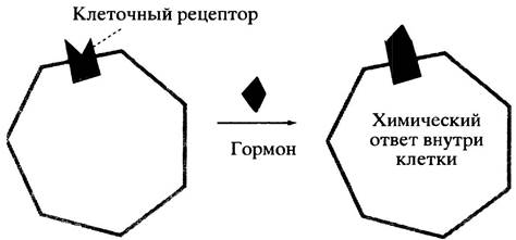
Hormones are rapidly cleared from the bloodstream; consequently, as soon as the gland halts hormone secretion, its concentration drops, hormone-receptor complexes dissociate, and the signal dissipates. However, while the hormone remains bound to its receptor, its presence induces specific chemical alterations within the cell, which constitute the hormonal response (Fig. 12.12).
Fig. 12.12. A hormone initiating Chemical Reactions in the cytoplasm by binding to a receptor on the cell surface
Modern concepts regarding the Nature of the hormonal response are rooted in the second messenger hypothesis.
WHAT IS A secondary messenger?
A hormone can be considered a primary messenger: by binding to its cellular receptor, it conveys a humoral signal to the cell. This interaction alters the level of a second messenger—an intracellular molecule that triggers specific chemical transformations within the cell in response to hormone-receptor binding.
What serves as The secondary messenger for glucagon, epinephrine, and norepinephrine?
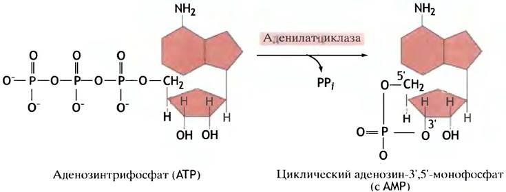
In the case of glucagon and epinephrine, the secondary messenger is cyclic adenosine-3',5'-monophosphate, or cyclic AMP (cAMP), which is synthesized from ATP by the enzyme adenylyl cyclase (Fig. 12.13).
Fig. 12.13. Synthesis of cyclic AMP from ATP, catalyzed by adenylyl cyclase
In turn, cAMP allosterically activates cAMP-dependent protein kinase, which phosphorylates the hydroxyl groups of serine or threonine residues in target proteins, thereby altering their activity. The cascade of regulatory reactions transmitting the hormonal signal is shown in Fig. 12.14.
Fig. 12.14. Stages of hormonal REGULATION OF METABOLISM. It is not shown here that cAMP is continuously degraded and protein dephosphorylation is carried out by phosphatases. The metabolic response is maintained as long as the hormone remains bound to the receptor

The mechanism by which cAMP activates protein kinase is illustrated in Fig. 12.15. In the absence of cAMP, the kinase exists as a tetramer formed by two catalytic and two Regulatory Subunits. Within the tetramer, the catalytic subunits are inactive. When cAMP binds to the regulatory subunits, the tetramer dissociates into monomers, releasing catalytically active subunits.
Fig. 12.15. Activation of cAMP-dependent protein kinase by cAMP. R and C represent the regulatory and catalytic subunits of the protein kinase, respectively

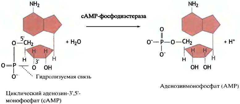
The enzyme cAMP phosphodiesterase hydrolyzes cAMP to AMP (Fig. 12.16). Therefore, the activation of protein kinase depends on the continuous production of cAMP, which ceases as soon as the hormone dissociates from the receptor. As already mentioned, hormones in the bloodstream are also continually degraded or cleared, meaning their presence depends on ongoing secretion. Thus, every stage of hormonal signal Transduction incorporates an inhibitory mechanism. When hormone levels drop, cAMP production stops, phosphorylated proteins are dephosphorylated by protein phosphatases, and the system returns to its initial state.
Fig. 12.16. Reaction catalyzed by cAMP phosphodiesterase. The enzyme received this name because it hydrolyzes the phosphodiester bond in the cAMP molecule
In this chapter, we do not discuss how a hormone triggers cAMP synthesis or how this synthesis terminates when the hormone leaves the receptor. Signal transduction issues
across cell membranes are examined in detail in Chapter 26. For now, it suffices to understand that the binding of glucagon and epinephrine to cell surface receptors leads to an increase in intracellular cAMP concentration.
Regulation of Receptor Number
The number of specific receptors on the cell surface is subject to regulation. Prolonged cell exposure to an agonist (any compound that binds to and activates a receptor) can lead to a reversible decrease in receptor number and a drop in cellular sensitivity to that agonist, resulting in a weaker response. This phenomenon is known as downregulation. Additionally, a receptor can be inactivated via phosphorylation by a cytoplasmic kinase, after which its binding to the hormone fails to stimulate cAMP synthesis. Because this type of phosphorylation is stimulated by cAMP, the intensity of the cellular response is regulated through a negative feedback loop.
All of these regulatory mechanisms occur with receptors for adrenaline, insulin, and glucagon.
How Does Insulin Regulate Metabolism?
Insulin affects Energy Metabolism in a manner diametrically opposed to glucagon (see Chapter 5). Insulin is a protein hormone that has been known for quite some time and studied perhaps more thoroughly than any other protein or hormone. This is due not only to its pivotal role in carbohydrate and lipid metabolism, but also to its therapeutic use in treating diabetes. Unfortunately, despite extensive efforts, The Mechanism of insulin action remains largely unclear. Insulin receptors have been identified on the outer surface of cells. More recently, it was discovered that these receptors span the plasma membrane, and their cytoplasmic domain functions as a tyrosine protein kinase. This kinase differs from the previously described protein kinases and is stimulated upon insulin binding (see Chapter 26). A characteristic feature of this kinase is that it phosphorylates tyrosine residues. The mechanism of insulin action is discussed in greater detail in Chapter 26.
REGULATION OF GLUCOSE and Lipid Uptake by Cells
Glucose cannot cross The Lipid Bilayer; therefore, a specialized membrane transport protein is present in cell membranes to mediate its passage. In the digestive tract, glucose is absorbed via Active Transport, whereas in all other tissues it enters cells through passive transport. This type of transmembrane transport relies on molecules moving down a concentration gradient and is termed Facilitated Diffusion (see p. 63).
In the Brain and liver, cellular glucose uptake is insulin-independent, but in muscle and fat cells it is accelerated by this hormone. Apparently, the cytoplasm of such cells contains inactive transport proteins. Insulin stimulates their conversion into an active form and their insertion into the membrane, thereby increasing membrane permeability to glucose (Fig. 12.17). Studies on fat cells have shown that the insertion of the transport protein is complete within just 7 minutes of insulin binding to its receptor. Once insulin is removed, the process reverses, and within 20–30 minutes the transport proteins exit the membrane and return to the cytoplasm.
Fig. 12.17. Insulin-mediated mobilization of glucose transporters in fat and certain other cells. This does not occur in liver and brain cells. Glucose Transport Across the plasma membrane proceeds via facilitated diffusion

The mechanism of this remarkable phenomenon is still unknown. It is possible that insulin stimulates the movement of the transport protein and its incorporation into the membrane, or alters the dynamic equilibrium between the membrane-bound and cytoplasmic pools of this protein.
Facilitated diffusion can only equalize glucose concentrations in the blood and cells. However, inside cells, glucose rapidly disappears—either by being converted into glycogen or by being consumed for metabolic needs. This prevents the equalization of the glucose concentration gradient and serves as the driving force for its transport into the cell.
The First stage of intracellular glucose conversion is its phosphorylation. This process is carried out by glucokinase in The Liver and by hexokinase in other tissues:
Glucose + ATP —> glucose-6-phosphate + ADP.
The Km value (for glucose) of glucokinase is much higher than that of hexokinase, meaning the affinity of the first enzyme for the substrate is lower (see p. 91). The profound significance of this difference becomes clear when we recall the glucostatic function of the liver. The liver does not need to take up glucose unless it is present in excess in the blood. When blood glucose is low, the liver produces it and releases it into the bloodstream. It would be illogical to simultaneously release glucose into the bloodstream and take it up from there. This does not happen because glucokinase does not phosphorylate glucose if its concentration is low compared to Km.
Another regulatory mechanism is provided for glucokinase. This enzyme is induced by insulin. The term induction refers to the acceleration of biosynthesis under The Influence of an inducer, in this case, insulin. The mechanism by which insulin accomplishes this is still unknown.
Fat metabolism begins with the uptake of fatty acids into cells rather than triglycerides themselves. Fatty acids easily cross membranes without the participation of any transport systems, as the lipid bilayer does not serve as a barrier for them. Therefore, the influx of fatty acids into the cell must be controlled. To do this, their levels in the blood must be regulated. Such regulation is carried out by glucagon, adrenaline, noradrenaline, and insulin acting on fat cells.
Having gained a general understanding of extracellular regulation, we can now proceed to examine the effects of hormones on individual metabolic pathways.
Regulation of Glycogen Synthesis and Breakdown by Extracellular Agents
Regulation of Glycogen Breakdown
We have already discussed the regulation of glycogen phosphorylase b in resting muscle and liver, which is based on the activation of the enzyme by AMP. This regulation ensures constant control of glucose levels against the Background of a stable consumption rate. However, There is a completely different regulatory system based on the conversion of phosphorylase b into phosphorylase a, whose activity does not depend on AMP. This conversion occurs as a result of the phosphorylation of the enzyme by protein kinase, which transfers the phosphate group of ATP to the hydroxyl group of one of the serine residues, thereby causing Conformational Changes in the enzyme molecule and its activation. It is important to remember the difference between phosphorylation—where a protein kinase transfers the phosphate group of ATP to the serine hydroxyl group—and phosphorolysis, which is a phosphorylase-catalyzed process of glycogen breakdown involving inorganic phosphate. In muscle cells, protein kinase is activated by adrenaline. This hormone, circulating in the blood, is recognized and bound by receptors On the surface of the muscle cell, thereby triggering the intracellular synthesis of cAMP. We will return to the mechanism by which cAMP stimulates the phosphorylation of phosphorylase b, but first, let us discuss the Physiological aspects of the entire process.
Adrenaline is produced in the adrenal glands in response to a nerve signal originating from the brain during an emergency situation that requires instant and active muscular activity (in English-language literature, such a situation is referred to as "fight or flight"). Adrenaline essentially hits the "alarm" button. A muscle cell has no time to wait until the indicator of decreased ATP-AMP concentration accumulates and intracellular positive feedback is triggered. The cell must instantly provide an unlimited supply of fuel for ATP generation so that the organism can cope with the impending danger. Below we will explain why glucose-6-phosphate, formed from the product of glycogen phosphorolysis (glucose-1-phosphate), is the optimal fuel in such a situation. Here we only note that due to the low ATP yield, a large amount of fuel must be kept in reserve to sustain glycolysis. This task is solved by the instant activation of glycogen phosphorylase.
Adrenaline also stimulates the release of glucose from the liver into the blood. Its purpose is to rapidly supply muscles with fuel in an emergency situation. Here too, The Effect of adrenaline is due to the rapid phosphorylation of glycogen phosphorylase.
Interestingly, potato tubers also contain a phosphorylase that breaks down starch. The degree of Homology between plant and muscle phosphorylase is very high: their Amino acid sequences are half identical. Nevertheless, potato phosphorylase is not activated by phosphorylation, presumably because potatoes do not need a "fight-or-flight" response and do not need to control blood glucose levels.
We should add that the liver also releases glucose into the blood in response to the binding of glucagon to receptors. This hormone is secreted by the pancreas when blood glucose levels drop and, like adrenaline, causes an increase in intracellular cAMP levels and the activation of glycogen phosphorylase. Muscle cells do not respond to the presence of glucagon because they lack specific receptors for it on their surface.
Mechanism of Regulation of Glycogen Synthesis and Breakdown by cAMP
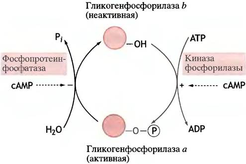
Glycogen phosphorylase exists in two forms: in form a, the serine hydroxyl is phosphorylated, whereas in form b, it is not. The interconversion scheme of both forms is shown in Fig. 12.18.
Fig. 12.18. Interconversion of glycogen phosphorylases a and b by the action of kinase and phosphatase. cAMP affects these transformations only indirectly. The hydroxyl group belongs to a serine residue of the protein
The activation by cAMP of phosphorylase kinase, which phosphorylates glycogen phosphorylase, is indirect. cAMP first activates protein kinase A (PKA; A stands for cAMP), which in turn phosphorylates phosphorylase kinase, converting it into an active state. The latter then phosphorylates glycogen phosphorylase b. The complete sequence of events unfolding after cell contact with the hormone is shown in the diagram:

Why such a complex regulatory mechanism? Each cell binds a small number of hormone molecules. Meanwhile, its response, especially in an emergency situation, must be rapid and powerful. Recall that a liver cell contains approximately 1 µmol of glucose residues (180 µg), i.e., ~ 6.02 • 1017 molecules. The binding of just a few hormone molecules to receptors must force the cell to process an astronomical number of glucose molecules, and do so very quickly. The same
holds true for the production of glucose-6-phosphate during energy generation in muscles.
Consequently, a weak signal—the binding of a hormone to a receptor—must be amplified to such an extent as to involve the maximum possible number of executioners of the command, namely enzyme molecules. Thus, the scheme discussed above is nothing other than a powerful regulatory cascade. Suppose 1 molecule of hormone activates 1 molecule of adenylate cyclase, which produces 100 molecules of cAMP per minute. This is already a hundredfold Amplification. If each cAMP molecule activates 1 molecule of protein kinase A, which phosphorylates phosphorylase kinase b with the same productivity, the amplification will be 100 • 100, and so on. In reality, hormonal activation of glycogen phosphorylase involves four stages of amplification. Cascade signal amplification is a general principle of hormone action.
Each molecule of glycogen phosphorylase attacks the end of one of the oligosaccharide chains of glycogen. If glycogen, like amylose, consisted of long linear chains, hormonal stimulation would be pointless, since the numerous molecules of activated glycogen phosphorylase would lack sufficient targets. This is presumably the reason for the extreme branching of glycogen (compared to starch), which allows it to undergo rapid breakdown in a critical situation. Unhurried plants do not need this and can afford to store glucose in the form of amylose.
Reversal of Phosphorylase Activation
All regulatory metabolic processes must be reversible. "Turning on" and "turning off" provides an additional layer of control over the regulated process. In the case of phosphorylase, the a form is converted back into the b form by a specific phosphatase (known as protein phosphatase I), which cleaves the phosphate group from the phosphorylated serine residue:

In the liver, this reaction is stimulated by free glucose through its allosteric effect on the a form.
During the hormonal response—the conversion of phosphorylase b into phosphorylase a—it is advantageous to suppress phosphatase activity so that it does not hinder the rapid accumulation of active glycogen phosphorylase molecules. As it turns out, many cells contain a protein known as phosphatase inhibitor I. It is phosphorylated by the exact same protein kinase A that phosphorylates phosphorylase kinase, thereby inhibiting the phosphatase. Thus, ultimately, cAMP not only "flips the switch" but also prevents it from being "turned off" prematurely.
During normal Muscle contraction (i.e., in situations not requiring cAMP regulation), phosphorylase kinase is allosterically activated by Ca2+ ions, the concentration of which spikes sharply in response to motor nerve signals (see Chapter 28). Because this activation of phosphorylase kinase is independent of phosphorylation, it subsides as soon as the initial relaxation signal arrives. The activation of the enzyme by Ca2+ ions is mediated by the regulatory protein calmodulin, which is present in almost all Eukaryotic cells. In the case of phosphorylase kinase, this small soluble protein serves as a tightly bound subunit of the enzyme. Calmodulin acts as a Ca2+ concentration detector: the binding of these ions induces conformational changes in calmodulin, enabling it to interact with a wide range of target proteins, including enzymes, and activate them.
Let us summarize the main points discussed above to cement the key elements of this complex regulatory system in our minds (Fig. 12.19).
Fig. 12.19. Regulation of Muscle phosphorylase kinase. The cAMP-independent mechanism of kinase activation by calcium ions is also shown. Calcium ions not only trigger muscle contraction but also supply it with energy. Ca2+ ions are required for the catalytic activity of the phosphorylated (active) kinase. They bind to calmodulin, which is a subunit of this enzyme, thereby activating it.

1. Phosphorylase is an enzyme that catalyzes The breakdown of glycogen via phosphorolysis. This reaction has nothing to do with phosphorylation—the transfer of a phosphate from ATP to the hydroxyl group of serine or threonine, which is catalyzed by protein kinase. Phosphatase carries out the hydrolytic Cleavage of phosphate from phosphorylated proteins.
2. In the liver and resting muscles, phosphorylase exists as the inactive, unphosphorylated form b, which is partially activated by cAMP. This activation is not accompanied by enzyme phosphorylation.
3. Adrenaline, acting on muscle and liver cells, as well as glucagon, acting on liver cells (but not muscle cells), cause an increase in the intracellular concentration of cAMP.
4. cAMP allosterically activates protein kinase, which in turn activates phosphorylase kinase, which specifically activates phosphorylase b. The latter is thereby converted into the active form a. The overall process can be viewed as an amplification cascade.
5. During normal muscle contraction, nerve impulses trigger an increase in the concentration of Ca2+ ions within muscle cells. These ions allosterically
activate (partially) phosphorylase kinase b. This results in the partial activation of phosphorylase. Unlike cAMP-induced phosphorylase kinase activation, Ca2+ activation does not involve its phosphorylation.
6. The conversion of active phosphorylase a back into the inactive form b is catalyzed by protein phosphatase I. Its activity is regulated by protein phosphatase inhibitor I, a protein that exhibits inhibitory properties only after being phosphorylated. This phosphorylation is carried out by the same cAMP-stimulated protein kinase that phosphorylates phosphorylase kinase b. Thus, the activation of phosphorylase is accompanied by the inhibition of phosphatase. In the liver, the conversion of phosphorylase form a to form b is activated by glucose.
How is glycogen synthase regulated?
The synthesis and breakdown of glycogen must be coordinated so that these two processes do not occur simultaneously; otherwise, a futile cycle would arise. As mentioned in the previous section, the activation of glycogen phosphorylase is a consequence of protein kinase A activation driven by cAMP. Glycogen synthase is directly phosphorylated by protein kinase A, but in this case, phosphorylation inactivates the enzyme. The regulatory cascade activating glycogen synthase is shorter, which makes sense since glycogen synthesis takes place under more stable physiological conditions than its breakdown during extreme situations.

We have already seen how phosphorylase a is inactivated. This is carried out by a phosphatase, which in turn is inhibited by a protein (protein phosphatase inhibitor) phosphorylated by protein kinase A. Something very similar occurs in the regulation of glycogen synthase. However, there are significant differences between the two regulatory pathways: 1) dephosphorylation of the synthase leads to its activation; 2) the protein kinase does not activate, but rather inhibits, the protein phosphatase inhibitor. This protein kinase is not identical to protein kinase A and, unlike the latter, is activated not by cAMP, but by insulin. Thus, insulin activates phosphatase I, which in turn activates glycogen synthase.
Remarkably, the exact same protein inhibitor participates in regulating both the synthesis and breakdown of glycogen; however, protein kinase A activates it, whereas the insulin-dependent kinase inactivates it.
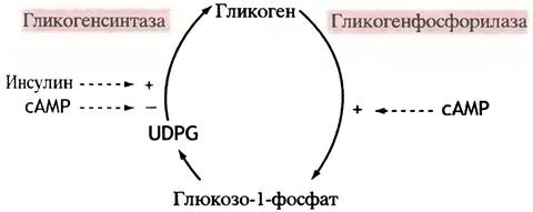
The regulatory mechanisms of glycogen synthase are illustrated in Fig. 12.20, and the General scheme of glycogen Metabolism regulation is shown in Fig. 12.21.
Fig. 12.20. Scheme of Reciprocal Regulation of glycogen phosphorylase and glycogen synthase. Enzyme molecules are indicated by colored circles: active forms are darker, inactive forms are lighter.

Fig. 12.21. Regulation of glycogen metabolism. The effects of insulin and cAMP on enzyme activities are indirect. UDРG - UDР-glucose
Regulation of glycolysis and gluconeogenesis by extracellular agents
It is logical to consider these two metabolic pathways together, as they largely involve the same enzymes. Glucagon signals the liver
that it needs to release glucose into the bloodstream either via Glycogenolysis (glycogen breakdown) or gluconeogenesis. Therefore, it makes sense that in the liver, cAMP turns on glycogen breakdown and gluconeogenesis (Fig. 12.22, a). It would be strange if cAMP simultaneously turned on glycolysis in the liver—a process that is the exact reverse of gluconeogenesis.
The situation in muscles is entirely different. There, The stimulation of cAMP synthesis by adrenaline serves as a command to boost energy production. Therefore, it is logical that cAMP in muscles promotes glycogen breakdown and activates glycolysis (unlike in the liver), since it is precisely through this process that glucose-6-phosphate is generated (Fig. 12.22, b), the further conversion of which leads to ATP synthesis.
Fig. 12.22. Differences in the regulatory response to glucagon and/or adrenaline signals in the liver (a) and muscle (b). Both hormones use cAMP as a secondary messenger. The term glycogenolysis here refers to glycogen breakdown

Thus, cAMP must inhibit glycolysis in the liver while activating it in muscles. The most crucial regulatory step is the reaction catalyzed by phosphofructokinase (see Fig. 8.7). In the liver, cAMP (in response to glucagon) inhibits phosphofructokinase, thereby suppressing glycolysis, whereas in muscles, cAMP (in response to adrenaline) accelerates glycolysis.
The effect of cAMP on phosphofructokinase in the liver deserves a separate discussion.
How does cAMP regulate phosphofructokinase activity?
This regulatory mechanism is well understood in the liver and is not too difficult to figure out. When blood glucose levels are low, glucagon levels are high, resulting in an increased intracellular concentration of cAMP in the liver. As a result, the cellular content of fructose-2,6-bisphosphate decreases. (No, that is not a typo!) We are talking precisely about this previously unfamiliar compound, rather than the well-known fructose-1,6-bisphosphate, which is formed during glycolysis with the participation of phosphofructokinase. Fructose-2,6-bisphosphate is an extremely potent allosteric activator of phosphofructokinase, and consequently of glycolysis as a whole. It is a true master regulatory molecule. Increase its concentration, and glycolysis accelerates; decrease it, and glycolysis slows down. Importantly, fructose-2,6-bisphosphate not only activates phosphofructokinase but also inhibits fructose-1,6-bisphosphatase (Fig. 12.23), one of the Key Enzymes of gluconeogenesis.
Fig. 12.23. Regulation of glycolysis by fructose-2,6-bisphosphate
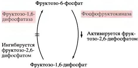
It is easy to guess that in the liver, cAMP must lower the level of 2,6-bisphosphate, thereby blocking glycolysis and activating gluconeogenesis. How is this achieved?
The Formation of fructose-2,6-bisphosphate is catalyzed by a specialized phosphofructokinase—PFK2. Following its discovery, the previously described phosphofructokinase came to be abbreviated as PFK1.
cAMP causes the inhibition of PFK2 (Fig. 12.24). This happens as follows: cAMP activates protein kinase, which phosphorylates PFK2 (Fig. 12.25). Phosphorylated PFK2 not only stops synthesizing fructose-2,6-bisphosphate but, conversely, actively hydrolyzes it (unphosphorylated PFK2 does not hydrolyze fructose-2,6-bisphosphate). Thus, phosphorylation changes not just the rate, but the very direction of the catalyzed reaction.
Fig. 12.24. Synthesis of fructose-2,6-bisphosphate by the second form of phosphofructokinase (PFK2) and the inhibition of the enzyme in the liver involving cAMP. This inhibition is indirect: cAMP activates protein kinase, which phosphorylates PFK1, thereby suppressing the synthesis of fructose-2,6-bisphosphate. Phosphorylated PFK1 hydrolyzes fructose-2,6-bisphosphate. Thus, cAMP decreases the level of fructose-2,6-bisphosphate in the liver

Fig. 12.25. Regulation of PFK2 activity in the liver via phosphorylation by a cAMP-dependent protein kinase. PFK2 is a bifunctional enzyme: in its unphosphorylated form, it synthesizes fructose-2,6-bisphosphate, whereas upon phosphorylation, it hydrolyzes it

What is good for the liver is bad for the muscles, since glycolysis—which supplies fuel during emergency exertion—cannot be suppressed there; consequently, phosphofructokinase cannot be inhibited. It is known that under the influence of adrenaline, the level of fructose-2,6-bisphosphate rises in muscles. This may be explained by the fact that cAMP accelerates glycogen breakdown, thereby increasing the content of fructose-6-phosphate, a substrate and activator of PFK2. However, this regulatory mechanism requires further study.
Glucagon regulates the activity not only of phosphofructokinase but also of pyruvate kinase, which catalyzes the final step of glycolysis. The glucagon-induced rise in intracellular cAMP concentration leads to the phosphorylation of pyruvate kinase in liver cells (but not in muscle cells), which is accompanied by its inactivation.
The rationale behind this is perfectly clear. If the blood needs to be enriched with glucose, the liver must stimulate gluconeogenesis and block glycolysis. Inhibiting pyruvate kinase, much like inhibiting phosphofructokinase, accomplishes the second of these tasks.
As noted repeatedly, muscles are engaged exclusively in glucose breakdown rather than synthesis; therefore, cAMP must not suppress glycolysis there. Indeed, an increase in cAMP levels does not lead to the phosphorylation of pyruvate kinase.
The glucagon-stimulated inhibition of liver pyruvate kinase is also important from the standpoint of gluconeogenesis regulation. The Synthesis of glucose from pyruvate involves two intermediate reactions:
Pyruvate + ATP + HCO3- —> Oxaloacetate + ADP + Pi + H-, (1)
Oxaloacetate + GTP <-> Phosphoenolpyruvate + GDP + CO2 (2)
It is easy to see that there is a potential risk of a futile cycle, since pyruvate kinase catalyzes the reaction:
Phosphoenolpyruvate + ADP —> Pyruvate + ATP.
Such a potential futile cycle could be described by the following scheme:

To prevent a futile cycle, pyruvate kinase must be inactivated during gluconeogenesis. Only then will all of the phosphoenolpyruvate
participate in gluconeogenesis reactions. In the liver, the enzyme is inactivated via a cAMP-dependent mechanism (Fig. 12.26). Thus, glucagon causes the inhibition of phosphofructokinase and the activation of fructose-1,6-diphosphatase, thereby stimulating gluconeogenesis and leading to an increase in blood glucose levels.
Fig. 12.26. Extrinsic Regulation of Carbohydrate Metabolism in the liver. Here, cAMP represents the Action of Hormones: glucagon and adrenaline. The effect of cAMP on enzyme activity is mediated everywhere by

Regulation of Lipid Metabolism by Extracellular Agents
Adipocytes face a choice: either store triglycerides or break down their reserves into Fatty Acids and release them into the blood. Insulin signals the former pathway, while glucagon signals the latter. However, in some animals—including humans—adrenaline and noradrenaline act similarly to glucagon. Adipose tissue is innervated by sympathetic nerve fibers, the endings of which release noradrenaline, whereas adrenaline is secreted by the adrenal glands.
The metabolic effects of all three hormones—glucagon, adrenaline, and noradrenaline—on fat cells are mediated by a common secondary
messenger, cAMP. The primary target of this cascade is hormone-sensitive lipase, which catalyzes the reaction:
triglyceride + H2O —> diglyceride + free fatty acid.
Diglycerides are subsequently degraded into glycerol and fatty acids. This hormone-sensitive lipase is activated by protein kinase-mediated phosphorylation and inactivated by protein phosphatase (Fig. 12.27). The binding of insulin to cell receptors blocks these effects.
Fig. 12.27. Activation of hormone-sensitive lipase in fat cells via cAMP-dependent phosphorylation. Insulin acts as an antagonist to catecholamines and glucagon, which promote an increase in cAMP concentration. Glucagon and adrenaline inhibit fat synthesis in the liver and fat cells, respectively, by preventing the dephosphorylation of acetyl-CoA carboxylase. Insulin activates the carboxylase

Insulin-stimulated glucose uptake promotes the conversion of excess glucose into fats by adipocytes. The key enzyme for fat synthesis is acetyl-CoA carboxylase, which catalyzes an irreversible reaction. It is inactivated by protein kinase phosphorylation—with AMP (not cAMP) acting as an allosteric activator—and reactivated by a phosphatase that removes the phosphate group. In turn, this phosphatase is inhibited by cAMP, the secondary messenger of glucagon and adrenaline.
Thus, if cellular ATP levels are low, AMP triggers the inactivation of carboxylase. During hypoglycemia (low blood glucose), glucagon maintains the carboxylase in an inactive state. Insulin exerts the opposite effect: it moderately stimulates carboxylase activity, thereby promoting lipid synthesis.
Chapter 12 Questions
1. What are the two principal Mechanisms for the reversible modification of enzyme activity?
2. How do changes in substrate concentration affect the reaction rates catalyzed by non-allosteric versus allosteric enzymes?
3. Why is the dependence of reaction rate on substrate concentration steeper for allosteric enzymes near Vmax/2 compared to standard enzymes?
4. How do allosteric effectors influence the enzymatic reaction rate at saturating substrate concentrations?
5. Describe the two models that explain homotropic cooperative substrate binding by an enzyme.
6. Why is allosteric regulation so widely used in cells?
7. What are the fundamental principles of intracellular and extracellular regulation?
8. Using the diagram, explain the concept of allosteric regulation in the metabolism of glycogen, glycolysis, and gluconeogenesis.
9. Pyruvate dehydrogenase is a key regulated enzyme. Typically, reaction products inhibit it. What three mechanisms are used to regulate pyruvate dehydrogenase activity?
10. How does intracellular regulation of fat synthesis and oxidation take place?
11. What determines the secretion of insulin and glucagon by the pancreas?
12. What is a secondary messenger? Name the secondary messenger for adrenaline and glucagon. How does a secondary messenger manage to affect cellular metabolism?
13. How does insulin regulate the rate of glucose uptake in fat cells?
14. How does сАМР activate The process of glycogen breakdown?
15. Glucagon activates liver phosphorylase via сАМР. Adrenaline activates phosphorylase in muscles in the exact same way. Explain why сАМР affects the process of glycolysis differently in the liver and muscles.
16. Some hormones that trigger different cellular effects use сАМР as a secondary messenger. How can a single substance regulate diverse processes?
17. Phosphofructokinase is a key regulatory enzyme. Name the main allosteric regulator of this enzyme. How is its level controlled in the liver?
18. Phosphoenolpyruvate is required for glucose synthesis in the liver. However, its production in the liver would be highly inefficient if phosphoenolpyruvate were dephosphorylated by pyruvate kinase to yield pyruvate. How is this futile cycle avoided? Why is this mechanism unsuitable for muscles?
19. How does glucagon induce the release of fatty acids from fat cells?
Last update: 06/08/2026
Editorial and Educational Adaptation: This material has been compiled based on the primary/original source text. The project team performed an editorial review, corrected technical inaccuracies, structured sections, and adapted the content for an educational format.
What was processed:
- elimination of formatting defects (OCR errors, structural breaks, corrupted characters);
- editorial organization of content;
- standardization of terminology in accordance with academic sources;
- verification of factual statements against the original source text.
All mentions of the author, publication year, and origin of the primary text have been preserved in accordance with the source.