BIOCHEMISTRY - L. Stryer - 1984
VOLUME 2
PART III. BIOSYNTHESIS OF MACROMOLECULAR PRECURSORS
CHAPTER 23. INTEGRATION OF METABOLISM
23.4. Key Junctions: Glucose 6-Phosphate, Pyruvate, and Acetyl CoA
The Factors Determining the flow of molecules in Metabolic pathways can be described in more detail by analyzing three key metabolites that stand at the crossroads of METABOLISM: glucose 6-phosphate, Pyruvate, and acetyl CoA. Each of these compounds has several alternative metabolic fates.
1. Glucose 6-phosphate. Glucose entering The Cell is rapidly phosphorylated to glucose 6-phosphate, which can then be stored as Glycogen, degraded to pyruvate, or converted into ribose 5-phosphate (Fig. 23.10). Glycogen is formed when glucose 6-phosphate and ATP are abundant. Conversely, when ATP and carbon skeletons are needed for biosynthetic reactions, glucose 6-phosphate enters The Glycolytic Pathway. Thus, The conversion of glucose 6-phosphate to Pyruvate can be either an anabolic or a catabolic process. A third pathway for glucose 6-phosphate is the Pentose Phosphate Pathway, which provides NADPH for reductive biosyntheses and ribose 5-phosphate for nucleotide synthesis. The relative amounts of these two products can vary over a wide range because of the remarkable versatility of this pathway, as discussed earlier (Section 15.6). Glucose 6-phosphate can be formed either by the mobilization of glycogen or from pyruvate and other noncarbohydrate precursors by Gluconeogenesis. As we shall see shortly, a low Blood-glucose level stimulates both Glycogenolysis and gluconeogenesis in The Liver and Kidneys. These Organs are distinguished by containing glucose 6-phosphatase, which enables glucose to be released into the blood.
Class="center">Figure 23.10. Metabolic fates of glucose 6-phosphate

2. Pyruvate. This three-carbon α-keto acid is also at a major metabolic crossroads (Fig. 23.11). Pyruvate is derived mainly from glucose 6-phosphate, lactate, and Alanine. Lactate is simply the reduced form of pyruvate. Pyruvate is readily reduced by Lactate dehydrogenase. This reaction regenerates NAD+, which allows Glycolysis to proceed continuously under anaerobic conditions. Lactate formed in active Tissues, such as contracting Muscle, is subsequently reoxidized to pyruvate. This process occurs mainly in the liver. The Significance of these interconversions is that they buy time and shift part of the metabolic burden of active muscle to the liver. Another readily reversible reaction in the Cytosol is the Transamination of the α-keto acid pyruvate to form the corresponding amino acid, alanine. In this way, several Amino Acids can enter the main metabolic pathways. Conversely, Some amino acids can be synthesized from carbohydrate precursors. Thus, transamination is a key reaction linking Amino Acid and Carbohydrate Metabolism. A third fate of pyruvate is its carboxylation inside Mitochondria to form oxaloacetate. This reaction and the subsequent conversion of oxaloacetate to phosphoenolpyruvate bypass an irreversible step of glycolysis and allow the Synthesis of glucose from pyruvate. Furthermore, the carboxylation of pyruvate is an important reaction for replenishing the intermediates of The Citric Acid Cycle. The activation of pyruvate carboxylase by acetyl CoA enhances oxaloacetate synthesis when The Citric Acid cycle slows down due to depletion of this metabolite. On the other hand, if the citric acid cycle is inhibited by an excess of ATP, the oxaloacetate synthesized from pyruvate enters the gluconeogenic pathway. A fourth major reaction of pyruvate is its oxidative decarboxylation to acetyl CoA. This irreversible reaction, which takes place in mitochondria, is a decisive step in metabolism: it commits the carbon atoms of CARBOHYDRATES and amino acids to oxidation in the citric acid cycle or to lipid synthesis. The pyruvate dehydrogenase complex, which catalyzes this irreversible step, is stringently regulated by numerous allosteric interactions and covalent modifications. Pyruvate is converted to acetyl CoA at a high rate only when the cell requires ATP or two-carbon fragments for lipid synthesis.
Figure 23.11. Metabolic fates of pyruvate and acetyl CoA
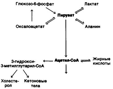
3. Acetyl CoA. The major sources of this activated two-carbon unit are The oxidative decarboxylation of pyruvate and the β-Oxidation of Fatty acids (Fig. 23.11). In addition, acetyl CoA is derived from ketogenic amino acids.
In contrast with many other molecules, the metabolic fate of acetyl CoA is highly restricted. The acetyl unit can be completely oxidized to CO2 in the citric acid cycle. Alternatively, three molecules of acetyl CoA can form 3-hydroxy-3-methylglutaryl CoA. This six-carbon compound is a precursor of Cholesterol and Ketone Bodies. A third major pathway for acetyl CoA is its export to the cytosol as citrate for fatty acid synthesis. It is important to emphasize once again that acetyl CoA cannot be converted into pyruvate in mammals. Consequently, mammals cannot convert Lipids into carbohydrates.
23.5. Metabolic Profiles of the Major Organs
Metabolism in the Brain, muscle, adipose tissue, and liver is highly specialized. Let us consider the differences among these organs in their use of fuel molecules.
1. Brain. Glucose is virtually the sole fuel for the human brain, except during prolonged starvation. The brain has no stores of fuel molecules and therefore requires a continuous supply of glucose. The brain consumes about 120 g of glucose per day, which corresponds to an ENERGY VALUE OF 420 kcal. In the resting state, the brain accounts for about 60% of the total glucose consumption of the body. During starvation, ketone bodies (acetoacetate and its reduced counterpart, 3-hydroxybutyrate) replace glucose as the brain's energy source. Acetoacetate is activated by The transfer of CoA from succinyl CoA, yielding acetoacetyl CoA (Fig. 23.12). Cleavage by thiolase then yields two molecules of acetyl CoA, which enter the citric acid cycle. Fatty acids cannot serve as fuel for the brain because they are bound to albumin and cannot cross the blood-brain barrier. Thus, ketone bodies are a transportable equivalent of fatty acids. We shall discuss below why The Use of ketone bodies rather than glucose as fuel is essential for minimizing protein breakdown during starvation.
Figure 23.12. Entry of ketone bodies into the citric acid cycle
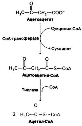
Figure 23.13. Metabolic exchange between muscle and liver

Table 23.1. Fuel reserves in a typical 70-kg man
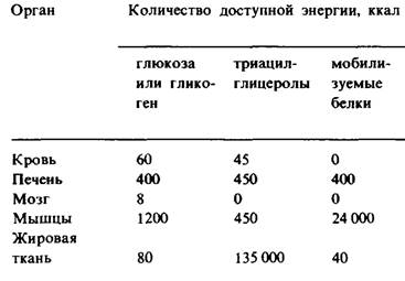
2. Muscle. The major fuels for muscle are glucose, fatty acids, and ketone bodies. Muscle differs from the brain in having a large store of glycogen (1200 kcal). About three-fourths of all glycogen in the body is stored in muscle (Table 23.1). After a meal, the glycogen content of muscle can reach 1%. This glycogen is readily converted into glucose 6-phosphate for use within muscle Cells. Like the brain, muscle lacks glucose 6-phosphatase, so it cannot export glucose. Instead, muscle retains glucose, which it prefers over other fuels during periods of high activity. In actively contracting Skeletal Muscle, The rate of glycolysis far exceeds that of the citric acid cycle. Most of the pyruvate formed under these conditions is reduced to lactate. Lactate flows to the liver, where it is converted into glucose. This process, known as the Cori cycle (Section 15.21), shifts part of the metabolic burden of muscle to the liver. In addition, large amounts of alanine are formed in actively working muscle by the transamination of pyruvate. Like lactate, alanine can be converted into glucose in the liver. The metabolism of resting muscle is organized quite differently. In it, Fatty acids are the major fuel. Ketone bodies can also serve as fuel for cardiac muscle. In fact, cardiac muscle prefers acetoacetate to glucose.
3. Adipose tissue. The triacylglycerol stores in adipose tissue represent a huge reservoir of metabolic energy. It amounts to 135,000 kcal in a typical 70-kg adult man. Adipose tissue is specialized for the Esterification of Fatty Acids and their release from triacylglycerols. In humans, the liver is the major site of fatty acid synthesis; therefore, a key biochemical function of adipose tissue is to activate these fatty acids and transfer the activated CoA derivatives to glycerol. Glycerol 3-phosphate, a key intermediate in this Biosynthesis (Section 20.1), is obtained by the reduction of dihydroxyacetone phosphate, which is formed from glucose via glycolysis. Adipose cells are unable to phosphorylate endogenous glycerol because they lack the necessary kinase. Therefore, adipose cells require glucose to synthesize triacylglycerols. Triacylglycerols are hydrolyzed to fatty acids and glycerol by lipases. The rate-limiting step in this process is the release of the first fatty acid from a triacylglycerol. This is catalyzed by a hormone-sensitive lipase, which can be reversibly phosphorylated. As in Glycogen Metabolism, cAMP acts as a messenger in a hormone-triggered amplifying cascade. Triacylglycerols in adipose tissue are continuously hydrolyzed and resynthesized. Glycerol formed by Hydrolysis is transferred to the liver. If glycerol 3-phosphate is abundant, fatty acids are re-esterified. However, if a lack of glucose creates a deficit of glycerol 3-phosphate, they are released into the Blood Plasma. Thus, the glucose concentration in adipose cells is the major factor determining the release of fatty acids into the blood.
Fig. 23.14. Synthesis and degradation of triacylglycerols in adipose tissue. Fatty acids enter adipose tissue in the form of very-low-density Lipoproteins (VLDL)
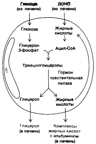
Fig. 23.15. Electron micrograph of a portion of a liver cell actively involved in the Synthesis and Secretion of very-low-density lipoprotein (VLDL) particles. The arrow points to a vesicle releasing its contents—VLDL particles
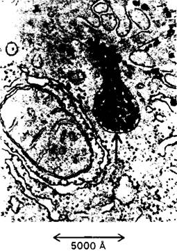
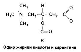
4. The Liver. The METABOLIC ACTIVITY OF the liver provides fuel for the brain, muscle, and other peripheral organs. Nutrients absorbed in the intestine travel primarily to the liver, allowing it to regulate the blood concentration of many metabolites. The liver takes up large amounts of glucose and converts it into glycogen. Thus, it can store up to 400 kcal. The liver can release glucose into the blood by breaking down stored glycogen or by performing gluconeogenesis. The major precursors for gluconeogenesis are lactate and alanine from muscle, glycerol from adipose tissue, and glucogenic amino acids from the diet. In addition, the liver plays a central role in regulating Lipid Metabolism. When fuels are abundant, fatty acids are synthesized in the liver, esterified, and secreted into the blood as very-low-density lipoproteins (VLDL) (Fig. 23.15). These plasma lipoproteins are the primary source of fatty acids used by adipose tissue for triacylglycerol synthesis. Conversely, during starvation, the liver converts fatty acids into ketone bodies. How do liver cells choose between these mutually exclusive pathways? The choice depends on whether fatty acids must enter the mitochondrial matrix. Recall that long-chain fatty acids cross The inner mitochondrial membrane only when esterified to carnitine (Section 17.7). The enzyme catalyzing The formation of acylcarnitine on the outer surface of this membrane is inhibited by malonyl-CoA, the committed intermediate in fatty acid synthesis. Thus, when long-chain fatty acids are being synthesized, they are prevented from entering the mitochondrial matrix, the compartment where β-oxidation and ketone body formation occur. Instead, these fatty acids are incorporated into triacylglycerols and Phospholipids. Conversely, when fuels are scarce, the concentration of malonyl-CoA falls. Under these conditions, fatty acids released from adipose tissue enter the mitochondrial matrix for subsequent conversion into ketone bodies. How does the liver meet its own energy needs? For its own energy needs, the liver prefers keto acids derived from the degradation of amino acids over glucose. Indeed, the primary role of glycolysis in the liver is to provide building blocks for biosyntheses. Furthermore, the liver cannot use acetoacetate as a fuel because it lacks the transferase required to activate it to acetyl-CoA. Thus, the liver spares the very fuels it exports to muscle and brain—a truly altruistic organ!
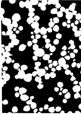
Fig. 23.16. Electron micrograph of very-low-density lipoprotein (VLDL) particles. These particles range in diameter from 300 to 800 Å; they transport triacylglycerols from the liver to adipose tissue
Last update: 06/08/2026
Editorial and Educational Adaptation: This material has been compiled based on the primary/original source text. The project team performed an editorial review, corrected technical inaccuracies, structured sections, and adapted the content for an educational format.
What was processed:
- elimination of formatting defects (OCR errors, structural breaks, corrupted characters);
- editorial organization of content;
- standardization of terminology in accordance with academic sources;
- verification of factual statements against the original source text.
All mentions of the author, publication year, and origin of the primary text have been preserved in accordance with the source.