BIOCHEMISTRY AND MOLECULAR BIOLOGY - W. ELLIOT - 2002
CHAPTER 9. THE ENERGY VALUE OF FATS
Fats are the primary energy source for ATP synthesis. They provide approximately half of the energy consumed by The Heart and resting skeletal Muscles. The amount of energy stored in the body as neutral fats in fat Cells is virtually unlimited, unlike the finite reserves stored as Glycogen. Furthermore, fats represent a more compact form of energy storage because they are less oxidized and hydrated compared to glycogen.
ATP is generated through The oxidation of both fats and glucose. Both pathways involve The Citric Acid Cycle and the Electron Transport Chain. The similarity between these two metabolic routes stems from the fact that both glucose and fat oxidation yield acetyl-CoA. In addition, fat degradation features a universal mechanism of Fatty acid oxidation through the sequential Cleavage of two-carbon units from the hydrocarbon chain as acetyl-CoA. This process generates NADH and FADH2, which are subsequently oxidized. One cannot fail to marvel at the efficiency of using the exact same biochemical machinery to harvest energy from vastly different dietary nutrients. Their oxidation pathways differ only in the initial stages, merging into a common stream within The Citric Acid cycle.
First and foremost, the following points deserve attention.
1. Before oxidation can begin, fats must undergo Hydrolysis to release Fatty acids. This process also produces glycerol from triglycerides; however, the metabolic fate of glycerol differs from that of fatty acids: it is phosphorylated and oxidized to dihydroxyacetone phosphate. Thus, during starvation, the glycerol moiety of triglycerides can be utilized by the Liver for Gluconeogenesis (see Chapter 11).
2. Peripheral Tissues extract fatty acids from the Blood, into which they are secreted by fat cells in response to elevated Glucagon levels. These acids also enter cells from chylomicrons and VLDL attacked by lipoprotein lipase (see p. 93).
3. Free Fatty acids are transported in the blood as anions reversibly bound to serum albumin. They readily cross Cell membranes, meaning their intracellular concentration is directly dependent on their levels in the bloodstream. As free fatty acids are cleared from the blood, their constant concentration is maintained through the dissociation of albumin complexes.
4. Fatty acids are broken down via the successive cleavage of two-carbon units in the form of acetyl-CoA.
5. Throughout their conversion into acetyl-CoA, fatty acids constantly exist in the form of acyl-CoA. Therefore, the initial stage of their METABOLISM, known as fatty acid activation, consists of the synthesis of a fatty acid acyl-CoA derivative.
Mechanism of Acetyl-CoA Formation from Fatty Acids
Activation of Fatty Acids via the Synthesis of Their Acyl-CoA Derivatives
The term activation reflects the fact that fatty acid thioesters are high-energy, reactive compounds. The activation reaction proceeds as follows:
RCOО- + АТР + CoA-SH —> RCO-S-CoA + AMP + РРi
∆G°' = -0.9 kJ • mol-1.
The change in Free energy during this process is small (due to the high energy of the thioester bond); however, the action of pyrophosphatase drives the overall process to be strongly exergonic and irreversible (∆G°' = -32.5 kJ • mol-1).
The fatty acid activation reaction is catalyzed by three related Enzymes—fatty acyl-CoA synthetases—which specialize in short-, medium-, and long-chain fatty acids.
Transport of Fatty Acid CoA Derivatives into Mitochondria
Fatty acids are activated on the outer mitochondrial membrane, which is topologically continuous with the Cytoplasm; however, their conversion into acetyl-CoA
takes place exclusively within the mitochondrial matrix. The acyl group is transported into the mitochondria without CoA via a specialized transport system, where it interacts with CoA-SH once again inside the mitochondria to regenerate acyl-CoA. This transport mechanism preserves the high-energy Nature of the acyl moiety, eliminating The Need for re-activation on the other side of the membrane. This is made possible because The transfer of the fatty acid residue into the mitochondria is mediated by the molecule carnitine:
Class="center">
Although esters are typically not high-energy compounds, The Structure of carnitine is such that the acyl-carnitine bond is indeed a high-energy linkage. This is likely why evolution selected carnitine as the acyl-carrying molecule. Acyl carnitine derivatives cross into the matrix, where the reverse reaction occurs: the acyl group is transferred from carnitine to the thiol group of free CoA, and carnitine returns to the Cytosol to fetch another fatty acid residue.

The pathway of transmembrane fatty acid transport into mitochondria is illustrated in Fig. 9.1. The two acyl group transfer reactions—onto carnitine and onto mitochondrial CoA—are catalyzed by two distinct enzymes: the first, operating on the outer side of the mitochondrion, is carnitine acyltransferase I, while the second, taking place in the matrix, is carnitine acyltransferase II. Certain genetic defects involve a deficiency in carnitine or carnitine acyltransferase, leading to Muscle pain and lipid accumulation in muscles.
Fig. 9.1. Mechanism of transport of long-chain fatty acid residues into mitochondria for subsequent oxidation. The transfer of acyl residues from acylcarnitine to CoA does not require Energy Expenditure

Conversion of acyl-CoA to acetyl-CoA within the mitochondria
This process involves four sequential reactions, three of which are similar to stages we have already encountered in the citric acid cycle
The conversion of succinate (Sp) to oxaloacetate: dehydrogenation using an FAD-dependent enzyme, Hydration, and NAD+-dependent dehydrogenation. Recall the sequential transformations Succinate -> Fumarate -> Malate -> Oxaloacetate in the citric acid cycle. The corresponding reactions involving acyl-CoA derivatives are shown in Fig. 9.2.
Fig. 9.2. Four sequential reactions by which the hydrocarbon chain of fatty acid CoA derivatives is shortened. The chain is shortened by two carbon atoms, which are released as the acetyl group of acetyl-CoA

A key reaction in Lipid Metabolism is the interaction between CoA-SH and β-ketoacyl-CoA, during which the hydrocarbon chain of the fatty acid residue is cleaved. Its two-carbon fragment is split off as acetyl-CoA, leaving a fatty acid residue shortened by two carbon atoms (acyl-CoA). The enzyme that catalyzes this reaction is called thiolase, because the initial acyl-CoA molecule is attacked by the thiol group of free CoA. Natural long-chain fatty acids contain an even number of carbon atoms, so the successive cleavage of two-carbon fragments sooner or later leads to The formation of butyryl-CoA, which is then converted into (3-acetoacetyl-CoA. The cleavage of this compound into two acetyl-CoA molecules completes The process of fatty acid chain shortening:

Thus, at each step of fatty acid breakdown, a β-ketoacyl-CoA with an increasingly shortened hydrocarbon chain is formed. This provides the rationale for naming the entire process described above β-Oxidation of Fatty acids. The NADH and FADH2 produced during β-oxidation then donate their electrons to the Mitochondrial Electron Transport chain (see Fig. 8.19).
Energy yield from fatty acid oxidation
A palmitic acid molecule (C16) is converted into 8 molecules of acetyl-CoA, additionally generating 7 molecules of FADH and an equal number of FADH2. Acetyl-CoA is subsequently oxidized in the citric acid cycle, while NADH and FADH2 are oxidized in the mitochondrial electron transport chain (see Chapter 8). The oxidation of NADH yields 2.5 ATP molecules, and the oxidation of FADH2 yields 1.5 molecules (since NADH is formed in the matrix, the shuttle mechanism is not involved in transporting it into the mitochondria).
Thus, the oxidation of 1 molecule of palmitic acid leads to the synthesis of 106 mol of ATP from ADP and Pi (taking into account the consumption of two ATP molecules for the formation of palmitoyl-CoA and the synthesis of one GTP molecule for each acetyl-CoA in the citric acid cycle). This means that the efficiency of the process is 33%; this fraction of free energy is converted into useful work in the form of ATP synthesis. Our calculation results can also be presented differently: the oxidation of 256 g of palmitic acid yields 45 kg of ATP. Of course, such an amount of ATP is never present at once; it is rapidly hydrolyzed to meet the Organism's energy demands.
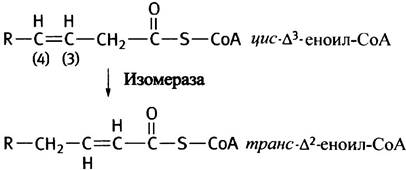
Olive oil is liquid because it has a high content of monounsaturated fats—triglycerides and Phospholipids containing Unsaturated fatty acids with one or more double bonds in their hydrocarbon chain. An example is palmitoleic acid, which features a double bond between the ninth and tenth carbon atoms. Up to the double bond, the chain of such an acid is shortened by normal oxidation to form cis-∆3-enoyl-CoA:

The double bond in this compound prevents acyl-CoA dehydrogenase from introducing another adjacent double bond between the second and third C atoms. β-Oxidation could end here; however, the enzyme isomerase shifts the double bond to the required position and forms the trans-isomer, which then serves as a substrate for enoyl-CoA hydratase (see Fig. 9.2).
This clever fix solves The problem of Biological Oxidation of monounsaturated fatty acids. Polyunsaturated fatty acids present additional challenges. Consider, for example, linoleic acid with its two double bonds (∆9 and ∆12). The Fate of one of them (∆9) during oxidation is as described above. The other bond (∆12) is reduced at the appropriate moment by a special enzyme, enabling The breakdown of linoleic acid via the β-oxidation mechanism (though the actual details are somewhat more complex).
Does the acetyl-CoA formed during β-oxidation always enter the citric acid cycle?
Up to this point, we have considered the situation where all synthesized acetyl-CoA enters the citric acid cycle. This is normally the case in all tissues, but emergency situations can arise in the liver where this does not happen.
Sometimes lipid metabolism becomes the primary energy source for the organism (see Chapter 5). This state occurs, for instance, during starvation, when all glycogen reserves are depleted. In diabetics, glucose deprivation occurs not from a lack of glucose, but due to the inability to utilize it. Be that as it may, the suppression of Glycolysis prompts fat cells to release fatty acids, and the liver to use them for synthesizing excessive amounts of acetyl-CoA—excessive in the sense that it exceeds the demands of the citric acid cycle. The situation is further aggravated by the fact that the cessation of Carbohydrate Metabolism leads to a deficiency of oxaloacetate, which is required to produce citrate from acetyl-CoA (see p. 121).
In this situation, within liver cells, two acetyl groups combine to form acetoacetate (CH3COCH2COO-), which is partially reduced to β-hydroxybutyrate (CH3CHOHCH2COO-). These two substances—Ketone Bodies—are released into the blood.
How is acetoacetate formed from acetyl-CoA?
Acetoacetyl-CoA is formed from acetyl-CoA by the Reversal of the reaction catalyzed by ketoacyl-CoA thiolase:
2CH3CO-S-CoA <-> CH3COCH2CO-SCoA + CoA-SH
It might seem that free acetoacetate should be formed As a result of acetoacetyl-CoA hydrolysis (which would shift the equilibrium of the given reaction toward product formation). Instead, acetoacetyl-CoA reacts with a third acetyl-CoA molecule to yield 3-hydroxy-3-methylglutaryl-CoA (HMG-CoA), which is then cleaved into acetoacetate and acetyl-CoA (Fig. 9.3).
Fig. 9.3. Formation of Ketone bodies in liver mitochondria during excessive fat oxidation (starvation, diabetes). 3-Hydroxy-3-methylglutaryl-CoA (HMG-CoA) serves as a Cholesterol precursor and, in this case, is synthesized in the cytoplasm via a reaction catalyzed by hydroxymethylglutaryl-CoA synthase.

Why acetoacetate synthesis proceeds in this manner remains unclear. Hydroxymethylglutaryl-CoA, synthesized by many animal cells, is a precursor of cholesterol—an essential component of The Plasma Membrane (see p. 58). Ketone bodies are produced in mitochondria (where fats are converted into acetyl-CoA). Moreover, the synthesis of hydroxymethylglutaryl-CoA required for cholesterol production is localized in the cytoplasm, which contains a specialized enzyme—hydroxymethylglutaryl-CoA synthase—attached to The Endoplasmic reticulum membrane.
Peripheral tissues can utilize acetoacetate to generate energy. In the mitochondria, it is converted into acetoacetyl-CoA through An acyl group transfer reaction with succinyl-CoA:

Acetoacetyl-CoA is subsequently cleaved by thiolase (in the presence of free CoA) into two molecules of acetyl-CoA. Upon preliminary dehydrogenation, β-hydroxybutyrate is also converted into acetoacetate.
Oxidation of Odd-Chain Fatty Acids
A small proportion of dietary fatty acids (e.g., of plant origin) contains an odd number of carbon atoms. The β-oxidation of such acids yields a five-carbon β-ketoacyl-CoA rather than acetoacetyl-CoA as the penultimate product. Cleavage of this intermediate by thiolase produces acetyl-CoA and three-carbon propionyl-CoA:

Propionyl-CoA is converted into succinyl-CoA and enters the citric acid cycle. The corresponding reactions are shown below. Epimerase catalyzes the Conversion of the D-configuration of methylmalonyl-CoA into the L-configuration:

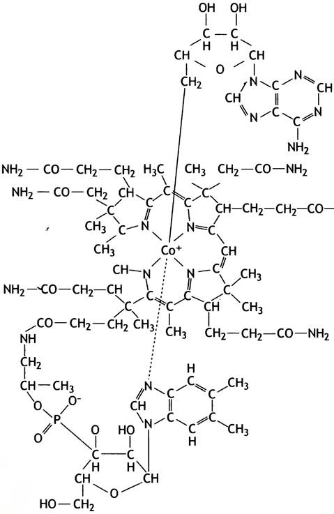
The final reaction (the isomerization of methylmalonyl-CoA) is notable for involving the most structurally complex coenzyme—deoxyadenosylcobalamin, a vitamin B12 derivative (Fig. 9.4).
Fig. 9.4. Structure of deoxyadenosylcobalamin
Propionate is also produced during the degradation of four Amino Acids (valine, isoleucine, Methionine, and Threonine), as well as from the cholesterol side chain.
Deficiency of methylmalonyl-CoA mutase or impaired coenzyme synthesis from vitamin B12 leads to fatal acidosis.
Peroxisomal Fatty Acid Oxidation
Peroxisomes are small, membrane-bounded vesicles found in many animal cells, which are discussed in detail in Chapter 16. It is worth noting
that they also oxidize fatty acids, although the bulk of fatty acids undergo oxidation in mitochondria. Peroxisomal β-oxidation of fatty acids likewise produces FADН2; however, this is oxidized not in the mitochondria, but within the peroxisomes themselves through direct interaction with oxygen, yielding hydrogen peroxide and heat. The Functions of peroxisomes are less well understood than those of other intracellular Organelles. It is hypothesized that one of their roles is the metabolism of fatty acids containing more than 18 carbon atoms, which are not oxidized by the mitochondrial system.
So far, we have focused on energy generation from glucose and fats. This leaves the third major dietary component: amino acids, and it would seem logical to discuss their transformations next. However, it is more practical to move directly from glucose and fat metabolism to their synthesis and its regulation in the body, especially since the generation of energy from amino acids yields intermediates of glycolysis or the citric acid cycle at early metabolic stages, meaning these pathways lack distinct peculiarities. Furthermore, the most important aspects of Amino acid metabolism are unrelated to energy production.
Questions for Chapter 9
1. Peripheral tissues obtain fatty acids from the blood. Describe three possible pathways for their delivery.
2. Which cells in the body do not use fatty acids as an energy source?
3. What is the initial stage of fatty acid degradation, and where does it take place? Where in Eukaryotic cells are fatty acids converted into acetyl-CoA, and how do they get there?
4. What do the oxidation reactions of fatty acids to acetyl-CoA have in common with certain reactions in the citric acid cycle?
5. What is the ATP yield from the oxidation of palmitic acid? Explain your answer.
6. Explain how a monounsaturated fat (∆9) is oxidized to acetyl-CoA.
7. Does acetyl-CoA produced during fatty acid breakdown always enter the citric acid cycle?
8. Hydroxymethylglutaryl-CoA is an intermediate produced during the synthesis of acetoacetate and cholesterol. Where do these processes take place?
Last update: 06/08/2026
Editorial and Educational Adaptation: This material has been compiled based on the primary/original source text. The project team performed an editorial review, corrected technical inaccuracies, structured sections, and adapted the content for an educational format.
What was processed:
- elimination of formatting defects (OCR errors, structural breaks, corrupted characters);
- editorial organization of content;
- standardization of terminology in accordance with academic sources;
- verification of factual statements against the original source text.
All mentions of the author, publication year, and origin of the primary text have been preserved in accordance with the source.