Review of Medical Physiology - William F. Ganong 2002
Endocrine System, Metabolism, and Reproduction
Energy Balance, Metabolism, and Nutrition
Lipid Metabolism
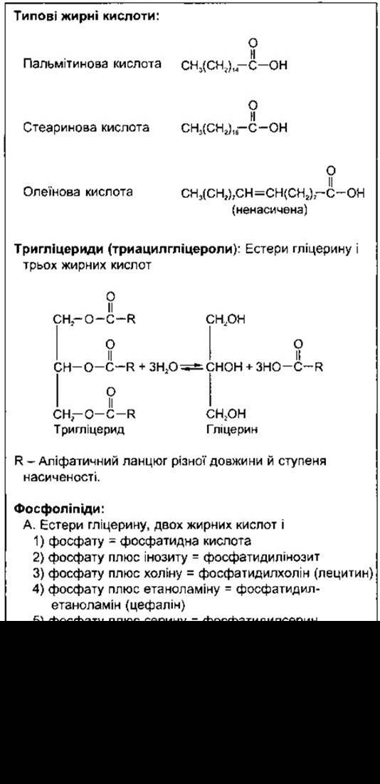
Biologically important lipids include Fatty acids and their derivatives, neutral fats (triglycerides), Phospholipids, and related compounds and sterols. Triglycerides consist of three fatty acids attached to glycerol (Table 17-4). Naturally occurring fatty acids contain an even number of carbon atoms. They can be saturated (lacking double bonds) and unsaturated (hydrogenated, with varying numbers of double bonds). Phospholipids are integral components of Cell membranes. Sterols are formed from various Steroid Hormones and Cholesterol.
Fatty acid oxidation and Synthesis
In the body, Fatty acids are broken down into acetyl-CoA, which enters The Citric Acid Cycle. The primary breakdown occurs in the Mitochondria via ß-Oxidation. Fatty acid oxidation begins with their activation (Fig. 17-24), a reaction that takes place both inside and outside the mitochondria. Medium- and short-chain fatty acids enter the mitochondria without activation, whereas long-chain fatty acids must be ester-linked to carnitine before they can cross The inner mitochondrial membrane. Carnitine is ß-hydroxy-y-trimethylammonium butyrate, which is synthesized in the body from Lysine and Methionine. Translocase transports the fatty acid-carnitine ester into the mitochondrial matrix in exchange for free carnitine. In the matrix, the ester is transferred to CoA, forming an activated fatty acid molecule available for ß-oxidation and supplying carnitine for further exchange. Beta-Oxidation involves the repeated Cleavage of two-carbon fragments from the fatty acid (see Fig. 17-24). The energy yield of this process is quite high. For instance, the Catabolism of 1 mole of a six-carbon fatty acid in The Citric Acid cycle to CO2 and H2O yields 48 moles of ATP, whereas the catabolism of the six-carbon carbohydrate glucose yields only 38 moles. Deficient ß-Oxidation of Fatty acids can be caused by carnitine deficiency or Genetic Defects in translocase or Other Enzymes involved in The transport of long-chain fatty acids into the mitochondria. Such a condition causes cardiomyopathy and also leads to hypoketotic hypoglycemia with coma—a serious and often fatal condition that arises after fasting when glucose reserves are depleted due to insufficient fatty acid oxidation to supply energy, while Ketone Bodies (see below) are not produced in sufficient quantities owing to a shortage of CoA in the Liver.
Class="center">Table 17-4. Lipids
Many Tissues are capable of synthesizing fatty acids from acetyl-CoA. Some syntheses of long-chain fatty acids from short-chain ones occur in the mitochondria by a simple Reversal of the reactions shown in Fig. 17-24. However, most fatty acids are synthesized de novo from acetyl-CoA via various pathways located primarily outside the mitochondria in microsomes. The steps of these pathways are illustrated in Fig. 17-25.
For unknown reasons, fatty acid synthesis in all Cells ceases when the chain length reaches 16 carbon atoms. Only small amounts of 12- or 14-carbon fatty acids are produced, and no acids with a carbon chain length exceeding 16 atoms are formed. Elongation of the carbon chain to 18 or more C atoms (stearic acid) takes place in the microsomes of liver cells. Fatty acids combine with glycerol to form neutral fats; this process occurs notably in fat depots, with the actual joining taking place in the mitochondria.
Ketone Bodies
In many tissues, acetyl-CoA residues condense to form acetoacetyl-CoA (Fig. 17-26). In the liver, which (unlike other tissues) contains deacylase, free acetoacetate is produced. This ß-keto acid is converted into ß-hydroxybutyrate and acetone, and since these compounds are poorly metabolized in the liver, they diffuse into the Circulatory system. Acetoacetate in the liver also arises from The formation of 3-hydroxy-3-methylglutaryl-CoA (see Fig. 17-26), and this pathway is more important than deacylation. Acetoacetate, ß-hydroxybutyrate, and acetone are termed ketone bodies. Tissues other than the liver transfer CoA from succinyl-CoA to acetoacetate and metabolize "active" acetoacetate to CO2 and H2O via the citric acid cycle. Other pathways for the METABOLISM of ketone bodies are also known. Under certain conditions, ketones serve as an important source of energy. Acetone is excreted in the urine and exhaled breath.
The normal Blood level of ketone bodies in humans is low (about 1 mg/dL), and because these bodies are normally metabolized as rapidly as they are formed, less than 1 mg is excreted over 24 hours. However, if the influx of acetyl-CoA into the citric acid cycle decreases due to poor supply of glucose metabolites or fails to increase with an increased supply of acetyl groups, acetyl-CoA accumulates, The rate of acetoacetyl-CoA Condensation rises, and more acetoacetate is produced in the liver. The capacity of tissues to oxidize ketone bodies quickly reaches its limit, and these bodies accumulate in the circulatory system (ketonemia). Two of the three ketone bodies—acetoacetate and ß-hydroxybutyrate—are anions of moderately strong acids (acetoacetic and ß-hydroxybutyric acids) and possess buffering properties, counteracting the pH shift that would otherwise occur. However, the buffering capacity can be exceeded, and the metabolic acidosis that develops in conditions such as diabetic ketonemia can be severe and even fatal.
Three conditions lead to deficient intracellular glucose supply: starvation, Diabetes Mellitus, and a high-fat, low-carbohydrate diet. In diabetes, cellular glucose uptake is reduced. If most dietary calories come from fats, a carbohydrate deficit arises because there is no major pathway for converting fats into CARBOHYDRATES. Liver cells become engorged with fat, which damages them and displaces Glycogen. In all these states, ketonemia develops primarily due to the overproduction of ketone bodies.
The odor of acetone on the breath of children after vomiting results from starvation ketonemia. Parenteral administration of relatively small amounts of glucose prevents ketonemia, which is why this carbohydrate is termed antiketogenic.
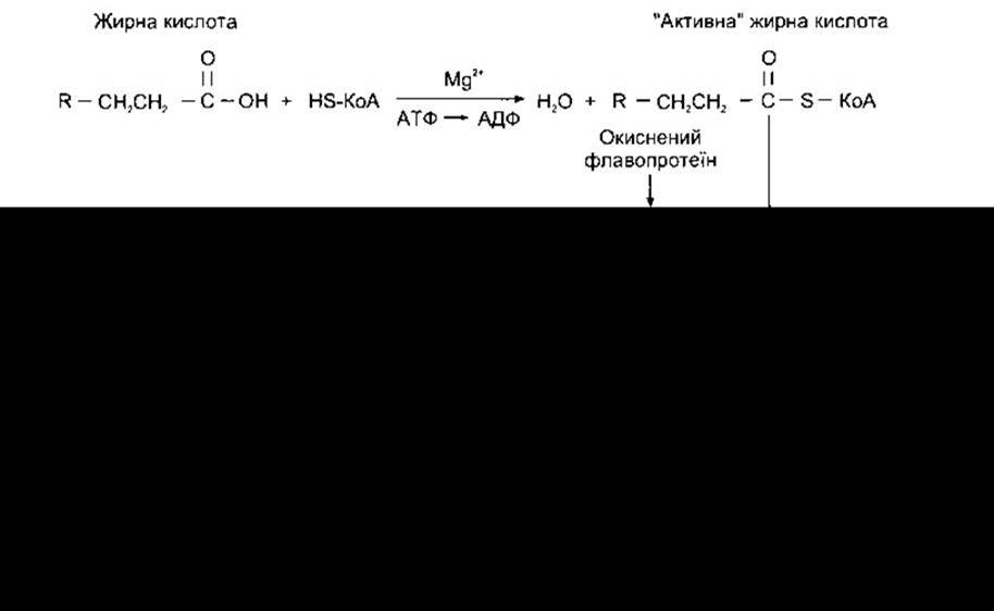
Figure 17-24. Fatty acid oxidation. This process of simultaneous cleavage of two-carbon fragments is repeated until the end of the chain.
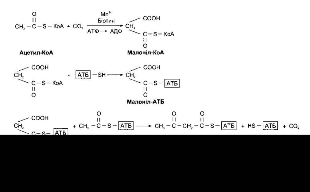
Figure 17-25. Synthesis of fatty acids via pathways found in microsomes. ACP stands for acyl carrier protein, which is part of the enzyme complex to which acyl residues attach during fatty acid synthesis. The process shown here repeats, whereby one acetyl-ACP complex is added to butyryl-ACP to form a six-carbon fatty acid derivative, then another acetyl-ACP complex to form an eight-carbon compound, and so on.
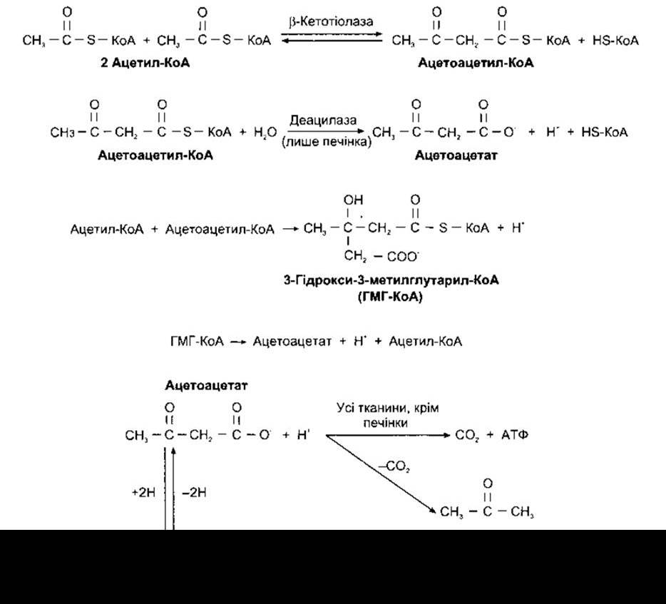
Figure 17-26. Formation and metabolism of ketone bodies. Note that There are two pathways for the formation of acetoacetate.
Cellular Lipids
Cellular lipids are divided into two main types: structural lipids, which are constituents of membranes and other cell parts, and neutral fat, stored by adipocytes in fat depots. Neutral fat is oxidized during starvation, whereas structural lipids are conserved. Naturally, fat reserves vary in size, yet in non-obese individuals they account for 15% of body weight in men and 21% in women. Far from being inert masses as once believed, they are active, dynamic tissues undergoing constant breakdown and resynthesis. In adipose tissue, glucose is metabolized to fatty acids, and neutral fats are synthesized. Conversely, neutral fats are broken down, and free fatty acids are released into the circulatory system.
Brown Fat
A third—specialized—type of lipid is brown fat, which constitutes only a small percentage of total body lipids. Brown fat, predominantly found in infants but also present in adults, is located between the shoulder blades, on the back of the neck, along the major Blood Vessels in the Thorax and abdomen, and occasionally in other sites. In brown fat depots, fat cells as well as blood vessels have a marked sympathetic innervation, in contrast to white fat depots, where only some fat cells are innervated, while the primary sympathetic innervation is restricted to blood vessels. Furthermore, ordinary fat cells contain a single large droplet of white fat, whereas brown fat cells consist of multiple smaller droplets. Brown fat Cells also contain numerous mitochondria. These mitochondria possess the standard inner proton conductance that generates ATP (Oxidative Phosphorylation, see above) as well as an alternative, non-ATP-generating proton conductance. This short-circuit conductance depends on a 32 kDa uncoupling protein now designated UCP1. This protein uncouples oxidative processes from ATP synthesis, resulting in greater heat production (Fig. 17-27). A second protein, UCP2, located in both white and brown adipose tissues, has recently been discovered, and evidence exists for several additional such Proteins. Activation of the sympathetic innervation of brown adipose tissue releases norepinephrine, which, acting on ß3-adrenergic receptors, stimulates lipolysis, and enhanced fatty acid oxidation in mitochondria increases thermogenesis. Consequently, changes in sympathetic activity in brown fat alter the efficiency with which food is assimilated and energy is produced. Variations in UCP protein expression may also influence metabolic efficiency.
There is evidence that brown fat Functions in this manner in animals and possibly humans adapted to cold environments, who exhibit enhanced heat production in brown fat along with accelerated blood flow. Neural stimulation of brown fat increases following food consumption, which likewise enhances heat production. Notably, postprandial thermogenesis is driven by two factors: the acute specific dynamic action (see above) resulting from food assimilation, and a somewhat smaller increase in heat generation by brown fat. The dependence of brown fat mass on food intake is discussed in Chapter 14.
Plasma Lipids and Lipid Transport
Most lipids are relatively insoluble in Water and do not circulate in a free form. Free fatty acids are bound to albumin, whereas cholesterol, triglycerides, and phospholipids are transported as lipoprotein complexes. These complexes significantly increase lipid solubility. There are six families of Lipoproteins (Table 17-5), classified according to their size and lipid content. The density of these lipoproteins (and consequently their sedimentation rate in an ultracentrifuge) is inversely proportional to their lipid content. In general, lipoproteins consist of a Hydrophobic core of triglycerides and cholesteryl esters surrounded by a monolayer of phospholipids and proteins (Fig. 17-28). The pathways by which lipoproteins travel from the intestine to the liver (the exogenous pathway) and by which lipids are transferred to and from tissues (the endogenous pathway) are illustrated in Fig. 17-29. The protein components of lipoproteins are called apoproteins. The principal apoproteins are APO E, APO C, and APO B (see Fig. 17-29). Two forms of APO B are known: the low-molecular-weight form, APO B-48, which is characteristic of the exogenous pathway transporting dietary absorbed lipids (see below), and the high-molecular-weight form, APO B-100, which is characteristic of the endogenous transport pathway.
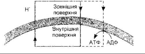
Fig. 17-27. Proton Transport Across the mitochondrial membrane in brown adipose tissue. Proton efflux occurs via The electron transport system, as in other mitochondria. In addition to the inward movement of protons that drives ATP synthesis, There is a non-ATP-generating proton "leak" back into the mitochondrial matrix. As a result, fat metabolism and ATP production are uncoupled. Compare with Fig. 17-7.
Chylomicrons are formed in the intestinal mucosa during the absorption of dietary fat Digestion products (see Chapter 25). They are very large lipoprotein complexes that enter the circulatory system via the lymphatic vessels. Following a meal, the blood contains so many of these particles that the plasma takes on a milky appearance (lipemia). Chylomicrons are cleared from the Circulation through the action of lipoprotein lipase, which is anchored to the capillary endothelial surface. This enzyme catalyzes The breakdown of triglycerides in chylomicrons into free fatty acids and glycerol, which are then taken up by adipocytes and re-esterified. Alternatively, free fatty acids remain in the bloodstream bound to albumin. Lipoprotein lipase, which requires heparin as a cofactor, also removes triglycerides from circulating very-low-density lipoproteins (VLDLs) (see below). Chylomicrons and VLDLs contain APO C, a complex of proteins that dissociate from them within capillaries. One of the components of this complex, apolipoprotein C-II, activates lipoprotein lipase.
Table 17-5. Major lipoproteins. Plasma lipids include these components plus free fatty acids derived from adipose tissue that circulate bound to albumin
Lipoprotein |
Size, nm |
Composition, % |
Origin |
||||
protein |
free cholesterol |
cholesteryl esters |
triglyceride |
phospholipids |
|||
Chylomicrons |
75-100 |
2 |
2 |
3 |
90 |
3 |
Intestine |
Chylomicron remnants |
30-80 |
... |
... |
... |
... |
... |
Capillaries |
Very-low-density lipoproteins |
30-80 |
8 |
4 |
16 |
55 |
17 |
Liver and intestine |
Intermediate-density lipoproteins |
25-40 |
10 |
5 |
25 |
40 |
20 |
VLDL |
Low-density lipoproteins |
20 |
20 |
7 |
46 |
6 |
21 |
IDL |
High-density lipoproteins |
7,5-10 |
50 |
4 |
16 |
5 |
25 |
Liver and intestine |
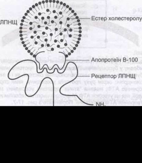
Fig. 17-28. Cytology/cytology/92.html">SCHEMATIC Structure OF a low-density lipoprotein, the LDL receptor, and the binding of LDL to the receptor via APO B-100.
Triglyceride-depleted chylomicrons remain in the bloodstream as cholesterol-rich lipoproteins termed chylomicron remnants, measuring 30-80 nm in diameter. These remnants are taken up by the liver, where they bind to chylomicron remnant and LDL receptors, initiating receptor-mediated endocytosis (see Chapter 1) followed by lysosomal degradation.
Chylomicrons and their remnants constitute the transport system for exogenous dietary lipids (see Fig. 17-29). There is also an endogenous system consisting of VLDLs, intermediate-density lipoproteins (IDLs), low-density lipoproteins (LDLs), and high-density lipoproteins (HDLs), which transport triglycerides and cholesterol throughout the body. VLDLs are synthesized in The Liver and transport triglycerides, formed from fatty acids and carbohydrates, to peripheral tissues. When triglycerides are abundant, VLDLs are converted into IDLs, which release phospholipids and acquire cholesteryl esters of HDL origin through the action of the plasma enzyme lecithin-cholesterol acyltransferase (LCAT; see Fig. 17-29). Some IDLs are cleared by the liver; others lose additional triglycerides and protein, presumably within the hepatic sinusoids, and are converted into LDLs. During this conversion, they lose APO E, while APO B-100 is retained.
LDLs supply tissues with cholesterol—an essential component of cell membranes that is also used by specialized Endocrine glands for the synthesis of steroid hormones. In the liver and most extrahepatic tissues, LDL uptake occurs via receptor-mediated endocytosis in coated pits (see Chapter 1). The receptors recognize the LDL component APO B-100 (see Fig. 17-28); they also bind to APO E, but not to APO B-48.
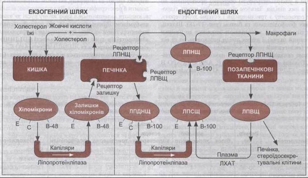
Fig. 17-29. Simplified diagram of the human lipoprotein lipid transport system. In the exogenous system, triglyceride-rich dietary chylomicrons are converted by lipoprotein lipase into chylomicron remnants rich in cholesteryl esters. In the endogenous system, the liver secretes triglyceride-rich VLDLs, which are converted into IDLs and subsequently into cholesteryl ester-rich LDLs. LCAT, lecithin-cholesterol acyltransferase. Letters on chylomicrons, chylomicron remnants, VLDLs, IDLs, and LDLs indicate the major apoproteins found within them. One-third of IDLs are taken up by macrophages and other cells via alternative mechanisms.
Human LDL receptors belong to a family of receptors that internalize macromolecules into cells via endocytosis in clathrin-coated pits (see Chapter 1). This large, complex molecule comprises a Cysteine-rich domain of 292 amino acid residues that binds LDL; a region of approximately 400 amino acid residues homologous to the precursor of the epidermal growth factor; a Serine- and Threonine-rich domain of 58 Amino Acids that serves as a site for glycosylation; a hydrophobic transmembrane domain of 22 amino acid residues; and a cytoplasmic tail of 50 amino acid residues (see Fig. 17-28). The Gene encoding this protein contains 18 exons, 13 of which encode protein sequences homologous to sequences found in other proteins. Thus, the LDL receptor is a mosaic protein built from exons that also encode other proteins.
During receptor-mediated endocytosis, each coated pit pinches off to form a coated vesicle and subsequently an endosome. Ion-selective pumps in the endosomal membranes lower the pH within these Organelles. In the case of the LDL receptor (but not the chylomicron remnant receptor), this acidification triggers the dissociation of LDL receptors, which are then recycled back to The Plasma Membrane (Fig. 17-30). Endosomes subsequently fuse with Lysosomes, and the cholesterol generated from cholesteryl esters by the action of lysosomal acid lipase becomes available for cellular needs (see Fig. 17-30). Intracellular cholesterol suppresses endogenous cholesterol synthesis by inhibiting HMG-CoA reductase (see below), stimulates the Esterification of excess liberated cholesterol, and inhibits the synthesis of new LDL receptors. Together, these reactions provide a feedback mechanism for regulating cellular cholesterol Homeostasis.
LDL uptake is also mediated by lower-affinity systems in macrophages and certain other cells. In addition, macrophages preferentially take up oxidatively modified LDLs. However, oxidation can also occur within macrophages themselves. In animals, large doses of antioxidants such as vitamin E slow the progression of atherosclerosis, although results from similar trials in humans remain inconclusive. The receptors for modified LDL on macrophages and related cells are called scavenger receptors. They are distinct from the receptors on other cell types and exhibit a higher affinity for modified LDLs. When macrophages become overloaded with oxidized LDLs, they transform into "foam cells," which can be found in early atherosclerotic lesions in blood vessels. Under steady-state conditions, cholesterol continuously enters and leaves cells. It is believed to be exported from cells by one of the ABC cassette proteins (see Chapter 1) and taken up by HDLs. These lipoproteins are synthesized in the liver and intestine. An individual HDL receptor has been identified and cloned. It was first discovered in steroid hormone-producing endocrine glands and the liver. The HDL system transports cholesterol back to the liver, where it is excreted in the Bile, thereby reducing plasma cholesterol levels.
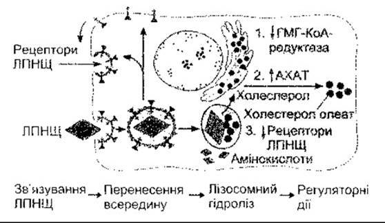
Fig. 17-30. Cellular uptake and metabolism of cholesterol. LDLs bind to receptors and are internalized via receptor-mediated endocytosis into low-pH endosomes. Receptors dissociate and are recycled to the membrane. Cholesteryl esters enter lysosomes, where free cholesterol is released for cellular processes. Cholesterol also inhibits HMG-CoA reductase (1), is partially converted into another cholesteryl ester by the enzyme acyl-CoA:cholesterol acyltransferase (ACAT) (2), and suppresses the formation of LDL receptors (3) (courtesy of MS Brown).
Apoprotein E is synthesized by Cells of the Brain, Spleen, Lungs, Adrenal Glands, Ovaries, and Kidneys, as well as the liver. Its concentration increases markedly following nerve injury, as it plays a role in nerve regeneration. The apolipoprotein E gene exists in Selection/30.html">The population as three alleles: APO-2, APO-3, and APO-4. The APO-4 allele is less common than APO-2 and APO-3, but it is more prevalent in patients with Alzheimer's disease (see Chapter 16) and is believed to contribute to its Pathogenesis.
Metabolism of Free Fatty Acids
Free fatty acids (FFAs) are delivered to adipocytes and other tissues by chylomicrons and VLDLs (see above). They are also synthesized within adipose depots, where they are stored. FFAs circulate bound to albumin and serve as a major energy source for many Organs. They are actively utilized by The Heart, and presumably all tissues, including the brain, can oxidize FFAs to CO2 and H2O.
The supply of FFAs to tissues is regulated by two lipases. As noted, lipoprotein lipase located on the capillary endothelial surface hydrolyzes triglycerides in chylomicrons and VLDLs, yielding FFAs and glycerol, which are then reassembled into new triglycerides within fat cells. Intracellular hormone-sensitive lipase in adipocytes catalyzes the breakdown of stored triglycerides into glycerol and fatty acids, which are subsequently released into the circulatory system.
Hormone-sensitive lipase is converted from an inactive to an active form via cAMP-dependent protein kinase A (Fig. 17-31). Adipocyte adenylate cyclase is activated by Glucagon as well as catecholamines (norepinephrine and epinephrine) acting via a $\beta$-adrenergic receptor. The pharmacological profile of this receptor is atypical; for example, it is resistant to propranolol and other drugs that block $\beta_1$ and $\beta_2$ receptors, suggesting it is a $\beta_3$ receptor. Efforts are underway to develop a selective $\beta_3$ agonist for potential use in the Treatment of obesity. ACTH, TSH, LH, serotonin, and vasopressin also increase lipolysis via cAMP, though The Physiological Role of these substances in regulating lipolysis remains incompletely understood.
Growth Hormone, glucocorticoids, and THYROID HORMONES also enhance The activity of hormone-sensitive lipase, albeit by slowing down new Protein Synthesis. It is suggested that growth hormone stimulates The production of a protein that enhances the capacity of catecholamines to activate cAMP, whereas cortisol promotes the synthesis of a protein that potentiates cAMP action. Conversely, Insulin and prostaglandin E decrease hormone-sensitive lipase activity, presumably by inhibiting cAMP formation.
Hormone-sensitive lipase activity increases during fasting and stress and decreases in the fed state and in response to insulin. Conversely, nutritional intake increases lipoprotein lipase activity, whereas fasting and stress decrease it.
The precursor for steroid hormones and bile acids is cholesterol which, as already noted, is an important structural component of cell membranes (see Chapter 1). It is found exclusively in animals. Similar sterols occur in plants, but under normal conditions they are not absorbed in the gastrointestinal tract. Most dietary cholesterol is found in egg yolks and animal fat. Cholesterol absorption takes place in the intestine, where it is incorporated into chylomicrons formed in the mucosa. After chylomicrons are depleted of triglycerides in adipose tissue, chylomicron remnants deliver cholesterol to the liver. The liver and other tissues also synthesize cholesterol. Part of the hepatic cholesterol is excreted into bile, both in free form and as bile acids, and is reabsorbed in the intestines. Most of the cholesterol in the liver is incorporated into VLDL and circulates in lipoprotein complexes (see above).

Fig. 17-31. Mechanism by which catecholamines increase hormone-sensitive lipase activity in adipose tissue. The beta receptor is likely a P3-adrenergic receptor.
The Biosynthesis of Cholesterol from acetate is shown in Fig. 17-32. Cholesterol regulates its own synthesis via a feedback mechanism by inhibiting HMG-CoA reductase, the enzyme that converts 3-hydroxy-3-methylglutaryl-coenzyme A into mevalonic acid. Consequently, when dietary cholesterol intake is high, hepatic cholesterol synthesis decreases, and vice versa. However, feedback compensation is incomplete, as a diet low in cholesterol and saturated fats leads to only a slight reduction in blood cholesterol levels.
Plasma cholesterol levels are lowered by thyroid hormones, which increase the number of LDL receptors in the liver, and by estrogens, which raise plasma HDL concentrations and decrease LDL levels. Estrogens enhance the catabolism of circulating LDL, likely by increasing the number of hepatic LDL receptors. Plasma cholesterol levels are elevated in biliary obstruction and untreated diabetes mellitus. When intestinal reabsorption of bile acids is impaired by resins such as colestipol, more cholesterol is required for bile acid synthesis. However, the reduction in plasma cholesterol is relatively small due to a compensatory increase in cholesterol synthesis. Lovastatin and related statins inhibit cholesterol synthesis by directly inhibiting HMG-CoA reductase (see Fig. 17-32). Another drug widely used to lower plasma cholesterol is the vitamin niacin; in large doses, it inhibits the mobilization of free fatty acids from peripheral fat depots and thereby reduces hepatic VLDL synthesis. Another drug, clofibrate, acts via a complex mechanism by increasing fatty acid oxidation in the liver and Muscles while decreasing lipoprotein secretion by the liver. Despite this, statins are currently well-known, widely used, highly effective drugs.
Link to Atherosclerosis
Cholesterol plays a critical role in the Etiology AND PATHOGENESIS of atherosclerosis. This extremely common disease causes myocardial infarction, cerebral thrombosis, ischemic Gangrene of the extremities, and other serious conditions. It results from cholesterol infiltration and the appearance of foam cells in affected arterial walls. This is accompanied by a complex cascade of changes involving platelets, macrophages, smooth Muscle cells, and growth factors that form proliferative lesions which eventually calcify. These changes deform blood vessels and make them rigid. Non-specialists often refer to this condition as arteriosclerosis, but formally, arteriosclerosis is a broader term meaning the loss of elasticity or hardening of Arteries from any cause. In individuals with elevated cholesterol levels, this drives The Development of atherosclerosis and its complications. Normal plasma cholesterol is 120–200 mg/dL, but in men there is a clear, tight positive correlation between CORONARY HEART DISEASE mortality and plasma cholesterol levels (above 180 mg/100 mL). Furthermore, it is now established that lowering plasma cholesterol through diet or medication slows down and even reverses atherosclerotic lesions and their complications. Interestingly, the reduction in myocardial infarctions directly correlates with the degree of coronary artery stenosis. It has also been proven that lowering plasma cholesterol can prevent the rupture of atherosclerotic plaques, including those that frequently initiate thrombus formation (see Chapter 32).
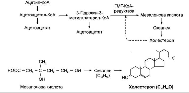
Fig. 17-32. Biosynthesis of cholesterol. Six molecules of mevalonic acid condense to form squalene, which is then hydroxylated and converted into cholesterol. The dashed arrow indicates feedback inhibition by cholesterol of HMG-CoA reductase, the enzyme that catalyzes the formation of mevalonic acid.
Plasma cholesterol levels increase during food intake and in certain conditions, including familial hypercholesterolemia caused by various Mutations in the LDL receptor. When examining patients, alongside cholesterol levels, it is also valuable to analyze plasma LDL and HDL levels. Individuals with elevated LDL levels experience a higher-than-normal rate of disease and complications, whereas those with elevated HDL levels show the opposite trend. Interestingly, women—who have a lower incidence of myocardial infarction than men—exhibit higher HDL levels. Moreover, HDL levels increase in physically trained individuals and those who consume one or two alcoholic drinks (100 mL) daily, while they decrease in smokers, obese individuals, and those with a sedentary lifestyle. Moderate alcohol consumption reduces the frequency of myocardial infarctions, whereas obesity and smoking are risk factors that increase it.
There is evidence that elevated levels of IDL, chylomicron remnants, and triglycerides also lead to atherosclerosis, whereas elevated levels of VLDL and chylomicrons do not. Other factors contributing to atherosclerosis are discussed in Chapter 32.
Animals deprived of dietary fats cease to grow, develop Skin and Kidney lesions, and become infertile. Supplementing their diet with linolenic, linoleic, and arachidonic acids cures all deficiency symptoms. These three polyunsaturated fatty acids are termed essential fatty acids due to their physiological effects. Similar deficiency symptoms have not been definitively documented in humans, but there is reason to believe that certain unsaturated fats are essential dietary components, particularly in children. Although the body can dehydrogenate fats, it cannot synthesize carbon chains with the double-bond arrangement characteristic of essential fatty acids.
One reason—and perhaps the primary one—why essential fatty acids are required for health is that they serve as precursors for Prostaglandins, prostacyclins, thromboxanes, lipoxins, Leukotrienes, and related compounds (Fig. 17-33). These compounds are called eicosanoids because they are derived from a 20-carbon (eicosa-) polyunsaturated fatty acid, arachidonic acid (arachidonate), as well as 20-carbon derivatives of linoleic and linolenic acids. Notably, they are formed from arachidonic acid via three distinct groups of enzymes (see Fig. 17-33).
Prostaglandins are a family of 20-carbon Unsaturated fatty acids containing a cyclopentane ring. Although initially isolated from the Prostate Gland, they are now known to be synthesized in most, if not all, body organs. The structures of several of them are shown in Fig. 17-34.
Prostaglandins (PGs) are divided into groups—such as PGE and PGF—based on the configuration of the cyclopentane ring. The number of unsaturated bonds in the side chains is indicated by a subscript, for example, the group E prostaglandin shown in Fig. 17-34 is prostaglandin PGE2.

Fig. 17-33. Major biologically active compounds derived from arachidonic acid; HETE, hydroxyeicosatetraenoic acid; DiHETE, dihydroxyeicosatetraenoic acid; EET, epoxyeicosatrienoic acid.
The precursor for various other prostaglandins, thromboxanes, and prostacyclin is prostaglandin H2 (PGH2). Various enzymes are involved in this synthesis (see Fig. 17-34). PGH2 is produced from arachidonic acid by cyclooxygenases (COX). There are two isoforms of cyclooxygenase encoded by different genes: cyclooxygenase-1 (COX-1) and cyclooxygenase-2 (COX-2). COX-1 is constitutively expressed, whereas COX-2 is induced by growth factors, cytokines, and tumor promoters.
Known prostaglandin receptors are serpentine receptors acting via heterotrimeric G proteins. Four PGE2 receptors have been isolated and designated EP 1 through EP 4 (Table 17-6), alongside prostacyclin and PGF2α receptors.
The effects of prostaglandins are diverse. Many of them are described in chapters dedicated to the specific systems in which they play a prominent role. They are particularly crucial in the FEMALE REPRODUCTIVE CYCLE, parturition, The Cardiovascular system, inflammatory responses, and pain signaling. Emerging evidence suggests they also participate in carcinogenesis, The regulation of apoptosis, and angiogenesis. Platelets synthesize thromboxane A2, which induces platelet aggregation and acts as a vasoconstrictor. The thromboxane A receptor is a typical G protein-coupled serpentine receptor (see Table 17-6); it acts via phosphatidylinositol to open Ca2+-activated Cl- channels. Thromboxane B2 is a metabolite of thromboxane A2. Prostacyclin, by contrast, inhibits platelet aggregation and acts as a vasodilator. It is produced by endothelial and smooth muscle cells in blood vessel walls. The release of thromboxane A2 into the blood by platelets at the site of a vascular injury triggers thrombus formation, whereas the release of prostacyclin in adjacent vessel areas serves to localize the thrombus and keep other vascular segments patent. Because the inhibitory effect of aspirin on platelets lasts longer than its effect on blood vessel walls, it exerts a valuable anticoagulant effect for the Prevention of myocardial complications and stroke (see Chapter 31).

Fig. 17-34. Metabolism of arachidonic acid mediated by cyclooxygenase-1 (COX-1) and cyclooxygenase-2 (COX-2).
Arachidonic acid is also converted into 5-hydroperoxyeicosatetraenoic acid (5-HPETE) via 5-lipoxygenase, which is involved in the activation of 5-lipoxygenase-activating protein (FLAP). 5-HPETE is subsequently converted into leukotrienes (LTs). Four of these leukotrienes are aminolipids containing amino acids: leukotriene C4 (LTC4) contains the tripeptide Glutathione, LTD4 contains Glycine and cysteine, LTE4 contains cysteine, and LTF4 contains cysteine and glutamic acid (Fig. 17-35). Additionally, arachidonic acid is converted into lipoxins via 15-HPETE (see Fig. 17-35).
Leukotrienes, thromboxanes, lipoxins, and prostaglandins are referred to as local hormones. They have a short half-life, are inactivated in many different tissues, and typically exert their effects locally in the tissues where they are synthesized.
Anti-inflammatory Steroids, such as cortisol, inhibit the release of arachidonic acid from its phospholipid stores via phospholipase A2, thereby slowing the production of all its derivatives (see Fig. 17-33). Nonsteroidal anti-inflammatory drugs (NSAIDs), such as aspirin and indomethacin, inhibit cyclooxygenases without affecting the lipoxygenase or CYP-enzyme pathways (see Fig. 17-34). Inhibition of COX-1 in experimental animals causes gastrointestinal and renal abnormalities, whereas inhibition of COX-2 relieves pain, inflammation, and fever. Aspirin, indomethacin, and most other clinically available NSAIDs block both COX-1 and COX-2 simultaneously; therefore, developing selective COX-2 inhibitors is crucial for minimizing gastrointestinal and renal side effects when treating pain, inflammation, and Arthritis.
Leukotrienes serve as key Transmitters in allergic responses and inflammation. Their release is triggered by the binding of specific allergens to IgE Antibodies On the surface of mast cells. Leukotrienes cause bronchoconstriction, constrict arterioles, increase vascular permeability, and recruit neutrophils and eosinophils to sites of inflammation. Knockout mice with a disrupted 5-lipoxygenase gene exhibit normal development and overall health, yet show resistance to certain forms of inflammation. In humans, asthma, psoriasis, adult respiratory distress syndrome, allergic rhinitis, Crohn's disease, and Ulcerative Colitis have been linked to the pathway involving this gene.
Table 17-6. Prostaglandin and Thromboxane Receptors
PGI2 |
IP |
PGE2 |
EP 1, EP2, EP3, EP 4 |
PGF2a |
FP |
Thromboxane A |
TP |
Two cysteinyl leukotriene receptors, CysLT1 and CysLT2, have been characterized pharmacologically, although their structures remain unknown. The leukotriene B4 receptor, BLT, is a G protein-coupled serpentine receptor. The CysLT1 receptor mediates bronchoconstriction, chemotaxis, and increased vascular permeability; the CysLT2 receptor mediates pulmonary vascular smooth Muscle contraction; and the BLT receptor primarily mediates chemotaxis. The involvement of these receptors in asthma is discussed in Chapter 37.
Lipoxin A causes vasodilation of small blood vessels, and both lipoxin A and lipoxin B attenuate the cytotoxic activity of natural killer cells (see Chapter 27). However, their precise physiological significance remains unclear.
It should be noted that 12-HETE, several dihydroxy derivatives of eicosatetraenoic acid (DHETs), and several epoxyeicosatrienoic acids (EETs) are produced from arachidonic acid by cytochrome P450 (CYP) Monooxygenases (see Fig. 17-33). While the precise roles of these products are not fully understood, DHETs and EETs influence renal salt and water excretion, which may be of significant physiological importance. Cytochrome P450 is a remarkable superfamily of over 300 enzymes that catalyze oxidation, epoxidation, aliphatic hydroxylation, and other reactions. In mammals, they are involved not only in eicosanoid metabolism but also in steroid hormone synthesis, drug metabolism, and fatty acid oxidation. They are classified into families and subfamilies based on Sequence Homology (CYP1, CYP2, CYP3, etc.), with 12 families identified in humans.
Obesity
Obesity is the most common and costly nutritional disorder in developed countries. It currently affects 33% of the adult population and continues to spread. It is associated with an increased risk of atherosclerosis, diabetes, and Gallbladder disease. Normal body fat reserves account for 12–18% of total body weight in men and 18–25% in women. Obesity is diagnosed when these values exceed 20% in men and 25% in women. Standard height and weight charts are also widely used. However, the metric most strongly correlated with body fat is the body mass index (BMI), which is calculated as body weight (in kilograms) divided by height squared (in meters). The normal BMI range is 20–25 kg/m2. Body weight increases and stabilizes during the third decade of life, rises slightly thereafter, and tends to decrease in advanced age. If caloric intake is not reduced with age, obesity develops. Furthermore, basal metabolic rate declines with age.
In humans, obesity has a strong genetic component, but it is also heavily influenced by environmental factors. For instance, in the United States, obesity is significantly less prevalent among women in higher socioeconomic groups than among those in lower groups. The relationship between obesity and diabetes is discussed in Chapter 19, and the control of food intake is covered in Chapter 14.
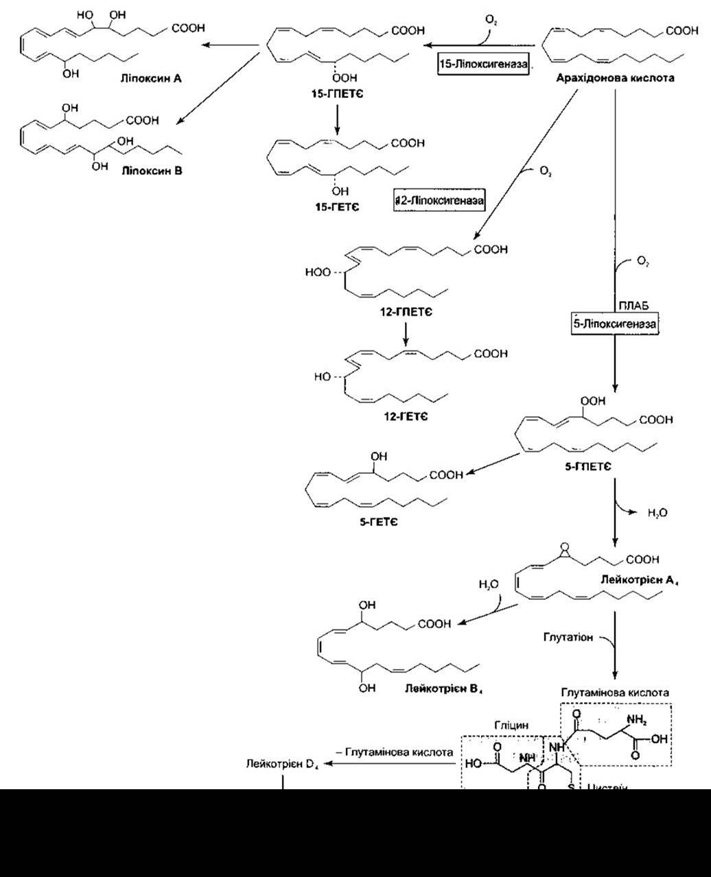
Fig. 17-35. Metabolism of arachidonic acid by lipoxygenases; FLAP — 5-lipoxygenase-activating protein.
Last update: 10/08/2026
Editorial and Educational Adaptation: This material has been compiled based on the primary/original source text. The project team performed an editorial review, corrected technical inaccuracies, structured sections, and adapted the content for an educational format.
What was processed:
- elimination of formatting defects (OCR errors, structural breaks, corrupted characters);
- editorial organization of content;
- standardization of terminology in accordance with academic sources;
- verification of factual statements against the original source text.
All mentions of the author, publication year, and origin of the primary text have been preserved in accordance with the source.