Biochemistry: The Chemical Reactions of Living Cells, Volume 2 - D. Metzler 1980
How electrons encounter oxygen, how ATP is formed in the process, and related phenomena.
Oxygenases and hydroxylases
Monooxygenases
Two classes of monooxygenases are known. Enzymes that require a cosubstrate [BH2 in equation (10-44)] In addition to the hydroxylated substrate are called external monooxygenases. The other group comprises internal monooxygenases, in which a part of the hydroxylated substrate's Structure acts as the cosubstrate. Many internal monooxygenases contain flavin Cofactors and lack metals.
a. Flavin-containing monooxygenases
First, recall that dihydroflavin reacts with O2 to form H2O2 (Ch. 8, Sec. I,7). Based on this fact, one might expect that monooxygenases catalyze the dehydrogenation of substrates using flavin cofactors, and that reduced flavins react with O2 to form H2O2. The resulting hydrogen peroxide is then used as a hydroxylating agent. A corresponding example has already been described in Ch. 8. Lactate oxygenase can convert lactate to Pyruvate via dehydrogenation and then oxidatively decarboxylate pyruvate to acetate using H2O2 [equation (8-67)]. One oxygen atom is transferred from O2 to the resulting acetate [139].
Another internal monooxygenase is Lysine oxygenase, a tetrameric FAD-containing protein composed of subunits with a Molecular Weight of ~61,000 [140]. Similar types of monooxygenases produced by Bacteria attack Arginine and other basic Amino Acids. Again, judging by the products formed [equation (10-49)], the removal of hydrogens is followed here by oxidative decarboxylation under the action of H2O2, just as in equation (8-67).
Class="center">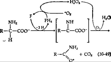
Following the Treatment of native lysine monooxygenase with various sulfhydryl-blocking Reagents, the modified enzyme yields an α-keto acid, ammonia, and H2O2 as products. These are precisely the products whose formation could have been predicted beforehand, assuming the hydrolytic Cleavage of the intermediate enclosed in parentheses in equation (10-49).
One way in which NADPH can be used as a cosubstrate is through the reduction of a flavoprotein enzyme. The reduced flavin can then react with O2 to form the hydroxylating agent. An example is 4-hydroxybenzoate hydroxylase, an enzyme that produces 3,4-dihydroxybenzoate as a product. NADH reacts only after the flavoprotein forms a complex with the substrate—evidence suggests that an oxygenated intermediate is formed in the process, possibly a hydroperoxide, as indicated in equation (10-50) (see also Ch. 8, Sec. I,7) [141].
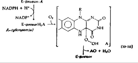
According to this mechanism, one of the two oxygen atoms of the hydroperoxide reacts with the aromatic substrate as efficiently as OH+ or a peroxy radical. Another suggestion [142] is that H2O can be eliminated from the hydroperoxide shown in equation (10-50) to form an oxaziridine, which can act as an active hydroxylating agent [equation (10-51)].
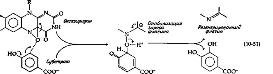
Attack by the oxaziridine on the nucleophilic center of the substrate will lead to The formation of the adduct shown. Subsequent elimination and tautomerization yield the product. An epoxide may also be formed as the initial product [equation (10-54)].
b. Reduced pteridines as cosubstrates
During the functioning of one group of hydroxylases, which includes phenylalanine hydroxylase from human Liver, the dihydro form of biopterin (p. 277) serves as a cosubstrate and is reduced by NADPH [equation (10-52)].
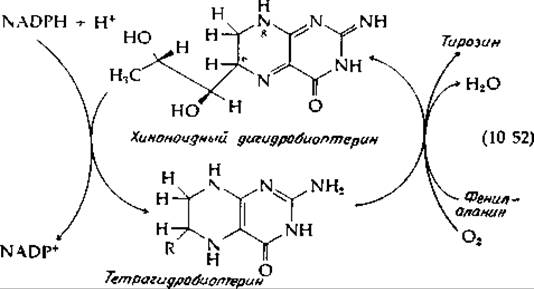
The resulting tetrahydrobiopterin is structurally similar to reduced flavin. The Mechanism of its interaction with O2 and the hydroxylation of phenylalanine to Tyrosine is likely fundamentally the same as in the case of 4-hydroxybenzoate hydroxylase. Dihydrobiopterin can exist as a series of isomers. The quinonoid form, represented in equation (10-52), is a tautomer of 7,8-dihydrobiopterin—this form is produced with the participation of Dihydrofolate Reductase (Ch. 8, Sec. L,2). The reader may perhaps be able to determine for themselves what arguments support the structure depicted in equation (10-52) [143, 144]. The reaction on the left side of equation (10-52) is catalyzed by pyridine nucleotide-dependent dihydropteridine reductase [145].
Phenylalanine hydroxylase is of particular interest because its absence is associated with one of the most thoroughly studied inherited biochemical disorders—phenylketonuria (Ch. 14, Sec. 3,5). Other pteridine-dependent hydroxylases are also known. For example, Brain Tryptophan hydroxylase produces 5-hydroxytryptophan, which is the first step in the Synthesis of the neurotransmitter 5-hydroxytryptamine (Ch. 16, Sec. B,4) [146].
c. Hydroxylation-induced migration
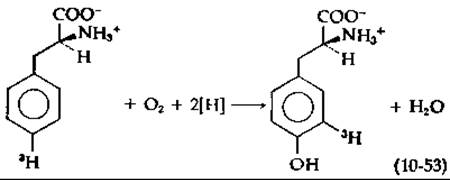
The general outcome of the enzymatic hydroxylation of Aromatic Compounds is the intramolecular migration of a hydrogen atom, a substituent atom, or a group [equation (10-53)] [147]. Termed the NIH shift (named after the researchers who discovered it at the National Institutes of Health laboratories), this migration provides insight into the possible mechanisms of hydroxylation. In equation (10-53), The addition of a hydroxyl group causes the displacement of a tritium atom. This migration can be visualized as the result of an electrophilic attack on the aromatic system, for instance, by an OH+ ion [equation (10-54)];
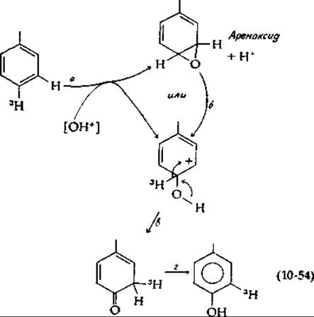
Such an attack can result in the formation of an epoxide (an arenoxide, step a) or directly a carbonium ion with the structure depicted in equation (10-54). Arenoxides have been prepared and shown to convert (likely via a carbonium ion intermediate, step b) into the final products while exhibiting the NIH shift [148, 149]. Thus, either Carbonium ions or arenoxides are logically the initial products of attack on aromatic substrates. These results suggest that the hydroxylating agent is electrophilic in nature—a property apparently shared by the previously discussed hydroperoxides, superoxide anions, oxaziridine intermediates, and the O2 molecule coordinated to a metal ion.
An example of an NIH shift involving the migration of a larger substituent is the hydroxylation of p-hydroxyphenylpyruvic acid [equation (10-55)], a key step in tyrosine METABOLISM.
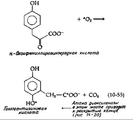
This reaction should be compared with the reaction given in equation (8-67). Undoubtedly, both proceed via a similar mechanism. In the first step, activated oxygen may attach first to the carbonyl group, and then the other end of the O2 molecule attaches to the aromatic ring via an electrophilic attack. Note that this attack is facilitated by the electron-donating Properties of the p-hydroxyl group. Subsequent decarboxylation cleaves the O—O bond, just as in equation (8-67). Simultaneously, as in equation (10-54), a hydroxylated carbonium ion is formed, in which the NIH shift takes place. Although the enzyme in question is a dioxygenase, its MECHANISM OF ACTION appears similar to that of monooxygenases.
d. α-Ketoglutarate as a decarboxylated cosubstrate
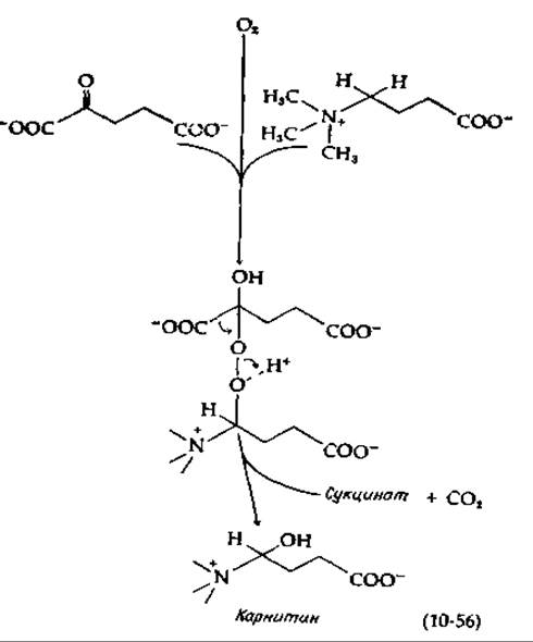
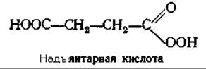
There is a group of monooxygenases capable of accepting hydrogen atoms from $\alpha$-ketoglutarate, which is decarboxylated in this process to succinate. Two of these enzymes are involved in the hydroxylation of lysine and Proline residues in the Collagen precursor, procollagen (Ch. 11, Sec. D,3). Another enzyme hydroxylates $\gamma$-butyrobetaine, converting it into carnitine (Ch. 9, Sec. A,6). All these enzymes contain Fe(II). The mechanism is likely similar to the one given in equation (10-55) [150, 151]: on the one hand, activated oxygen adds to the carbonyl group of $\alpha$-ketobutyrate (nucleophilic addition), and on the other hand, it carries out an electrophilic attack on the hydroxylated substrate [equation (10-56)]:
Another pathway is also possible: metal-bound oxygen can react with ketoglutarate, leading to its decarboxylation into persuccinic acid, which can act as a hydroxylating agent [151a]:
d. Ascorbic Acid as a Cosubstrate
Many hydroxylases require the presence of a reducing agent. Ascorbic acid (Vitamin C; see Box 10-F) is most frequently the most effective in this regard. This holds true for all the α-ketoglutarate-dependent hydroxylases discussed in the previous section. Ascorbic acid apparently serves to maintain the metal-containing catalyst in a reduced state. However, the enzyme dopamine-β-hydroxylase utilizes ascorbic acid as a true cosubstrate in the synthesis of norepinephrine [noradrenaline; Equation (10-57)] [152].
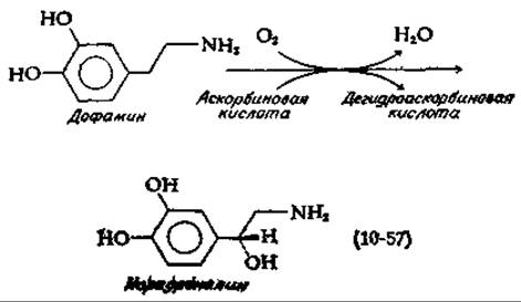
This reaction, which plays a crucial role in brain Neurons, also proceeds actively in the Adrenal Glands. It has long been known that the adrenals are particularly rich in ascorbic acid. The structure of the oxidized form of vitamin C, dehydroascorbic acid, is shown in Appendix 10-F. Dopamine β-hydroxylase contains several copper atoms, and it is believed that ascorbic acid reduces two copper atoms from the +2 to the +1 state. O2 then binds—presumably in a manner similar to its binding to hemocyanin (Sec. B,4). This results in the formation of a metal-coordinated O2-2 ion species, which is utilized in the hydroxylation reaction. It should also be mentioned that another copper-containing enzyme (ascorbic acid oxidase; Appendix 10-Z) acts on the vitamin C molecule.
Appendix 10-F
Vitamin C: ascorbic acid
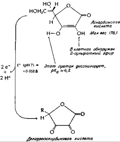
In ancient sailors suffering from scurvy, hemorrhages in the Skin, Gums, and joints served as precursors of imminent death. Although around the turn of the 18th century it was observed that the progression of the disease could be halted by eating citrus fruits, another 200 years passed before attempts were made to isolate vitamin C. Ascorbic acid was first obtained in crystalline form around 1930. Szent-Györgyia gives a very vivid and fascinating account of these investigations.
Among mammals, only humans and guinea pigs must obtain ascorbic acid from their diet; other species are able to synthesize it themselves. The requirement for vitamin C is high compared to other Vitamins. To prevent scurvy, 10 mg of the vitamin per day is required; however, even at this intake level, certain clinical symptoms are observed, such as fragility of small skin capillaries. In various "official" recommendations, the adopted dose of vitamin C ranges from 30 to 70 mg/day. Intense debate has recently surrounded Linus Pauling's recommendations to consume 0.25–10 g of ascorbic acid per dayb. Pauling and his followers maintain the view that ascorbic acid exerts a specific effect by preventing the common cold or mitigating its courseв. On the other hand, critics hold the opinion that high doses of this seemingly harmless compound may lead to obscure and dangerous side effects. Although ascorbic acid possesses antioxidant properties, in the presence of ferric ions it promotes the formation of free radicals, an excessively high content of which can lead to adverse consequences.
The biochemical Functions of ascorbic acid are not yet fully understood. Aside from its well-established reducing properties and its facile oxidation to dehydroascorbic acid (structure shown on p. 442), it is also known to be a weak acid capable of forming complexes with metals. Ascorbic acid is present in very high concentrations in the adrenal glands, where one of its functions may be to serve as a cosubstrate for dopamine-β-hydroxylase [equation (10-57)]. Ascorbic acid is likely involved in the hydroxylation of procollagenв (sec. J,2,d), and it is possible that the Prevention of the common cold is precisely due to enhanced collagen hydroxylation. Experiments with guinea pigs have shown that high levels of ascorbic acid result in more rapid wound healingг,д.
Ascorbic acid, together with Fe(II) and O2, serves as a potent non-enzymatic hydroxylating reagent for aromatic compoundsе,ж. Like hydroxylases, this reagent attacks nucleophilic sites (for example, in The conversion of phenylalanine to tyrosine). 18O2 oxygen atoms are incorporated into the hydroxylated products. Although H2O2 is formed in the reaction mixture, it cannot replace ascorbate. However, it remains unclear what bearing this system has on the biochemical functions of ascorbate.
а Szent-Gyоrgyi A., Annu. Rev. Biochem., 32, 1–14 (1963).
б Pauling L., Vitamin C and the Common Cold, Freeman, San Francisco, California, 1970
в Barnes M. J., Kodicek E., Vitam. Horm. (N. Y.), 30, 1–43 (1972).
г Yew M.-L. S., PNAS, 70, 969–972 (1973).
д Harwood R., Grant M. E., Jackson D. S., BJ, 142, 641–651 (1974).
е Ullrich V., Staudinger H., Block K., Hayaishi O., eds., Biological and Chemical aspects of Oxygenases, pp. 235–249, Maruzen, Tokyo, 1966.
ж Hamilton G. A., Workman R. J., Woo L., JACS, 86, 3391–3392 (1964).
e. Cytochrome P-450-catalyzed hydroxylation
The final class of hydroxylases is formed by Hemoproteins generally designated as cytochrome P-4501). These Proteins are involved in the hydroxylation of Steroids [153] and alkanes [154, 155], as well as the hydroxylation of the methylene group of camphor [156]. They also participate in the metabolism of various drug compounds [135, 157, 158]. The corresponding reactions are characterized by the following features: the hydroxyl group is introduced without inversion of configuration [equation (10-58)].
1) Cytochrome P-450 is so named because in its reduced form it forms a pigment whose carbon monoxide complex absorbs at 450 nm.
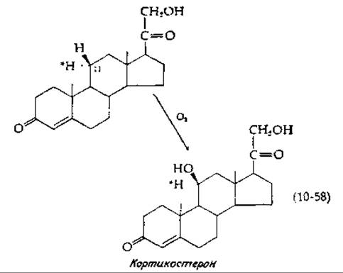
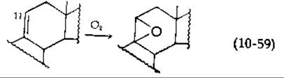
This equation illustrates the 11ß-hydroxylation of a steroid, a vital step in The Biosynthesis of Steroid Hormones. Another characteristic feature of the 11ß-hydroxylase system is that the same enzyme catalyzing the reaction described by equation (10-58) converts an unsaturated derivative into an epoxide [equation (10-59)].
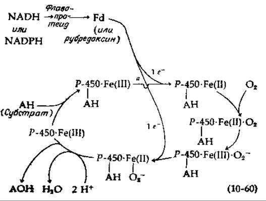
The functioning of cytochrome P-450 hydroxylases involves The transfer of electrons from NADH or NADPH through a flavoprotein to ferredoxin (Fd) or rubredoxin. Apparently, one of these latter Non-heme iron proteins subsequently reduces the iron within the substrate-cytochrome P-450 complex from the Fe (III) state to the Fe (II) state [equation (10-60), reaction a]. Oxygen then binds to the ferrous iron, oxidizing it to the ferric state. At this point, an additional electron enters from the Electron Transport Chain. The coordinatively bound oxygen O-2 or O2-2~ attacks the substrate, and cytochrome P-450 is released in the Fe (III) state. Although the detailed mechanism of this process remains unknown, a very similar reaction sequence must operate in all such cases.
Several forms of cytochrome P-450 from mammalian liver are known [135]. All of them are tightly bound to the membranes of the Endoplasmic reticulum and are difficult to solubilize. Unlike Other Enzymes, these do not require a rubredoxin-like protein as an intermediate carrier, as they can react directly with the flavoprotein. An interesting feature of these enzymes is their inducibility. The administration of various drugs, such as phenobarbital and many Other Compounds, can trigger up to a 20-fold increase in cytochrome P-450 activity. Aromatic Hydrocarbons induce a hydroxylase distinct from the one induced by barbiturates. The Role of cytochrome P-450 is often to convert a drug or other foreign compound into a readily excretory form. However, the outcome is not always beneficial; for instance, 3-methylcholanthrene, a potent inducer of cytochrome P-450, is converted via hydroxylation into a powerful carcinogen [159].
Addendum 10-3
Copper-Containing Proteins
Despite the diverse BIOLOGICAL FUNCTIONS OF copper, it was not recognized as an essential dietary component until 1924. Copper is so widely distributed in foods that human copper deficiency has virtually never been reported. Deficiency is occasionally observed in animals—sometimes because Zn2+ inhibits Cu2+ absorption, and sometimes due to the binding of copper by molybdate into an inert complex. There are copper-deficient desert regions in Australia where neither plants nor animals can survive. Animals suffering from copper deficiency develop skeletal abnormalities, Hair depigmentation, and impaired Hemoglobin synthesis. Cytochrome oxidase activity in these animals is very low. Furthermore, the Elastin protein in arterial walls exhibits poor cross-linking, rendering the Arteries fragile.
An adult human ingests approximately ~2–5 mg of copper per day, of which about 30% is absorbed. The total copper content in The Human Body is roughly ~100 mg, with both absorption and excretion (via Bile) being tightly regulated. Because excess copper is toxic, this regulation is critical. Wilson's disease is a well-known pathological condition characterized by the accumulation of copper in The Liver and brain. The normal copper concentration in these Tissues is about ~10-4 g-atom/L. For most chelate centers, the divalent copper ion exhibits the strongest metal-ion binding affinity (Table 4-2), which explains why copper in living Cells exists almost entirely in complexed forms with proteins.
Copper ions form the active centers of A wide variety of catalystsa-g. Like iron, the copper ion serves as a site for interaction with O2. Its ability to undergo reversible reduction enables it to function in various oxidation-reduction processes. The simplest function of copper proteins—akin to cytochrome c—is acting as a single-electron carrier. Bacterial azurins are bright blue, low-molecular-weight proteins believed to function in Electron Transport Chains. The "blue protein" from Pseudomonas aeruginosa has a molecular weight of 16,300 and contains a single Cu2+ ion per molecule. The absorption spectrum of this bright blue protein is shown in Fig. 13-8. The blue color is characteristic of the Cu2+ ion; it is observed in the hydrated Cu(H2O2+4 ion and even more intensely in the Cu(NH3)2+4 ion. This color, arising from an internal d-d electron transition within the copper atom, is further intensified in copper peptide chelates, one of which is depicted in equation (4-38). In these striking blue proteins, the intensity of the (d-d) absorption bands is an order of magnitude higher than in simple compounds—a phenomenon not yet fully understood. Coordination with one or more sulfur atoms from Methionine side chains remains a distinct possibilityd.
Low-molecular-weight blue proteins include stellacyanin, a copper-containing mucoprotein from the Japanese lacquer treee. Composed of 108 amino acid residues, this peptide contains 20% carbohydrate and a single copper atom. Plastocyanin, initially isolated from the alga Chlorella, was subsequently found in all green plants. It is believed to function in The electron transport chain connecting the two light-absorbing Photosystems (Chapter 13, Section D, 6).
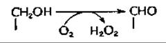
Most copper-containing proteins react with O2. Sometimes this reaction is reversible, as in the case of the oxygen carrier hemocyanin (Chapter 10, Section B, 4). More frequently, however, oxygen enters into a chemical reaction in an "activated" state. One group of enzymes couples the dehydrogenation of organic substrates with the reduction of oxygen to H2O2. For instance, galactose oxidase (from Polyporus) catalyzes the Conversion of the 6-hydroxymethyl group of galactose into an aldehyde groupf,g:
The large polypeptide chain of this enzyme, with a molecular weight of 68,000, contains a single copper atom. Unlike the blue proteins, galactose oxidase has a dark green tint. Although neither oxygen nor galactose alone alters the enzyme's spectrum, their simultaneous presence induces a spectral change. It is hypothesized that galactose and O2 bind to copper, which may cycle between the Cu(I) and Cu(III) states during this process. A similar mechanism likely operates in tyrosinase (see below)g. Galactose oxidase has been utilized to modify Glycoproteins on the outer Cell membrane surface. Exposed galactosyl or N-acetylgalactosaminyl residues are oxidized by the enzyme to the corresponding C6-aldehydes, which are then mildly reduced using tritiated sodium borohydridee.
Amino Oxidases containing both Cu2+ and Flavin Coenzymes are functionally similar to amino acid oxidases (Table 8-4). One such amino oxidase converts ε-amino groups of lysine side chains into aldehyde groups in Collagen and elastin (Chapter 11, Section D, 3). Another copper-containing enzyme is urate oxidase, which induces the decarboxylation of its substrate (Fig. 14-33).
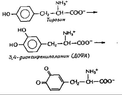
Certain copper-containing enzymes, such as dopamine-ß-hydroxylase [equation (10-57)], are typical hydroxylases. Among the copper-dependent hydroxylases, tyrosinase (also known as polyphenol oxidase) is widely distributed; it catalyzes a two-step reaction involving hydroxylation followed by dehydrogenation. First identified in Fungi, this enzyme has also been isolated from the Japanese lacquer tree. It is abundant in plant tissues, where it causes the browning of bruised or harvested fruit. In animals, tyrosinase participates in the synthesis of dihydroxyphenylalanine (DOPA) and the Formation of the dark skin and hair pigment, melanin. A deficiency or blockage of this enzyme in melanocytes (melanin-producing cells) results in albinism. Tyrosinase from the fungus Polyporus has a molecular weight of ~120,000 and consists of four subunits, each containing one Cu(I) atom.
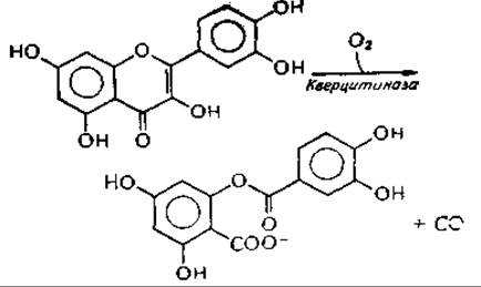
An unusual reaction is catalyzed by the copper-containing quercetinase from Aspergillus flavus.
This enzyme is classified as an oxygenase. The reaction leads to the formation of CO; the most probable mechanism involves the formation of a peroxide intermediateh,i.
Blue copper-containing oxidases are unusual catalysts in that they can reduce both atoms of molecular oxygen to H2O. In this respect, they resemble cytochrome oxidase (which also contains copper; Section B, 5), but they lack iron. Ascorbic acid oxidase from plant tissues converts ascorbate into dehydroascorbate (Addendum 10-J). Upon adding a substrate to this enzyme, its blue color fades, demonstrating that the copper is reduced to the (+1) state. Laccase, derived from Japanese lacquer tree sap or the fungus Polyporus, catalyzes substrate transformations similar to those of tyrosinase, but yields H2O instead of hydrogen peroxide. Unlike tyrosinase, the Polyporus enzyme is insensitive to CO and exhibits a molar extinction coefficient at 610 nm >1,000. Laccase has been shown to contain at least Three types of copper ions. One is blue, resembling the copper site in azurins, and binds oxygen. Another copper ion is non-blue and likely serves as an anion-binding center—a function possibly required to stabilize intermediate peroxide species. The remaining two Cu2+ ions form a diamagnetic pair that acts as a two-electron acceptor, acquiring electrons from the substrate and subsequently transferring them to oxygen, presumably via a peroxide intermediate.
Ceruloplasmin is a blue protein with a molecular weight of ~150,000, containing 8 Cu+ and 8 Cu2+ ions. As the primary copper-binding protein in Blood, it accounts for 3% of the body's total copper content. Ceruloplasmin appears to be involved in the systemic regulation of copper Homeostasis; for instance, its levels are depressed in copper storage disorders such as Wilson's disease. Furthermore, ceruloplasmin possesses enzymatic activity reminiscent of laccase, as it can also catalyze The oxidation of Fe2+ to Fe3+. This latter reaction is physiologically crucial because only Fe3+ can bind to the iron-transport protein transferrin (Addendum 14-D). Consequently, ceruloplasmin is sometimes referred to as ferroxidase.
A widely distributed group of proteins includes erythrocuprein, cerebrocuprein, etc., which were initially regarded merely as copper storage proteins. However, recent years have revealed that these proteins function as superoxide dismutases [equation (8-61)]. In eukaryotic Cytoplasm, these enzymes exist as dimers with a molecular weight of 31,300; each subunit contains one copper atom and one zinc atom. It is proposed that the copper is coordinated to three imidazole nitrogen atoms. Presumably, it undergoes sequential Oxidation and reduction during reactions with superoxide radicals. The crystal structure of this enzyme has been solved, which will soon provide a more detailed picture of the copper coordination environmentj.
In addition to these copper-zinc superoxide dismutases, Mitochondria and many bacteria contain manganese-dependent enzymes that perform the identical function (Addendum 13-A). E. coli cells possess both manganese- and iron-containing superoxide dismutasesk.
A cytoplasmic copper-binding proteinр has also been described, which resembles metallothionein (Supplement 7-E) both in its high Cysteine content and in several other properties.
а Frieden Е., Sсi. Am., 218, 103—114 (1968).
б O’Dell В. L., Campbell В. J., Compr. Biochem., 21, 191—203 (1971).
в Peisach J., Aisen P., Blumberg W. E., eds., Biochemistry of Copper, Academic Press, New York, 1966.
г Malkin P., Malmström В. G., Adv. Enzymol., 33, 177—244 (1970).
д Jones T. E., Rorabacher D. B., Ochrymowycz L. A., JACS, 97, 7485—7486 (1975).
е Peisach J., Levine W. G., Blumberg W. E., JBC, 242, 2847—2858 (1967).
ж Etlinger M. J., Kosman D. J., Biochemistry, 13, 1247—1251 (1974).
з Dyrkacz G. D., Libby R. D., Hamilton G. H., JACS, 98, 626—628 (1976).
и Gahmberg C. G., JBC, 251, 510—515 (1976).
к Krishnamurty H. G., Simpson F. J., JBC, 245, 1467—1471 (1970).
л Vanneste W. H., Zuberbühler A. In: MOLECULAR MECHANISMS OF Oxygen Activation (O. Hayaishi, ed.), pp. 371—404, Academic Press, New York, 1974.
м Fee J. A., Struct. Bonding (Berlin), 23, 1—60 (1975).
н Weser U., Struct. Bonding (Berlin), 17, 1—65 (1973).
о Thomas K. A., Rubin В. H., Bier C. J., Richardson J. S., RichardsonD. C., JBC, 249, 5677—5683 (1974).
п Yost F. J., Jr., Fridovich I., JBC, 248, 4905—4908 (1973).
p Wingle D. R., Premakumar R., Wiley R. D., Rajagopalan К. V., ABB, 170, 253—266 (1975).
Last update: 06/08/2026
Editorial and Educational Adaptation: This material has been compiled based on the primary/original source text. The project team performed an editorial review, corrected technical inaccuracies, structured sections, and adapted the content for an educational format.
What was processed:
- elimination of formatting defects (OCR errors, structural breaks, corrupted characters);
- editorial organization of content;
- standardization of terminology in accordance with academic sources;
- verification of factual statements against the original source text.
All mentions of the author, publication year, and origin of the primary text have been preserved in accordance with the source.