Biochemistry - Chemical Reactions in Living Cells, Volume 3 - D. Metzler 1980
Light in Biology
Photosynthesis
Electron Transport Chains in Chloroplasts
Let us now take a closer look at the processes involving the reduced electron acceptor A- [Eq. (13-26)] as well as the processes associated with the return of the electron to oxidized Chl+. The electrode potential of the P700 pigment, which is part of Photosystem I in METABOLISM/14.html">Chloroplasts (Fig. 13-18), is +0.43 V. A light quantum with a wavelength of 700 nm carries enough energy (1.77 eV) to photoreduce a carrier whose potential is 1.77 V more negative, i.e., -1.3 V. However, the probability of forming such a powerful reducing agent is very low. First, the capture of light energy cannot proceed with 100% efficiency1). If the photochemical reaction goes through a triplet state, a significant portion of the absorbed energy is lost. Furthermore, no carriers are known that possess such a negative redox potential. In addition, we do not know the electrode potential of the hypothetical carrier Z. Most authors estimate it to be from ~-0.55 to -0.6 V, which is close to the potential of ferredoxin; hence, it has been suggested that Z represents a bound ferredoxin molecule. According to more recent data, a special Fe-S protein acts in this role [99]. Be that as it may, it is clear that Z is capable of rapidly reducing ferredoxin, possibly with the participation of as-yet-unknown electron-carrying Proteins. Reduced chloroplast ferredoxin, being a single-electron carrier (Chap. 10, Sec. B), can transfer electrons via the flavoprotein Fd-NADP reductase to NADP+, resulting in The formation of NADPH.
For green plants, the terminal link of the Electron Transport Chain is clearly established, which cannot be said of Bacteria. In this case, it has not been possible to detect the photochemical synthesis of reduced intermediates with a potential value close to that of ferredoxin. It is believed that the electron acceptor is a quinone (most likely ubiquinone) [102]. Since the E0' of quinone is close to zero, the reduction of NADP+ requires The Use of a "reversed electron flow" driven by ATP. This flow reversal could occur through The oxidation of half of the photoreduced quinone in The electron transport chain by the oxidized Chl+ of reaction centers, which is coupled with ATP synthesis (cyclic Photophosphorylation).
In chloroplasts, the copper-containing protein plastocyanin (PC) can be considered a candidate for The Role of the donor that transfers electrons directly to P700 and occupies a position in the electron transport chain running from Photosystem II to photosystem I (Fig. 13-18). The important role of plastocyanin was revealed in studies with Scenedesmus Cells; under copper deficiency (Figs. 1-9), these cells are unable to photoreduce CO2 using H2, but the Hill reaction proceeds in them at a normal rate. At the other end of the chain is Q, the electron acceptor of photosystem II. Q stands for quencher; this acceptor quenches the fluorescence of P682 chlorophyll, i.e., chlorophyll a contained in the reaction center of photosystem II. Irradiation of chloroplasts with light of 650 nm wavelength activates photosystem II, but not photosystem I. Under these conditions, Q finds itself in a reduced state, and an increase in Chl* fluorescence is observed, which is probably due to the absence of the electron acceptor Q. If, however, photosystem I is activated by longer-wavelength red light, Q remains predominantly in the oxidized form, which leads to fluorescence quenching.
1) It is usually overlooked that light carries not only energy but also Entropy as well. An important consequence of this fact is that only 78% of the energy of sunlight with a wavelength of 700 nm can be used to perform chemical work (Knox R. S.: Biophys. J., 9, 1351, 1969).
The exact nature of compound Q is unknown, but most researchers believe it to be one of the plastoquinones (PQ). Plastoquinone A, which predominates in spinach chloroplasts, has the Structure shown in Fig. 10-8, with nine isoprenoid units in the side chain. Spinach chloroplasts contain at least six other plastoquinones. Plastoquinones C, hydroxylated at various positions of the side chain, are particularly widespread. In plastoquinone B, these hydroxyl groups are acylated. There are A number of other modifications, including differences in the number of isoprenoid units in the side chains [103, 104]. Thus, several different plastoquinones may function simultaneously in the electron transport chain. According to estimates, there are about five plastoquinone molecules per reaction center, so plastoquinones can serve as a kind of electron buffer between the two Photosynthetic Systems. Consistent with this, Q is thought to be a small pool of plastoquinones associated with the reaction center and separated from the larger pool by a DCMU-inhibited reaction.
It is generally accepted that in the chain between Photosystems II and I There are two electron carriers—cytochrome b559 and cytochrome f (the latter being a c-type cytochrome) [104a]. Although parallel pathways have been suggested [105], most investigators believe the carriers are arranged in the following sequence:
Class="center">
The sequence PQ→cytochrome b559→cytochrome f appears to be very similar to the mitochondrial carrier sequence [106] ubiquinone→cytochrome bT→cytochrome c1, which contains coupling site II for ATP synthesis (Fig. 10-11). As indicated in Fig. 13-18, the corresponding segment of the electron transport chain in chloroplasts is apparently also coupled with ATP synthesis.
The similarity between Cell/39.html">Mitochondrial and Chloroplast electron transport systems became even more evident when it was found that ATP synthesis requires the chloroplast coupling factor CF1, which is similar in properties to the mitochondrial protein F1 (Chap. 10, Sec. D, 8). Like the mitochondrial coupling factor, CF1 consists of subunits of five different types [107, 108]. Similar to Mitochondria, chloroplasts (in the light) also "pump" protons across membranes. However, in this case protons accumulate inside the thylakoids, whereas in mitochondria they are pumped outward. The coupling factor CF1 is located on the outer surface of the thylakoids, facing the stromal matrix, whereas factor F1 is on the inner side of the mitochondrial membrane. It is highly probable that chloroplasts and mitochondria utilize the same Mechanism of ATP synthesis.
It has been suggested that The transport of protons across thylakoid membranes is coupled with the cyclic Oxidation and reduction of plastoquinones (analogous to ubiquinone in mitochondria) and that photosystem II is localized inside the thylakoids. In that case, following the Cleavage of a Water molecule, two protons (one per electron) would remain inside the thylakoid, while the electrons would be driven by light across The Lipid Bilayer to the acceptor Q located on the outside. Similarly, it can be postulated that chlorophyll in photosystem II is localized on the inner side of the bilayer, and acceptor Z on the outside (Fig. 13-18). Since a proton is released during the externally occurring reduction of NADP+ to NADPH, a total of 1.5 protons is pumped for each electron passing through the Z-system [107, 109]. According to the chemiosmotic hypothesis (Chap. 10, Sec. D, 9, c), it is precisely this proton translocation, leading to the generation of a pH gradient and a Membrane Potential, that serves as the source of Free energy required for ATP synthesis.
The pathways by which cyclic photophosphorylation occurs in chloroplasts have not yet been established. It is believed that cytochrome b563 (cytochrome b6) participates in this process, but it remains unclear whether the electrons are directed further to plastoquinones or pass directly to cytochrome f.
Of great interest is the photophosphorylation of inorganic phosphate to pyrophosphate (PPi) carried out by chromatophores from R. rubrum. Evidence indicates that the PPi thus formed is subsequently involved in a number of different energy-dependent reactions taking place in chromatophores [110]. One such example is the formation of NADH via reverse electron flow. The immediate question arises: how can these data be reconciled with the view that PPi is always immediately hydrolyzed (Chap. 11, Sec. B, 2)? It is possible that not only in bacteria, but also in other organisms, PPi is sometimes utilized as an additional energy source.
As for the electron donor in photosystem II, there is compelling Evidence for the direct participation of two to four Mn2+ ions present in each of the reaction centers [111]. Therefore, the chain is often depicted as running from H2O through Mn2+ to P682. A special scheme has been proposed [112] illustrating the direct interaction between oxidized chlorophyll and a water hydroxyl ion coordinated to the metal:
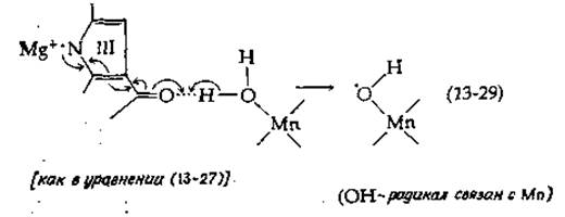
Much like the extremely complex mechanism of O2 reduction by cytochrome oxidase—a component of the electron transport chain in mitochondria (Chap. 10)—the four-electron dehydrogenation of two water molecules to form one O2 molecule in chloroplasts is difficult to represent as a simple scheme. In experiments studying oxygen evolution under periodically repeated light flashes, it was established that this process is based on a four-quantum mechanism in system II [112a]. The oxidizing equivalents at this end of the chain must somehow be stored until their number becomes sufficient for the release of an oxygen molecule. All these events take place at the metalloenzyme center (see Box 13-A), presumably involving two Mn2+ ions. Each Mn2+ ion, upon binding a radical according to scheme (13-29), can be oxidized to Mn3+, thereby supporting a second Light absorption event, which results in the binding of yet another OH radical. Ultimately, the formation of an O2 molecule can be represented schematically as follows:
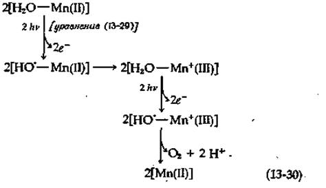
Another aspect associated with electron transfer at this end of the chain is the possible presence of a specialized site where the energy required for ATP synthesis is stored [113]. In this case, the process must involve more steps than indicated in scheme (13-30), making it even closer to the cytochrome oxidase system operating in reverse. Although the overall Nature of the processes occurring at the terminal stage of O2 formation is far from clear, research in this direction is somewhat facilitated by the discovery of specific inhibitors. For instance, hydroxylamine appears to block H2O oxidation without affecting Electron transfer from artificial Donors through photosystems II and I.
Since the incorporation of CO2 via The Calvin Cycle (Chap. 11, Sec. G, 2) is apparently the primary process in the formation of carbon-containing photosynthetic products, the source of reducing equivalents must be the cleavage of six water molecules accompanied by the evolution of O2. Otherwise, equation (13-25) would not be balanced. Nevertheless, data exist indicating that bicarbonate ions serve as the immediate source of oxygen in O2 formation [114]. Later experiments show that 18O from bicarbonate is not incorporated into O2, but bicarbonate nevertheless stimulates oxygen evolution [115], most likely acting as an allosteric effector.
Box 13-A
Manganese
Typically, the manganese content in Tissues is less than one part per million on a dry weight basis, which corresponds to an average concentration of 0.01 mM in fresh tissues (by comparison, Mg2+, which is present in large amounts in animal tissues, has a concentration of 10 mM). The manganese content in bones is slightly higher (3.5 ppm). Nevertheless, this element is an essential dietary componenta,b, and its deficiency leads to diseases with distinct symptoms, notably ovarian and testicular tissue degeneration, shortening and bending of limbs, and other skeletal deformities. The organic matrix in bones and Cartilage becomes noticeably depleted. The content of galactosamine, as well as hyaluronic acid and chondroitin sulfates, decreases in cartilage. This element is also crucial for plant growth.
Manganese is located in the middle of the first transition metal series. The stable Mn2+ ion has five 3d electrons with a maximum total spin. The less stable Mn3+ ion likely plays a major role in the functioning of certain Enzymes and may also participate in photosynthetic oxygen evolution [Scheme (13-30)].
A striking number of enzymes exhibit a specific requirement for Mn2+. These include galactosyl and N-acetylgalactosaminyl transferases involved in mucopolysaccharide synthesis (Chap. 12, Sec. B, 1), as well as lactose synthetase [Eq. (12-11a)]. Pyruvate carboxylase [Eq. (8-2)] contains four tightly bound Mn2+ ions, one for each biotin molecule. The manganese ion is required for the catalytic action of the enzyme at the transcarboxylation step; in addition, Mn2+ or Mg2+ is needed at the initial biotin carboxylation step.
Manganese is a component of the cherry-red superoxide dismutase from E. coli [Eq. (8-61); see also Box 10-3]. This enzyme, with a Molecular Weight of 40,000, contains two Mn(III) atoms. Similar enzymes have been isolated from chicken Liver mitochondria and from Yeast. The yeast enzyme is a tetramer; each subunit with a molecular weight of 24,000 contains one bound manganese atom1). A protein known as avimanganin appears to be an inactive form of the chicken enzyme. Interestingly, cytoplasmic superoxide dismutases from the same sources are Cu-Zn enzymes (Box 10-3)d. Manganese ions in superoxide dismutases are believed to undergo transitions between oxidation states II and III during the catalysis of the reaction described by equation (8-61). The same presumably applies to the manganese-containing protein (or proteins) in chloroplasts [Eq. (13-30)]. Another Mn2+-containing protein is concanavalin A (Fig. 5-7).
Many proteins that require Mg2+ can utilize Mn2+ instead, a circumstance frequently exploited by chemists in studying enzyme active sitese. The strong paramagnetism of Mn2+ allows the application of EPR spectroscopy (Box 5-B) as well as The Study of paramagnetic relaxation of NMR signals (Chap. 7, Sec. D, 7). Manganese can also substitute for Zn2+ in zinc-dependent enzymes, sometimes inducing interesting alterations in their catalytic propertiesf.
Perhaps the function of manganese lies in the Introduction/15.html">Regulation of enzyme Activity. For example, it is known that Glutamine Synthetase (Ch. 14, Sec. B, 2) in one of its states is active only in the presence of Mg2+, yet upon adenylylation, it tightly binds Mn2+. Many Nucleases and DNA polymerases alter their Specificity when Mg2+ is replaced by Mn2+. The Significance of these differences in vivo is difficult to assess at present, but they should be kept in mind.
Mn2+ accumulates inside bacterial spores (Ch. 16, Sec. C, 1). For Bacillus subtilis, the presence of Mn2+ is an absolutely essential requirement for the initiation of sporulation. During the logarithmic growth phase, bacteria grown in a medium containing 1 μM Mn2+ can accumulate it intracellularly up to a concentration of 0.2 mM; during sporulation, the concentration of Mn2+ becomes even highera.
a O’Dell B. L., Campbell B. J., Compr. Biochem., 21, 191–203 (1971).
b Leach R. M., Jr., in: Trace Element Metabolism in Animals-2 (W. G. Hoekstra et al., eds.), pp. 51–59, Univ. Park Press, Baltimore, Maryland, 1974.
c Baker A. P., Griggs L. J., Munro J. R., Finkelstein J. A., JBC, 248, 880–883 (1973).
d Villafranca J. J., Yost F. J., Fridovich I., JBC, 249, 3532–3536 (1974).
e Ravindranath S. D., Fridovich I., JBC, 250, 6107–6112 (1975).
f Mildvan A. S., Annu. Rev. Biochem., 43, 357–399 (1974).
g Haffner P. H., Goodsaid-Zalduondo F., Coleman J. E., JBC, 249, 6693–6695 (1974).
h Villafranca J. J., Wedler F. C., Biochemistry, 13, 3286–3291 (1974).
i Deuel T. F., Prusiner S., JBC, 249, 257–264 (1974).
Last update: 06/08/2026
Editorial and Educational Adaptation: This material has been compiled based on the primary/original source text. The project team performed an editorial review, corrected technical inaccuracies, structured sections, and adapted the content for an educational format.
What was processed:
- elimination of formatting defects (OCR errors, structural breaks, corrupted characters);
- editorial organization of content;
- standardization of terminology in accordance with academic sources;
- verification of factual statements against the original source text.
All mentions of the author, publication year, and origin of the primary text have been preserved in accordance with the source.