Plant Physiology - Musienko M.M. 2001
Photosynthesis: Physiological, Biochemical and Ecological Aspects
Photosystem II
This is a Functional and Structural entity of the photosynthetic unit capable of absorbing light, oxidizing Water, reducing plastoquinone, ensuring the asymmetric transmembrane distribution of electrical charges, generating a chemical potential, and producing hydrogen ions.
Although in recent years research into elucidating the quantitative and qualitative polypeptide composition of LHC II, the Components of the water-oxidizing complex, and the participants in light-induced electron transport has been progressing at a rather rapid pace, numerous questions regarding the COMPOSITION OF THE photosystem II complex still remain unresolved.
It is generally believed that the core of the PS II complex contains 5 hydrophobic Polypeptides. In addition, it contains up to 50 molecules of chlorophyll a associated with Proteins of 47 and 43 kD. It is hypothesized that the photosystem II reaction center (P680 and pheophytin), along with quinone-type acceptors, are localized in a complex consisting of two polypeptides with respective masses of 30 and 34 kD.
It has been proven that the smallest PS II core polypeptide, with a mass of 10 kD, is associated with cytochrome b559. This is possibly a tetramer. Despite its small size, it is presumed to span the entire bilayer of the thylakoid membrane. Overall, the precise role of cytochrome b559 remains unclear.
Furthermore, PS II has been found to contain three additional polypeptides with masses of 33 kD. Two of these are integral components of the PS II core: one acts as the apoprotein for the secondary quinone acceptor Q, while the other plays a specific role in water oxidation. The third has been identified as a peripheral membrane protein that is also related to water oxidation. Two additional peripheral Membrane Proteins of the PS II complex, with masses of 23 and 17 kD, are likewise involved in the water-splitting process.
Additionally, the composition of PS II includes polypeptides with masses of 29, 27, 26, and 25 kD, which constitute the native light-harvesting complex containing chlorophyll a and carotenoids. In most higher plant METABOLISM/14.html">Chloroplasts, the 26 kD polypeptide is the most abundant.
Primary electron transfer processes in photosystem II involve single-electron transfer within the RC II, a four-electron oxidation reaction of two water molecules, and the two-electron reduction of plastoquinone. Chlorophyll a (P680) serves as the primary donor. The question of whether the oxidized state P+680 is localized on a single chlorophyll a molecule or shared between two remains a subject of debate. The primary Charge Separation in RC II is an extremely rapid, light-induced electron transfer between P680 and pheophytin. The plastoquinone molecule QA, which is bound to a 47 kD polypeptide, acts as the acceptor. The reduction of the QA acceptor yields a semiquinone anion.
The PS II complex contains two different plastoquinones acting as sequentially operating electron acceptors: QA, which is likely bound to the 47 kD polypeptide, and QB, which is associated with the 33 kD polypeptide of the central core.
The secondary acceptor links the single-electron charge transfer processes in the RC with the reduction of free plastoquinone in such a way that electrons are transferred in pairs. The P680 pigment is located closer to the inner surface of the membrane, whereas the QA acceptor is positioned closer to the outer surface. Due to this asymmetric arrangement, photochemical charge separation in the RC can generate a transmembrane Electrochemical Potential difference.
Particularly critical reactions take place on the donor side of PS II. First, let us consider the reduction of P680. Component Z acts as the intermediate electron donor for this process. It Functions as a single-electron carrier that links RC II with the water-oxidizing complex. It remains unknown whether component Z interacts with the RC individually or in multiple copies, and if so, how they cooperate—sequentially or in parallel? Chemically, component Z is presumed to be a plastosemiquinone cation (PQH+).
The heavier CP 47 subunit of photosystem II exhibits photochemical activity not only as part of the intact complex but even in an isolated state. Overall, it encompasses the P680 of the reaction center, component Z, pheophytin as the primary acceptor of PS II, and a bound plastoquinone molecule QA as the secondary acceptor of PS II. The Amino Acid Sequence of this subunit has been established. This protein contains seven α-helical domains that span the thylakoid membrane. Among these, four domains contain pairs of Histidine residues responsible for binding chlorophyll molecules. A significant portion of this polypeptide, comprising 200 Amino Acids, forms a po-
lar region responsible for interacting with functional complexes—the Primary and secondary electron Donors for P680 (Figs. 54, 55).
Class="center">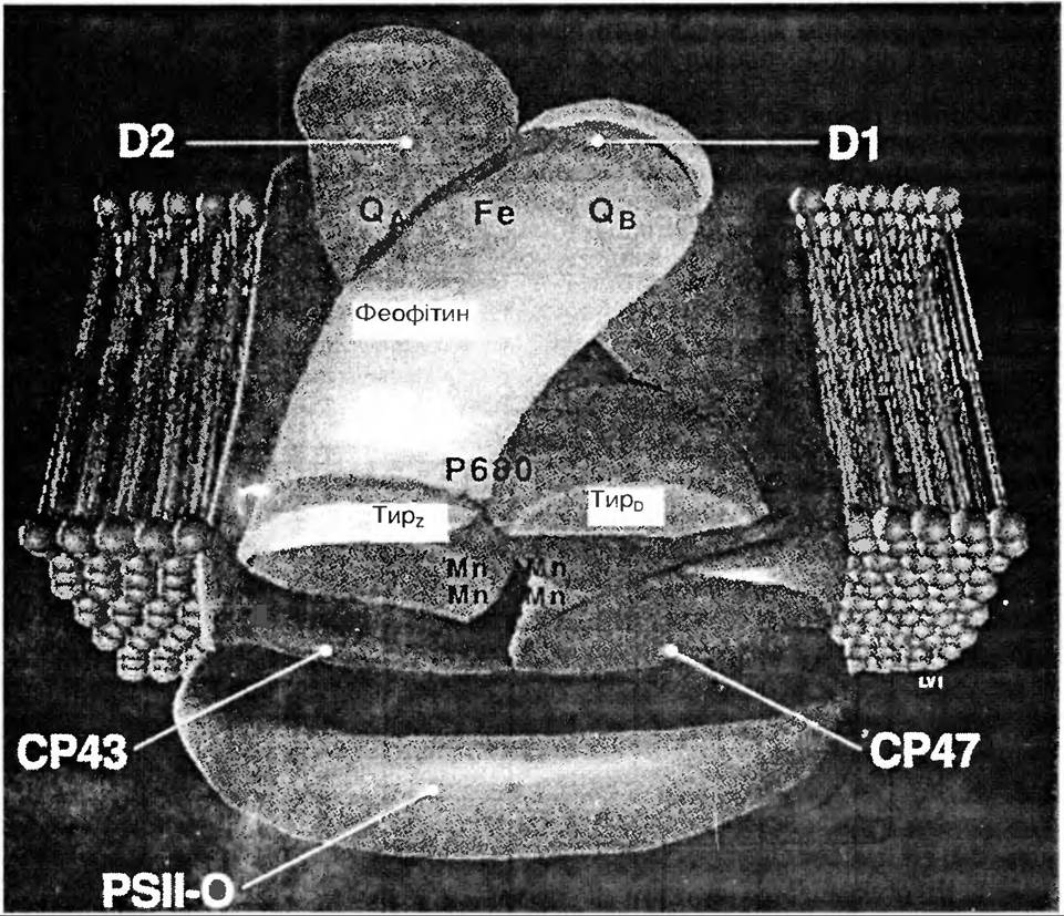
Fig. 54. Model of the central complex of PS II (D1 and D2 are polypeptides binding the CP47 and CP43 chlorophyll complexes)
The lighter PS II subunit, CP43, does not display photochemical activity in its isolated state. It primarily functions as an internal antenna of LHC II. Consequently, it is located
in close contact with the adjacent CP47 subunit to ensure high Efficiency of Energy migration to the PS II RC.
Water-Oxidizing Complex of Photosystem II.
The minimal oxygen-evolving complex of PS II was first isolated in 1985 (I. Kenchi et al., 1985). In addition to the 47, 43, 32, and 30 kD polypeptides and cytochrome b559, it contained an extrinsic 33 kD membrane protein, 3–4 manganese atoms, and 2 plastoquinone molecules.
It is believed that the central complex of PS II, which contains the 33 kD polypeptide and manganese ions, serves as the site of water splitting. Two extrinsic membrane proteins, of 23 and 16 kD, provide the necessary environment to ensure the tight binding of calcium and chloride ions. It has been proven that in the presence of the 23 kD protein, The amount of chloride ions required for oxygen evolution is reduced several-fold. Assuming the validity of the hypothesis that chlorine and calcium bind to the 33 kD protein, proteins 23 and 16 kD apparently provide the correct conformation of the 33 kD protein required to retain these ions.
Oxygen evolution processes are most closely linked to the presence of manganese within the water-oxidizing complex. It is hypothesized that there are at least two pools of manganese: one is loosely bound to the membrane and directly drives The formation of the O2 molecule, while the other is tightly bound to the membrane. The Functions of the latter are not yet fully understood, although data indicate its association with the light-harvesting chlorophyll a/b-protein complex.
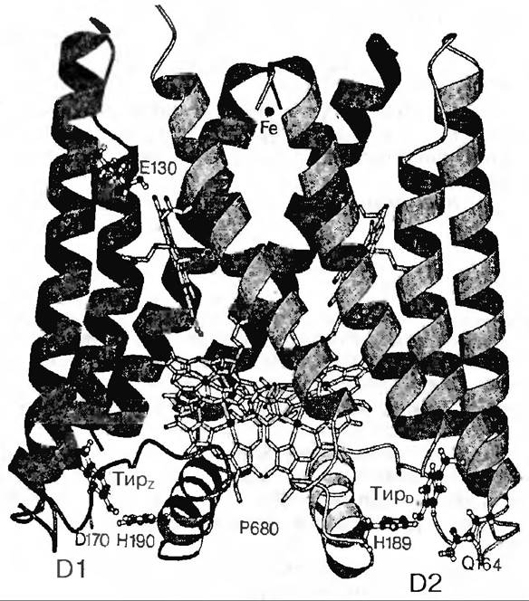
Fig. 55. Structural model of the PS II reaction center
It has been found that there are 4 atoms of manganese per 400 chlorophyll molecules, or in other words, per photosystem II unit. By extracting manganese from thylakoids using a Tris-buffer, a direct quantitative correlation was established between the amount of evolved oxygen and the loss of manganese ions. Therefore, a model was proposed according to which each oxygen-evolving center is functionally coupled to 4 manganese atoms. In isolated thylakoid fragments bearing PS II components, the Mn-containing center—housing exactly four manganese atoms—is indeed localized within the donor side. By sequentially releasing 4 electrons, these atoms are able to achieve a high oxidation state, thereby driving water oxidation, which culminates in the release of molecular oxygen:
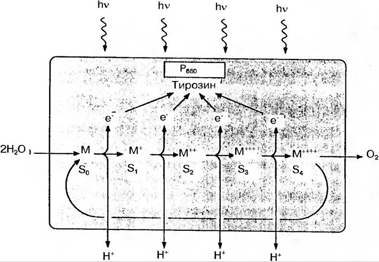
Thus, the P+680 pigment molecule of the PS II reaction center acts as a strong oxidant, accepting an electron (i.e., oxidizing the Z donor) and initiating the cyclic operation of the S-state system, which splits water (Fig. 56).
System S is likely the Mn-containing protein complex capable of accumulating four positive charges.
Z and the transmembrane cytochrome b559 act as intermediate components transferring electrons from the water-oxidizing complex to P680. Its center is possibly located closer to the inner side of the membrane, thereby ensuring active electron transport to the acceptor groups of Z and P680. Two functionally distinct forms of cytochrome b559 have been identified: low-potential and high-potential. Such heterogeneity may be due to differences in the microenvironment of two identical cytochrome b559 molecules.
In general, the minimal model scheme of photosystem II can be represented as follows:
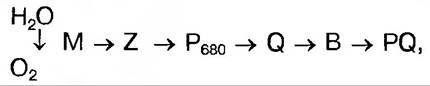
where M is an oxygen-evolving system-specific intermediate component capable of accumulating up to four charges, i.e., M+; M+2, M+3, M+4; P680 is the specific chlorophyll *a* molecule of the PS II reaction center, which undergoes light-driven oxidation; Q is the primary stable electron acceptor, which is a quinone molecule; Z is the electron donor for P680; B is the secondary electron acceptor, also a quinone-type molecule designated as QB, which links PS II with the plastoquinone pool PQ and PS I.
Thus, the macromolecular complex of PS II comprises at least seven distinct polypeptide subunits, whose coordinated action ensures the migration of absorbed light quanta as neutral excitation to the reaction center P680, charge separation and stabilization, plastoquinone reduction, water splitting with oxygen evolution, and proton release into the thylakoid lumen.
NADP Reduction. Ferredoxin is one of the key links in the photosynthetic electron transport system. During the successive reactions leading to NADP+ reduction, electrons are first transferred to the intermediate enzyme *ferredoxin-NADP reductase*. Only this chloroplast flavoprotein enzyme reduces NADP+. The presence of two catalytic centers enables this enzyme to interact with both ferredoxin and NADP+.
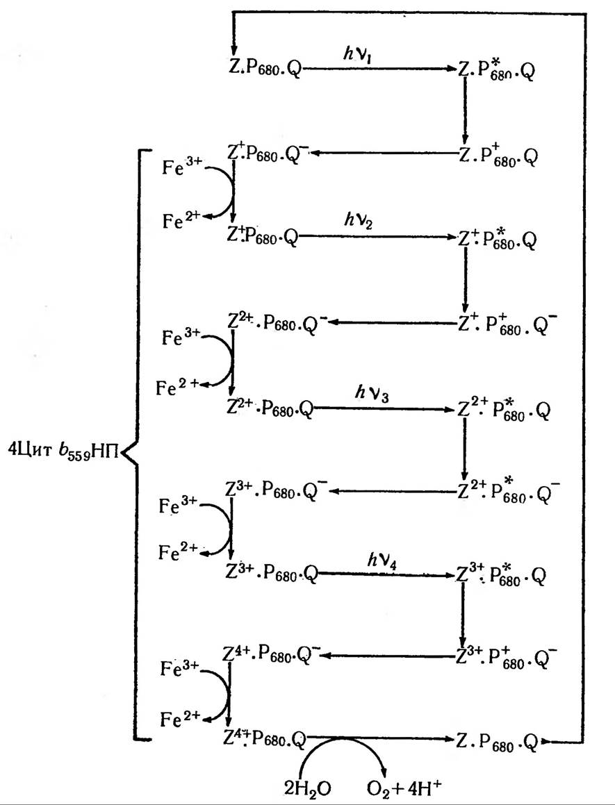
Fig. 56. The oxygen-evolving system functioning in PS II. Four positive charges must accumulate on the carrier Z, which donates electrons to P680, before an oxygen molecule is released.
Last update: 07/08/2026
Editorial and Educational Adaptation: This material has been compiled based on the primary/original source text. The project team performed an editorial review, corrected technical inaccuracies, structured sections, and adapted the content for an educational format.
What was processed:
- elimination of formatting defects (OCR errors, structural breaks, corrupted characters);
- editorial organization of content;
- standardization of terminology in accordance with academic sources;
- verification of factual statements against the original source text.
All mentions of the author, publication year, and origin of the primary text have been preserved in accordance with the source.