Human Biochemistry, Volume 1 - Murray R. 1993
Bioenergetics and Metabolism of Carbohydrates and Lipids
Transport and Storage of Lipids
Plasma Lipids and Lipoproteins
Composition
Extraction of plasma Lipids with an appropriate solvent followed by fractionation of the resulting extract demonstrates that Blood Plasma contains triacylglycerols, Phospholipids, Cholesterol, and cholesterol esters, as well as small amounts of unesterified long-chain Fatty acids (free fatty acids) that account for less than 5% of the total fatty acids present in plasma. Free fatty acids (FFAs) are the most metabolically active plasma lipids. The major lipid classes found in blood plasma are listed in Table 26.1.
Pure fat has a lower density than Water; consequently, the higher the lipid-to-protein ratio in Lipoproteins, the lower their density (Table 26.2). This principle forms the basis for separating plasma lipoproteins by ultracentrifugation. The flotation rate of each lipoprotein in a NaCl solution (specific gravity 1.063) can be expressed in Svedberg flotation units (Sf). One Sf unit is equal to 10-13 cm/s per dyne/g at 26 °C. Table 26.2 presents the composition of various lipoprotein fractions obtained by plasma centrifugation. Different lipid classes are present in most fractions. Because these fractions represent physiological components of plasma, a general chemical analysis of whole plasma for various lipids (with the exception of FFAs) provides little useful information.
Class="center">Table 26.1. Human blood plasma lipids
|
Lipids |
mg/100 ml |
|
|
Average |
Range |
|
|
Total amount |
570 |
360—820 |
|
Triacylglycerols |
142 |
80—180 1) |
|
Total phospholipids 2) |
215 |
123—390 |
|
Phosphatidylcholine |
50—200 |
|
|
Phosphatidylethanolamine |
50—130 |
|
|
Sphingomyelins |
15—35 |
|
|
Total cholesterol |
200 |
107—320 |
|
Free cholesterol (unesterified) |
55 |
26—106 |
|
Free fatty acids (unesterified) |
12 |
6—16 1) |
The total amount of fatty acids (calculated as stearic acid) ranges from 200 to 800 mg/100 ml, of which 45% are incorporated into triacylglycerols, 35% into phospholipids, 15% into cholesterol esters, and less than 5% exist as free fatty acids.
1) Varies depending on dietary intake.
2) Determined by phosphorus content. Lipid phosphorus × 25 = phospholipid as phosphatidylcholine (4% phosphorus).
Table 26.2. Composition of human blood plasma lipoproteins
|
Fraction |
Source |
Diameter, nm |
Density |
Flotation rate Sf |
Composition |
||||||
|
Protein content, % |
Total lipid content, % |
Percentage of total lipids |
|||||||||
|
Triacylglycerol |
Phospholipid |
Cholesterol ester |
Cholesterol (free) |
Free fatty acids |
|||||||
|
Chylomicrons |
Intestine |
100—1000 |
<0.96 |
>400 |
1—2 |
98—99 |
88 |
8 |
3 |
1 |
... |
|
Very low-density lipoproteins (VLDL) |
Liver and intestine |
30—90 |
0.96—1.006 |
20—400 |
7—10 |
90—93 |
56 |
20 |
15 |
8 |
1 |
|
Intermediate-density lipoproteins (IDL) |
VLDL and chylomicrons |
25—30 |
1.006—1.019 |
12—20 |
11 |
89 |
29 |
26 |
34 |
9 |
1 |
|
Low-density lipoproteins (LDL) |
20—25 |
1.019—1.063 |
2—12 |
21 |
79 |
13 |
28 |
48 |
10 |
1 |
|
|
High-density lipoproteins (HDL) HDL2 |
VLDL from liver and intestine? |
10—20 |
1.063—1.125 |
33 |
67 |
16 |
43 |
31 |
10 |
... |
|
|
HDL3 |
Chylomicrons? |
7.5—10 |
1.125—1.210 |
57 |
43 |
13 |
46 |
29 |
6 |
6 |
|
|
Albumin-FFA |
Adipose tissue |
>1.21 |
99 |
1 |
0 |
0 |
0 |
0 |
100 |
||
FFA—free fatty acids; VHDL (very high-density lipoproteins) represent a minor fraction with a density of 1.21—1.25.
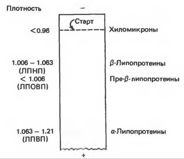
Fig. 26.1. Separation of plasma lipoproteins by Electrophoresis.
Alongside Methods based on differing densities, lipoproteins can also be separated by electrophoresis (Fig. 26.1) and identified more precisely by Immunoelectrophoresis. Aside from FFAs, four Major Groups of lipoproteins have been identified that are important both physiologically and in clinical Diagnosis: (1) chylomicrons, formed in the intestine during triacylglycerol absorption; (2) very low-density lipoproteins (VLDL or pre-β-lipoproteins), synthesized in the liver to transport triacylglycerols; (3) low-density lipoproteins (LDL or β-lipoproteins), representing the end product of VLDL Catabolism; and (4) high-density lipoproteins (HDL or α-lipoproteins), which participate in the METABOLISM of VLDL, chylomicrons, and cholesterol. The principal lipid of chylomicrons and VLDL is triacylglycerol, whereas the predominant lipids of LDL and HDL are cholesterol and phospholipids, respectively (Table 26.2).
The protein moiety of lipoproteins is termed apolipoprotein or apoprotein; it accounts for about 60% of some HDL particles, but only 1% of chylomicrons.
A typical lipoprotein, such as a chylomicron or VLDL, consists of a lipid core (composed mainly of nonpolar triacylglycerols and cholesterol esters) surrounded by a surface monolayer of more polar phospholipids, cholesterol, and apoproteins. Some apoproteins are integral Structural components of the lipoprotein and remain permanently associated with it, whereas others can be transferred to other lipoproteins (Fig. 26.2).
Apolipoproteins (Apoproteins)
Lipoproteins contain one or more Proteins or Polypeptides known as apoproteins. These proteins are designated by capital letters of the alphabet (A, B, C). For instance, the two major apoproteins of HDL are designated A-I and A-II. The primary apoprotein of LDL is apoprotein B, which is also a component of VLDL and chylomicrons. However, apoprotein B of chylomicrons (B-48) is smaller than apoprotein B in LDL or VLDL (B-100) and possesses a different Amino Acid Composition. B-48 is synthesized in the intestine, whereas B-100 is synthesized in the liver. (In rats, the liver apparently produces both B-100 and B-48.) Apoproteins C-I, C-II, and C-III are small polypeptides that can freely transfer from one lipoprotein to another (Table 26.3). The carbohydrate constituents, which account for approximately 5% of apoprotein B, include mannose, galactose, fucose, glucose, glucosamine, and sialic acid. Consequently, certain lipoproteins are also Glycoproteins. C-II is an essential activator of extrahepatic lipoprotein lipase and plays a key role in clearing triacylglycerols from the bloodstream. A-I in HDL serves as an activator of plasma lecithin:cholesterol acyltransferase, an enzyme responsible for the bulk of cholesterol Esterification in humans.
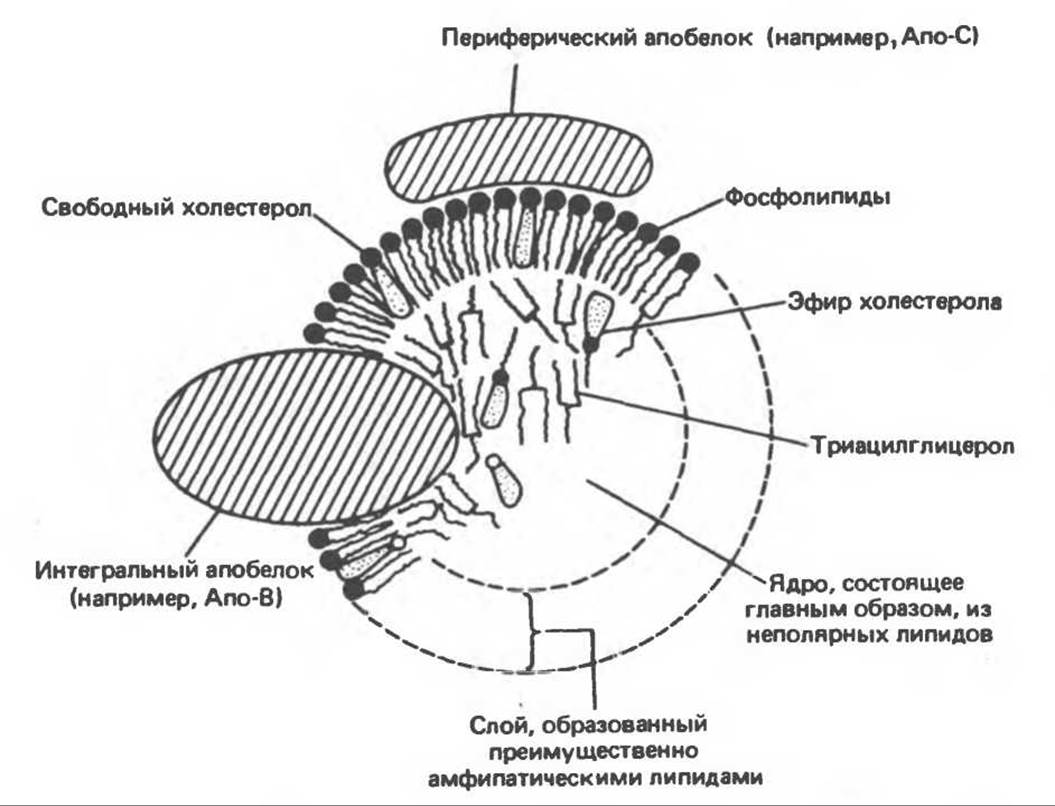
Fig. 26.2. Cytology/cytology/92.html">SCHEMATIC STRUCTURE OF a plasma lipoprotein. Note the similarity to The structure of Cell/30.html">The Plasma Membrane. Evidence suggests that small amounts of triacylglycerols and cholesterol esters are also located in the surface monolayer, while free cholesterol is present in the interior core.
Table 26.3. Apoproteins of human plasma lipoproteins
|
Apoproteins |
Lipoproteins |
Molecular weight |
Notes |
|
A-I |
HDL, chylomicrons |
28 300 |
Activator of lecithin:cholesterol acyltransferase (LCAT) |
|
A-II |
HDL, chylomicrons |
17 400 |
Composed of two identical monomers joined by a disulfide bridge |
|
B-100 |
LDL, VLDL, IDL |
350 000 |
Synthesized in the liver |
|
B-48 |
Chylomicrons, chylomicron remnants |
200 000 |
Synthesized in the intestine |
|
C-I |
VLDL, HDL |
7000 |
Putative activator of LCAT |
|
C-II |
VLDL, HDL, chylomicrons |
8800 |
Activator of extrahepatic lipoprotein lipase |
|
C-III |
VLDL, HDL, chylomicrons |
8750 |
Exists in several polymorphic forms depending on sialic acid content |
|
D |
HDL subfraction |
32 500 |
Possibly identical to the cholesterol ester transfer protein |
|
E (Arginine-rich) |
VLDL, HDL, chylomicrons, chylomicron remnants |
34 000 |
Present in excess in β-VLDL in patients with type III hyperlipoproteinemia. This is the only apoprotein found in HDL of animals in which hypercholesterolemia has been induced by a special diet |
In addition to apoproteins A, B, and C, several other apoproteins have been identified in plasma lipoproteins. One of these is apoprotein E isolated from VLDL (in which arginine accounts for 10% of the total Amino Acids), which normally comprises 5—10% of the total VLDL apoproteins. The proportion of apoprotein E in the broad β-VLDL electrophoretic fraction is elevated in patients with type III hyperlipoproteinemia.
Last update: 06/08/2026
Editorial and Educational Adaptation: This material has been compiled based on the primary/original source text. The project team performed an editorial review, corrected technical inaccuracies, structured sections, and adapted the content for an educational format.
What was processed:
- elimination of formatting defects (OCR errors, structural breaks, corrupted characters);
- editorial organization of content;
- standardization of terminology in accordance with academic sources;
- verification of factual statements against the original source text.
All mentions of the author, publication year, and origin of the primary text have been preserved in accordance with the source.