Amino Acids, Peptides and Proteins - T. Dévényi, J. Gergely 1976
Methods of Immunochemical Analysis
Protein Analysis by Gel Diffusion Methods
Immunoelectrophoresis
Principle of the method. Immunoelectrophoresis is a combination of electrophoretic Separation of Proteins with double diffusion and immunoprecipitation in gel. Following the electrophoretic separation of proteins in Agar gel, a specific immune serum diffuses toward the resulting fractions. This method allows proteins to be characterized not only by their migration rate in an electric field but also by their antigenic properties.
Applications. Immunoelectrophoresis is used to identify components of protein mixtures, primarily Blood serum. It is highly useful in the Diagnosis of paraproteinemias and immunodeficiency dysproteinemias. This method is also employed to monitor the purity of protein preparations (detection of impurities) and to analyze proteins from Tissues and Body Fluids.
Two variants of this method have become most widely used: 1) the Grabar–Williams macromethod [8, 9] and 2) the Scheidegger micromethod modification [21], the popularity of which is due to its speed and The ability to analyze small amounts of material.
THE GRABAR AND WILLIAMS MACROMETHOD OF IMMUNOELECTROPHORESIS [8, 9]
EQUIPMENT
1. Power supply. A power source capable of delivering a current up to 100 mA at a stabilized voltage of 100—150 V is required.
2. Electrophoresis apparatus. The apparatus consists of two plexiglass tanks measuring 400 x 40 x 70 mm, equipped with built-in platinum electrodes 400 mm long and 0.5—0.8 mm in diameter. At the end of each tank, there are outlet ports connected by a rubber hose. Each tank of the electrophoresis apparatus is filled with 1 L of buffer solution. During electrophoresis, a constant flow of buffer solution through the apparatus must be maintained. To achieve this, a 4—5 L bottle is placed above the level of the tanks, from which the buffer solution flows into the tanks and then drains through the outlets at the ends.
PROCEDURE
1. Preparation of the buffer solution. Immunoelectrophoresis of proteins is usually carried out in Michaelis buffer with a pH of 8.2 and an Ionic strength of 0.05.
Michaelis buffer is prepared as follows: 47.6 g of sodium diethylbarbiturate is dissolved in 3000 mL of distilled Water with a pH of 7, and acidified first to approximately pH 8.4 with 65 mL of 1 N HCl, and then carefully to pH 8.2. Distilled water is then added to a final volume of 4265 mL. If a buffer solution of lower ionic strength is required, further dilution is performed.
2. Preparation of the agar plate. A 1.5% agar gel is prepared using the buffer solution specified above (agar purification and gel preparation are described on p. 127).
Typically, immunoelectrophoresis is performed in an agar gel layer on 180 x 130 mm Glass plates, which must be thoroughly washed, degreased, dried, and placed strictly horizontally before the experiment; any deviation of the plate from the horizontal level leads to uneven thickness of the agar gel. The following technique is recommended: a glass cuvette of appropriate size is filled with 5% molten agar; after it solidifies, a horizontal surface is formed, on which the glass plate is placed. To ensure that the plate remains strictly horizontal, the cuvette should not be moved after the agar has solidified.
On both edges of the glass plate, strips of filter paper 40 mm wide, moistened with buffer solution, are applied so that they project 30 mm beyond the edge of the glass.
Then, glass rods or tubes 5 mm in diameter are placed parallel to each other on the glass surface at a distance of 31 mm apart; after the agar solidifies, troughs for introducing antisera will be formed in their place.
After melting 250—300 mL of agar, it is poured onto the prepared glass plate with filter paper strips to form a gel layer 3—4 mm thick. The agar should be poured carefully to avoid air bubbles. After the agar solidifies, the glass rods are removed using two pairs of tweezers, and wells for introducing the test samples are cut on both sides of each trough at a distance of 8 mm from it. Wells measuring 3 x 15 mm are cut using a scalpel, a blade, or a special glass or metal tool. The excised agar gel plugs are carefully removed from the wells by prying them from below or by aspirating them with a large-diameter needle (Fig. 26).
Then, the agar gel is trimmed along the contour of the glass plate and the filter paper strips so that the gel-coated strips can be bent downward at a right angle to the plane of the plate.
The prepared agar plate is mounted in the electrophoresis apparatus, and the gel-coated filter paper wicks are immersed in the buffer solution filling the tanks.
Molten agar is used to repair the integrity of the gel in areas of accidental damage to ensure uniform electrical conductivity across all sections.
3. Sample loading. A solution containing approximately 15 mg of the test protein (for example, 0.2 mL of blood serum when studying Serum proteins) is warmed to 40°C and mixed with 0.8 mL of 3% molten agar prepared in distilled water and cooled to 40°C. The resulting mixture is loaded into the sample well on the agar plate. The remaining free volume of the well is filled with 1.5% agar gel in buffer solution. Care should be taken to avoid air bubbles when filling the well.
Class="center">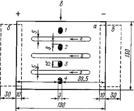
Fig. 26. Preparation of an agar plate for immunoelectrophoresis (macromethod).
a — agar gel layer; b — filter paper strips; c — sample wells (1, 2, 3, 4); d — troughs for immune serum. (Dimensions are in mm.)
4. Electrophoresis. At a voltage of 120 V, the electrophoretic separation of blood serum proteins lasts 4—5 h. The optimal voltage gradient is 3—6 V/cm. Under these conditions, electrophoresis can be carried out without special cooling while avoiding gel desiccation. As the current increases, a voltage drop occurs; therefore, the circuit voltage must be checked several times during the electrophoresis run.
5. Antibody addition. To avoid experimental failure, the optimal dilution of the immune serum should be determined beforehand, at which the clearest precipitation reaction with the test antigen (in this case, blood serum proteins) is observed.
Upon completion of electrophoresis, the filter paper wicks are removed, and the troughs in the agar gel are filled with immune serum appropriately diluted with saline. (Approximately 1 mL of serum is required to fill the trough.)
The agar plate is then placed in a humid chamber and left until precipitation bands form, which appear in the gel after 2–5 days.
6. Recording immunoelectrophoresis results. Precipitation bands can be recorded by contact printing directly from native preparations (see p. 135) or by photography after appropriate staining (see p. 135).
NOTES
1. After each electrophoresis run, it is recommended to reverse the electrode polarity.
2. Michaelis buffer is highly convenient for immunoelectrophoretic analysis of blood serum proteins. However, other Buffer solutions can also be used. In such cases, of course, the agar gel should be prepared using the same buffer solution in which the electrophoresis is performed.
SCHEIDEGGER'S MICROIMMUNOELECTROPHORESIS [21]
Apparatus. 1. Power supply (see p. 46).
2. Electrophoresis apparatus. The apparatus consists of two Plexiglas tanks measuring 250 x 100 x 35 mm, each divided by a partition into two compartments. A platinum wire electrode with a diameter of 0.5–0.8 mm is mounted in the outer compartment of each tank. Filter paper strips, moistened with buffer solution and immersed in it on both sides of the partition, serve as current conductors between the compartments.
A chamber for holding the agar plates, also made of Plexiglas, is positioned between the two buffer tanks. Along its upper inner edge on both sides, There is a 2 mm wide ledge on which the Microscope slides with agar gel are placed. The distance between the edges of the ledge is 76 mm. The agar plate chamber is closed with a Plexiglas lid, which protects the agar from drying out during electrophoresis. Each tank compartment is filled with 150 ml of buffer solution, which should preferably be changed before each experiment.
3. Device for cutting wells and troughs in agar gel. When performing Scheidegger's microimmunoelectrophoresis, the sample wells and the antiserum trough are cut into the agar gel using a special device. Fig. 27,A shows the layout of the wells and the trough. To cut the trough (a), two steel blades measuring 5 x 45 x 0.08 mm are mounted in the device (Fig. 27,B) at a distance of 1 mm from each other. To cut the wells (b), two segments of a hypodermic needle, also 5 mm high, are mounted in the device at a distance of 3 mm on either side of the outer surface of the blades. These needle segments are not centered exactly on the template but are offset from it by 5 mm. A microscope slide covered with a layer of agar gel is placed under the template of the device; the spring-loaded handle is pressed down, and by plunging the blades and needles into the gel, the outlines of the trough and wells are marked. After aspirating the agar gel pieces from the wells using a needle segment of appropriate size, the test samples are introduced into them. Electrophoresis is then performed, and only upon its completion is the gel removed from the trough to introduce the antiserum.
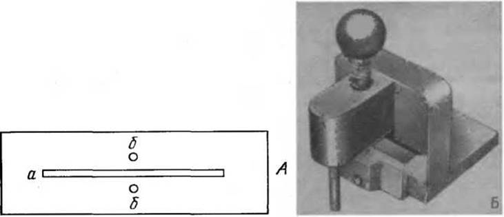
Fig. 27. A. Layout of the wells for Sample application (b) and the trough from which the immune serum diffuses (a). B. Device for cutting wells and troughs.
METHOD
1. Preparation of buffer solution. Microimmunoelectrophoresis is typically carried out in Michaelis buffer with a pH of 8.2 and an ionic strength of 0.1 μ.
Stock solution: 194.2 g of sodium acetate and 294.2 g of veronal are dissolved in 10 l of distilled water.
By diluting the stock solution with distilled water (8 : 9.6) and adjusting the pH to 8.2 with 0.1 N HCl, a buffer solution with an ionic strength of 0.15 μ is obtained. To obtain a buffer solution with an ionic strength of 0.1 μ, it should be diluted 1.5-fold with distilled water.
2. Preparation of microscope slides. The slides onto which the agar gel layer is applied must be thoroughly washed and degreased. They should be washed with sulfuric acid containing potassium dichromate, then rinsed free of acid with running tap water and distilled water, and stored in ethanol. Before use, the slides are wiped dry, ensuring that no dust particles remain On the surface.
3. Preparation of the agar gel layer. Dry microscope slides are placed on a horizontal surface (see p. 138), and using a pipette with a sufficiently wide tip, 2 ml of 1.5% molten agar prepared in buffer solution is poured onto The surface of each slide (for agar purification and gel preparation, see p. 127). It is essential to ensure that the agar gel layer is uniform, covers the entire surface of the slide, and is free of air bubbles. As soon as the agar solidifies, the plates are transferred to a humid chamber and stored in a refrigerator. Most often, agar plates are prepared 24 hours before the experiment.
The layout of sample wells and the antiserum trough should be marked immediately before electrophoresis, as described on p. 141.
4. Sample application. The test protein solution, such as blood serum, is introduced into the sample well using a capillary pipette, filling it to the brim. A volume of 0.001 ml of solution is sufficient for electrophoresis.
5. Electrophoresis. After the test samples are applied, the agar plates are placed in the chamber of the electrophoresis apparatus. Contact between the agar gel and the buffer tanks of the apparatus is established using filter paper strips moistened with buffer solution. The paper wicks should overlap the gel by approximately 15 mm at both ends of the agar plate.
The chamber containing the agar plates is closed with the lid, and the current is turned on. If the voltage measured at the edges of the agar plate reaches 45 V, the electrophoretic separation of proteins will be completed within 45–60 min.
6. Antiserum application. Upon completion of electrophoresis, the previously marked trough is cleared of agar gel and filled with specific immune serum using a capillary pipette or a tuberculin syringe (it is recommended to determine its optimal dilution beforehand). Approximately 0.05 ml of immune serum is required to fill the trough. Diffusion takes place in a humid chamber at room Temperature for 16–24 h.
7. Recording microimmunoelectrophoresis results. Precipitation bands can be recorded by contact printing directly from the native preparation (see p. 135) or by photography after staining (see p. 135).
NOTES
1. The Agar gel electrophoresis apparatus manufactured by Labor Müszeripari Müvek (Hungary), model Labor 59952, has proven highly reliable. Electrophoresis can be performed on ten agar plates simultaneously in the central chamber of this apparatus. The instrument is supplied complete with a leveling table (which ensures a uniform agar layer), staining troughs with inserts, a gel cutter for making wells and troughs, and a micropipette. The apparatus is easy to operate and is equally suitable for both electrophoresis and immunoelectrophoresis.
2. In the absence of a commercial well and trough cutter for agar gel, a simple homemade punch can be fashioned by mounting two razor blades and a pair of injection needles (with their sharp tips sawn off) into a cork stopper, According to the dimensions specified on page 141.
3. The easiest way to remove the sharp tips of injection needles is to insert a stylet into the needle and grind the tip on a rotating grinding stone.
4. The electrophoretic separation of serum Proteins can be easily monitored by staining the blood serum with Congo red. The dye binds to albumin and migrates along with it. It should be noted that staining slightly increases the migration rate of albumin.
5. If the concentration of the protein mixture under study is relatively low (below 3%), it is recommended to increase the sample well volume to accommodate a larger amount of material. Larger wells can be easily made using suitable glass tubes with a diameter of 2–4 mm.
6. By punching two sample wells on a single agar plate, we can simultaneously analyze two different proteins or protein mixtures and compare their immunoelectrophoretic characteristics. This is particularly important for blood serum protein analysis, where the test serum is placed in one well and normal serum in the other. Under these conditions, it is easier to evaluate the immunoelectrophoretic Features of the test serum. This approach is highly useful when determining whether the mixture under study contains a specific protein (for example, whether a tissue extract contains blood serum proteins, or which serum components are present in a protein preparation isolated from urine).
7. Immunoelectrophoretic characterization of serum proteins. Immunoelectrophoresis is used for blood serum protein analysis more frequently than other Methods. Its popularity stems from the fact that, using a very small volume of serum, one can characterize 15–20 protein fractions instead of the 5 accessible by zone electrophoresis. The number of individual protein fractions detected by immunoelectrophoresis depends on the quality of the antiserum used.

Fig. 28. Immunoelectrophoresis of human blood serum.
Rabbit antiserum against human serum proteins, manufactured by Behringwerke AG (FRG), served as the antibody source.
The upper part of Fig. 28 shows an immunoelectrophoregram of human blood serum. The diagram below serves as a guide for interpreting the immunoelectrophoregrams of any human serum; it illustrates the relative positions of its constituent protein fractions.
8. Identification of individual protein fractions.
a) A protein solution of known composition is loaded into one of the wells as a control and subjected to electrophoresis. For example, if one needs to determine the presence of albumin in the protein mixture under study, a solution of pure albumin is used as a control.
b) Specific immune antisera are used for identification. For example, the presence of albumin in the protein mixture under study can also be determined using a specific anti-albumin antiserum. If the protein mixture contains albumin, a single precipitin arc corresponding to this protein should appear on the immunoelectrophoregram.
c) Fraction identification can also be performed using specific staining. For example, if Lipoproteins need to be identified in a protein mixture, the resulting immunoelectrophoregram is stained with Dyes that specifically detect Lipids. In this case, only the precipitin arcs containing lipoproteins should be stained.
9. Methods for specific staining of precipitin arcs. The previously described staining methods for proteins and lipoproteins are fully suitable for staining precipitin arcs in immunoelectrophoresis. Several specific staining methods that allow the identification of certain protein fractions are described below.
a) Staining of Glycoproteins [22]. Solutions: I. 1% periodic acid in 50% ethanol; II. A mixture of 50 ml of 0.01 M p-phenylenediamine, 50 ml of 0.01 M a-naphthol, and 10 ml of 10% hydrogen peroxide.
Procedure. Immerse the dried agar plate in solution I for 15 min, then wash with distilled water for 15 min, and stain with solution II until a light violet tint appears. After that, rinse the preparation with running tap water for 10 min and dry at 37°C.
b) Staining of ceruloplasmin. Solutions: I. Dissolve 200 mg of alizarin blue in 100 ml of acetic acid at 55°C and dilute 10-fold with 70% acetic acid; II. 70% acetic acid.
Procedure. Stain the dried agar plate in solution I for 20 min, then wash in solution II for 30 min.
e) Staining of haptoglobin. Dissolve 2.0 g of benzidine1 in 20 ml of acetic acid with heating. After cooling, bring the final volume of the solution to 100 ml with distilled water, add 0.5–1.0 g of activated charcoal, shake, let stand for 15 min, and then filter into a dark glass bottle. Immediately before staining, add a few drops of hydrogen peroxide to the benzidine solution.
After staining for 15 min, wash the agar plate with distilled water.
10. The shape of the precipitin arcs allows one to assess the homogeneity or heterogeneity of the proteins under study. Homogeneous proteins yield symmetrical, regularly shaped precipitin arcs (for example, among serum proteins, albumin produces such an arc). A heterogeneous population of molecules typically produces an elongated, asymmetrical precipitin arc. This is characteristic of IMMUNOGLOBULINS in normal human serum (IgG, IgM, IgA).
Immunoglobulins of different classes share many common antigenic determinants but differ in their electrophoretic mobility; consequently, they form an elongated, asymmetrical precipitin arc during immunoelectrophoresis. This phenomenon is of particular importance in the diagnosis of so-called monoclonal gammopathies. In these diseases (such as multiple myeloma or Waldenström's macroglobulinemia), a clone of malignantly proliferating plasma Cells typically produces a homogeneous monoclonal immunoglobulin. A pathological increase in the population of these immunoglobulin molecules results in a characteristic symmetrical precipitin arc during immunoelectrophoresis. In Fig. 29, which shows serum immunoelectrophoregrams from IgA- and IgG-type myelomas, it can also be observed that a significant increase in the concentration of individual proteins in monoclonal gammopathies leads to the dissolution of precipitin arcs in antigen excess. This phenomenon can lead to misinterpretation of immunoelectrophoregrams, as the dissolution of the precipitate is perceived as the absence of the given protein fraction, whereas it is actually present in large excess. In such cases, antigen excess should be eliminated by a 5- to 10-fold dilution of the high-protein test serum. Following this, the symmetrical precipitin arc characteristic of monoclonal gammopathies, which differs from the precipitin arc of normal immunoglobulins, should appear on the immunoelectrophoregram (Fig. 29).
1 Benzidine is carcinogenic; therefore, working with it requires appropriate safety precautions.
11. In certain cases, especially in the immunoelectrophoretic analysis of low-molecular-weight proteins, it is convenient to use the antiserum application method proposed by Backhaus in 1967 [2]. After applying a layer of agar gel onto a glass plate, it is cut longitudinally into two equal halves, and one of them is removed. Then, approximately 5 mm from the cut line, a sample well is made in the gel, into which the test material is poured, and electrophoresis is performed in the usual manner. Upon completion of the electrophoresis, molten agar is mixed with specific antiserum and poured onto the vacant half of the glass. Precipitin bands form partly in the remaining half of the agar gel and partly in the gel applied to the glass after electrophoresis. In conventional immunoelectrophoresis, low-molecular-weight proteins (e.g., Bence-Jones proteins) can migrate a considerable distance from the well due to rapid diffusion; if they enter the antiserum trough, no precipitin line will form. The Backhaus method avoids such difficulties because the antiserum trough is simply absent, and the precipitin bands of low-molecular-weight proteins appear in the agar gel layer applied after electrophoresis.

Fig. 29. Immunoelectrophoresis of blood serum in monoclonal immunoglobulinopathies.
A. IgG myeloma; B. IgA myeloma. Patient serum is in the upper wells; normal human serum is in the lower wells.
12. The immunoelectrophoresis method allows comparing not only protein mixtures (Antigens) using a given antiserum, but also antisera using a given antigen. For such an analysis, two parallel troughs are cut into the agar gel plate, and a sample well is made between them at a distance of 3 mm from the edge of each trough. A solution of a specific protein is introduced into the well, and after electrophoresis, the troughs are filled with the antisera being compared. The precipitin bands that appear on both sides of the applied sample allow for the comparison of the studied antisera.
13. Preparing antiserum of the required quality often encounters serious difficulties. Host animals differ significantly in their ability to synthesize Antibodies; along with interspecies differences, there are substantial individual variations in the immunoreactivity of host animals within the same species. Therefore, it is recommended to immunize several animals with the same antigen simultaneously; this increases the probability of obtaining a high-quality antiserum that yields the required number of precipitin bands.
14. The immunoelectrophoresis methods described above are applicable only for qualitative analysis.
15. The resolving power of immunoelectrophoresis far exceeds that of zone electrophoresis, but it should be remembered that, like other immunodiffusion methods, immunoelectrophoresis detects the minimum number of reacting antigen-antibody systems.
16. If electroendosmosis in the agar gel during electrophoresis is a serious obstacle to immunoelectrophoretic analysis, it is recommended to replace the agar gel with agarose gel.
17. Radioimmunoelectrophoresis. If an antigen or antibody is labeled with a radioactive isotope, immunoelectrophoresis can be used to analyze extremely small amounts of material. It is known that the antigenic properties of IgG do not change when the IgG molecule, acting as an antibody, is part of an immune complex. If we have an appropriate radiolabeled anti-IgG antiserum, we can use it to detect IgG in immune complexes (for example, in precipitates). Radioimmunoelectrophoresis consists of the following stages:
a) First, the antigen under study (e.g., human serum albumin) is subjected to electrophoresis in the usual manner. Then, the antiserum trough is filled with the appropriate antiserum (e.g., rabbit antiserum to human serum albumin) and left for Diffusion and the formation of precipitin bands.
b) As soon as the precipitin bands form, the antiserum trough is filled with 131I-labeled antiserum to rabbit y-globulin (e.g., obtained from a guinea pig) and left again for diffusion.
в) The isotope-labeled antiserum reacts with the rabbit y-globulin that is part of the precipitin bands (immune complexes). The isotope-containing precipitates can then be detected using autoradiography.
Radioimmunoelectrophoresis can also be performed using antigens labeled with radioactive isotopes. The procedure is similar to the one just described, but after immunoelectrophoresis, the isotope-labeled antigen (in the example above, human serum 131I-albumin) is poured into the antiserum trough and left for diffusion.
Last update: 06/08/2026
Editorial and Educational Adaptation: This material has been compiled based on the primary/original source text. The project team performed an editorial review, corrected technical inaccuracies, structured sections, and adapted the content for an educational format.
What was processed:
- elimination of formatting defects (OCR errors, structural breaks, corrupted characters);
- editorial organization of content;
- standardization of terminology in accordance with academic sources;
- verification of factual statements against the original source text.
All mentions of the author, publication year, and origin of the primary text have been preserved in accordance with the source.