BIOCHEMISTRY AND MOLECULAR BIOLOGY - W. ELLIOTT - 2002
CHAPTER 10. TRANSITION FROM CATABOLISM TO ANABOLISM. SYNTHESIS OF FATS AND RELATED COMPOUNDS IN THE ORGANISM
In previous chapters on METABOLISM, we discussed catabolic Reactions Involving the breakdown of fats and CARBOHYDRATES. Another key metabolic function is the synthesis of molecules, or anabolism. The best place to start exploring anabolism is with the synthesis of fats.
Fat synthesis occurs primarily from excess carbohydrates that are not utilized to replenish Glycogen stores. Certain Amino Acids also contribute to this process. Excess dietary intake further promotes fat accumulation. Fat represents the most compact form of energy storage in the body; we would have to increase enormously in size if we stored energy exclusively as glycogen.
Mechanism of Fat Synthesis
During metabolism, Fatty acids are converted into acetyl-CoA. Conversely, acetyl-CoA is converted back into fatty acids (the fact that fatty acids are synthesized from acetyl-CoA, a two-carbon unit, explains why naturally occurring fatty acids almost always have an even number of carbon atoms). Excess carbohydrate intake leads to fat deposition because glucose is broken down into Pyruvate, which is then converted into acetyl-CoA. This latter reaction, catalyzed by pyruvate dehydrogenase, is irreversible. Consequently, unlike Bacteria and plants—which can carry out such conversions—animal Cells cannot convert acetyl-CoA into pyruvate, and therefore fats cannot be converted into glucose.
Any metabolic pathway includes at least one step that operates via different biochemical reactions in the forward and reverse directions. This holds true for both the degradation and synthesis of fats. Their respective metabolic pathways overlap in places while diverging in others. As a result, both processes are thermodynamically favorable, irreversible, and subject to distinct regulation.
For the synthesis of fatty acids from acetyl-CoA to become thermodynamically favorable, it must incorporate an irreversible step that requires additional
energy. Already at the initial stage of fatty acid synthesis—The formation of malonyl-CoA from acetyl-CoA and CO2—the energy of ATP Hydrolysis is utilized. Subsequently, the fatty acid molecule loses CO2.
At first glance, this might seem pointless, but this mechanism confers thermodynamic irreversibility on the process, resolving an energetic rather than a purely chemical problem. Malonic acid has the Structure HOOC-CH2-COOH, and malonyl-CoA is HOOC-CH2-CO-S-CoA. The reaction shown below is catalyzed by acetyl-CoA carboxylase:
Class="center">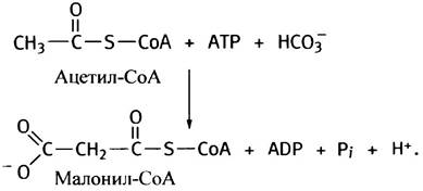
The prosthetic group of this enzyme is biotin. As you already know, all carboxylases utilize the energy of ATP hydrolysis to introduce CO2 into substrates. An intermediate in this reaction is "activated CO2"—carboxybiotin (see p. 123).
Thus, synthesizing fatty acids requires the sequential addition of two-carbon units to acetyl-CoA. The active donor of these units is malonyl-CoA, although the malonyl moiety itself contains three carbon atoms rather than two. Before examining fatty acid synthesis in greater detail, however, we need to clarify what an acyl carrier protein, or ACP, is.
Acyl Carrier Protein and β-Ketoacyl Synthase
All catabolic transformations involve thioesters formed with CoA rather than free fatty acids. Similarly, all reactions in fatty acid synthesis involve thioesters, but instead of CoA, they utilize a simpler molecule that can be viewed as half of CoA. Let us recall The structure of CoA.
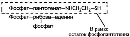
The highlighted portion of the molecule is the thiol-containing "carrier"—4-phosphopantetheine—which is employed in fatty acid synthesis. Unlike acetyl-CoA, it Functions not in a free state, but bound to ACP. ACP can be thought of as a protein integrated into CoA, or conversely, as a giant CoA molecule in which the AMP fragment is replaced by a protein.
Another enzyme, or more precisely, a functional domain—β-ketoacyl synthase—also contains a reactive thiol group. This group belongs to a Cysteine residue located in the active center of the enzyme. In mammals, both ACP and β-ketoacyl synthase are components of a large multifunctional complex.
Mechanism of Synthesis of CoA-Derived Fatty Acids
Let us consider the case where both thiol groups in the CoA-synthase complex are free (Fig. 10.1, a).
Fig. 10.1. Fatty acid synthesis. Although animal β-ketoacyl synthase is a domain of a large protein molecule, it is depicted here as a separate protein. Following seven successful cycles, palmityl-ACP is formed, which is then hydrolyzed to yield free palmitate

The acetyl group (CH3CO-) is transferred from acetyl-CoA to the thiol group of ACP by a specific transferase (Fig. 10.1, b). Next, it is transferred to the thiol group of β-ketoacyl synthase, thereby freeing its binding site on ACP once again (Fig. 10.1, c). This site is then occupied by a malonyl residue transferred from malonyl-CoA by another transferase, forming malonyl-ACP (Fig. 10.1, d). Following this, the synthase catalyzes The transfer of the acetyl group to the malonyl group, yielding β-ketoacyl-ACP and CO2. In this specific case, the product is β-ketobutyryl-ACP (Fig. 10.1, e). Remaining within the protein complex, the latter is reduced to butyryl-ACP (Fig. 10.1, f). Ultimately, the system transitions to a state (Fig. 10.1, g) analogous to that shown in Fig. 10.1, c: the thiol group of ACP is free, while the synthase is acylated with a saturated fatty acid residue. The sole difference is that the acetyl residue (see Fig. 10.1, c) has now been replaced by a butyryl residue (see Fig. 10.1, g). If all the reactions described above occur once more, the butyryl residue is converted into a hexanoyl residue. Five additional cycles of the process lead to the formation of palmitoyl-ACP; subsequently, the action of a hydrolase enzyme releases palmitic acid (C16) into solution:
СН3(СН2)14СО-S-АПБ + Н2О —> СН3(СН2)14СОО- + АПБ-SH.
The reaction catalyzed by β-ketoacyl-ACP synthase is irreversible due to the decarboxylation step involved. Because of this stage, the entire fatty acid synthesis pathway acts like a one-way street.
If There is a need to synthesize a fatty acid containing more than 16 carbon atoms, such as stearic acid (C18), chain elongation of palmitic acid is carried out by other enzyme systems.
Organization of Fatty Acid Synthesis
The sequence of reactions described may seem simple to some and complex to others, but one thing is certain: this animal enzyme assembly is remarkably organized. In E. coli cells, the same sequence of reactions is catalyzed by separate Enzymes, so that after each reaction the products dissociate into solution and diffuse to the next enzyme. In animals, all stages of fatty acid synthesis following the formation of malonyl-CoA take place
within a single giant protein complex consisting of multiple subunits. Each subunit possesses enzymatic activity, but typically functions only as part of the complex (in fact, two such multienzyme complexes are combined into a dimer). The growing fatty acid chain binds alternately to the thiol groups of ACP and β-ketoacyl synthase without ever leaving the complex until synthesis is completed with the formation of palmitate. Both chain elongation and the reduction of keto groups occur while the substrates are bound to ACP. The long, flexible arm of 4'-phosphopantetheine is presumably required to allow various intermediates to interact with the appropriate catalytic centers of the multienzyme complex. This organization accelerates synthesis by avoiding multiple rounds of product dissociation and diffusion, and by facilitating its binding to the next enzyme.
Reduction Stages in Fatty Acid Synthesis
In the sequence of events leading to the elongation of the fatty acid chain attached to ACP, an important role is played by the reduction of β-ketoacyl residues, which proceeds in three stages (Fig. 10.2).
Fig. 10.2. Reduction stages of fatty acid synthesis

The reducing agent here is NADPH (not NADH!), which serves as an electron carrier.
What is NADP+?
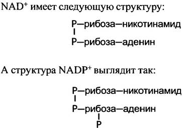
The additional phosphate group in the NADPH molecule is attached to the 2'-hydroxyl group of the ribose linked to adenine:
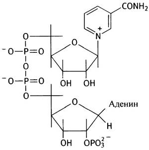
This phosphate group has virtually no effect on the Redox Potential of the molecule and is actually used as a signal for protein identification and recognition of the molecule. NAD+-dependent enzymes do not react with NADP+, and vice versa (with rare exceptions).
For a long time, it was thought that The Use of NADPH in fatty acid synthesis was nothing more than a quirk of nature, a biological oddity. However, it gradually became clear that the parallel existence of the two pyridine Coenzymes plays a crucial role in separating metabolic pathways. To understand this, one must remember that energy production can be viewed as The oxidation of reducing equivalents, whereas fat synthesis represents the consumption of reducing equivalents. These are diametrically opposed goals! The Cell separates these tasks without compartmentalizing them spatially: oxidative branches of metabolism use NAD+ as an electron carrier, while reductive branches utilize NADP+ for this purpose. The reduction of these chemically similar compounds proceeds differently. NAD+ is reduced during Glycolysis, in the pyruvate dehydrogenase reaction, in The Citric Acid Cycle, and during fat oxidation. But how is NADP+ reduced? This will be discussed in the next section.
Where are fatty acids synthesized?
The primary site of fatty acid synthesis is The Liver and, to a lesser extent, adipose tissue, although fats are also produced by many other Organs, such as the Mammary Glands during Lactation.
Within the cell, palmitate synthesis from acetyl-CoA is localized in the Cytosol, whereas Fatty acid oxidation takes place in the Mitochondria. The main source of acetyl-CoA for fatty acid synthesis is the pyruvate dehydrogenase reaction occurring in the mitochondrial matrix (see p. 116). Acetyl-CoA cannot cross the mitochondrial membrane on its own, so a mechanism must exist to transport acetyl groups from the mitochondria to the cytosol, the site of fatty acid synthesis. Inside the mitochondria, acetyl-CoA is converted into citrate (see p. 119), which is transported out of the mitochondria into the cytosol by a specific transport system. In the cytosol, citrate is cleaved by the enzyme citrate lyase into acetyl-CoA and oxaloacetate. This reaction is coupled with ATP hydrolysis, making it virtually irreversible:
Citrate + ATP + CoA-SH + H2O —> Acetyl-CoA + Oxaloacetate + ADP + Pi.
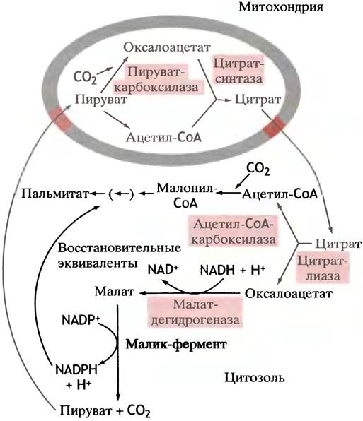
Oxaloacetate cannot return to the mitochondrion on its own because the mitochondrial membrane is impermeable to it, and no specific transporters exist. Instead, it is reduced to malate by NADH (note: NADH, not NADPH!). Malate then undergoes oxidative decarboxylation to pyruvate via a specific malic enzyme that utilizes NADP+ (not NAD+) as a coenzyme. The resulting NADPH participates in fatty acid synthesis (Fig. 10.3).
Fig. 10.3. Reduction of NADP+ for fatty acid synthesis. The net effect of both reactions is the transfer of reducing equivalents from NADH to NADP+. The pyruvate generated in the Cytoplasm enters the mitochondria. The source of oxaloacetate is shown in Fig. 10.4
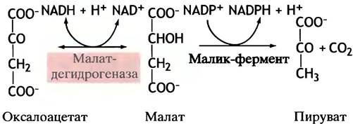
Fig. 10.4. Diagram illustrating the Water/144.html">Origin of the acetyl groups (acetyl-CoA) and reducing equivalents (NADPH + H+) required for fatty acid synthesis. The action of citrate lyase is coupled with the Cleavage of ATP to ADP and Pi
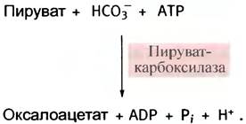
Pyruvate can now be transported back into the mitochondria, where it is converted back into oxaloacetate by pyruvate carboxylase (see p. 120):
In fact, citrate leaves the mitochondria only when its concentration in the matrix is sufficiently high. This occurs when carbohydrates are in excess; otherwise, it is not exported to the cytosol.
Thus, the citrate mechanism not only shuttles acetyl groups out of the mitochondria, but also provides for the generation of NADPH, which is utilized in
fatty acid synthesis. The reduction of oxaloacetate to malate in the cytoplasm by NADH, coupled with the oxidation of malate to pyruvate via NADP+, together constitute an elegant mechanism for transferring electrons from the NADH pool to the NADPH pool, which is required for reductive biosynthetic reactions. In vitro, citrate is able to stimulate the initial step of fat synthesis catalyzed by acetyl-CoA carboxylase, yielding malonyl-CoA. However, the physiological significance of this activation remains unclear for now. The regulation of this enzyme's activity is discussed in Chapter 12.
As can be seen from the pathway scheme, each molecule of acetyl-CoA formed in the cytosol from citrate is accompanied by one molecule of NADPH. However, each cycle of fatty acid synthase requires two molecules of NADPH (see Fig. 10.2). The process compensating for this NADPH deficit will be described in Chapter 13, as it belongs to a different branch of metabolism—the Pentose Phosphate Pathway.
Synthesis of Unsaturated fatty acids
Unsaturated fatty acids are required by the Organism both for the synthesis of membrane Polar Lipids and for several other purposes. The liver possesses a specialized enzyme system capable of introducing a single double bond into the middle of the stearic acid chain, converting it into oleic acid.
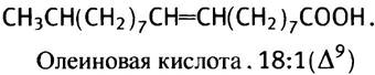
However, this enzyme system is unable to introduce an additional double bond between the central double bond and the methyl end of the molecule. Therefore, it cannot be utilized by the animal organism for the synthesis of linoleic acid, which contains two double bonds, or linolenic acid, which contains three.
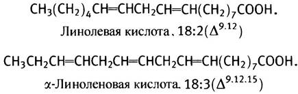
Since these acids are essential for the synthesis of Membrane Lipids and Eicosanoids, their sole source is the diet (plants possess enzymes that synthesize polyunsaturated fatty acids with double bonds near the terminal end of the molecule). Meanwhile, the liver can further elongate linoleic acid and introduce additional double bonds into it. This is how arachidonic C20-acid (20:4, ∆5,8,11,14) with four double bonds is synthesized, as well as C22- and C24-acids, which are constituents of neural tissue lipids. All of these conversions proceed via acyl-CoA derivatives.
Synthesis of Triglycerides and Membrane Lipids from Fatty Acids
The organism synthesizes neutral fats for energy storage. To form an ester bond between glycerol and fatty acids, the latter must be activated by coupling with acyl-CoA; this reaction is catalyzed by acyl-CoA synthetase:
RСООН + СоА-SН-АТР —> RСО-S-СоА + АМР + РРi.
The Free energy of hydrolysis for standard carboxylic acid esters is lower than that of the corresponding thioesters; therefore, such activation of fatty acids renders triglyceride synthesis an exergonic process.
Note that the acceptor of acyl groups is not glycerol itself, but glycerol-3-phosphate, which is formed by the reduction of dihydroxyacetone phosphate, an intermediate of glycolysis.

In the liver (but not in adipocytes), glycerol-3-phosphate is also produced via the direct phosphorylation of glycerol. All stages of triglyceride synthesis are illustrated in Fig. 10.5.
Fig. 10.5. Synthesis of triglycerides (neutral fats) from glycerol-3-phosphate

Synthesis of Glycerophospholipids
Cell membranes contain Phospholipids, whose structural backbone is formed by a glycerol moiety.
Phosphoglycerides differ only in The Nature of the alcohol residue linked to phosphatidic acid via a phosphoester bond.
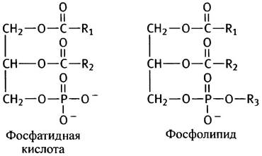
R3 can be a Serine, ethanolamine, Choline, Inositol, or diacylglycerol residue. Phosphatidylethanolamine and phosphatidylcholine predominate in eukaryotic membranes, so we will examine their synthesis first. Ethanolamine and choline have the following structures:
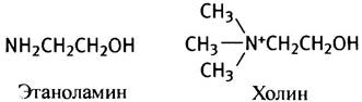
Both of these compounds are alcohols and can be designated as R-OH. To participate in phospholipid synthesis, these alcohols are first "activated" via a two-step process. In the first step, they are phosphorylated by ATP:

In the second step, the phosphorylated alcohols react with cytidine triphosphate (CTP), an ATP analog in which the adenine residue is replaced by a cytosine (cytidine is cytosine riboside). CTP is present in all cells because it is required for RNA and DNA Synthesis (see Chapter 21). One can only guess why CTP, rather than ATP, is used in the phosphorylation of alcohols. Whether this is an evolutionary "quirk" or a design whose purpose we do not yet understand remains unknown. In any case, the reaction of CTP with phosphoesters closely parallels the formation of UDP-glucose from glucose-1-phosphate and UTP (see p. 87):
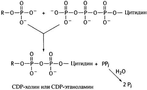
The hydrolysis of pyrophosphate drives this reaction to completion, making it irreversible and highly exergonic. The final reaction synthesizing phosphatidylethanolamine and phosphatidylcholine involves a 1,2-diglyceride, which is formed from phosphatidic acid through the action of a phosphatase (see p. 54).
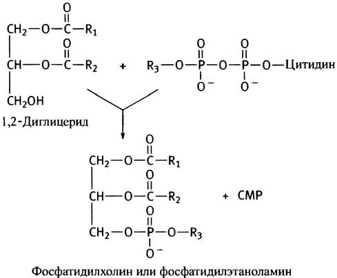
Thus, The pathway of phosphoglyceride synthesis described above boils down to the activation of the alcohol polar HEAD group by CTP, followed by the transfer of the phosphorylated alcohol to another alcohol, a diglyceride. Another energetically feasible pathway involves the activation of the diglyceride and the transfer of the diglyceride phosphate residue to choline or ethanolamine. This is how phosphatidylinositol and cardiolipin are synthesized in Eukaryotic cells (see p. 55). In the latter case, a second diglyceride molecule acts as the alcohol.

A schematic Overview of both glycerophospholipid synthesis pathways is presented in Fig. 10.6. In reality, the situation is considerably more complex due to various interconversions of phospholipids. For instance, they can exchange serine and ethanolamine residues, phosphatidylserine can be decarboxylated to form phosphatidylethanolamine, and the latter can be methylated to yield phosphatidylcholine. All of these processes occur to varying degrees across different organisms.
Fig. 10.6. Two pathways of phosphoglyceride synthesis. ROH represents an alcohol whose residue serves as the polar head group in the synthesized phospholipid. In mammals, cardiolipin and phosphatidylinositol are synthesized via pathway a, whereas phosphatidylcholine, phosphatidylethanolamine, and phosphatidylserine are synthesized via pathway b. Phospholipid synthesis in bacteria proceeds via pathway a

Site of Membrane Lipid Synthesis
In animal cells, the synthesis of membrane lipids takes place On the surface of the smooth Endoplasmic reticulum. The synthesized lipids are transported to their destinations either via membrane vesicles budding off from the Golgi apparatus or with the help of carrier Proteins (the precise mechanism of this transport is not yet fully understood).
Synthesis of Prostaglandins and Related Compounds
The name of this group of compounds, eicosanoids, originates from the Greek numeral for "twenty." This corresponds to the exact number of carbon atoms in their molecules, The Biosynthesis of which begins with polyunsaturated fatty acids. Although eicosanoids are present in the body in minute amounts, their range of physiological functions is remarkably broad.
Eicosanoids are divided into three main groups named after the cells in which they were first discovered: prostaglandins, thromboxanes, and Leukotrienes. Prostaglandins were originally found in semen. Initially, they were thought to originate from the Prostate Gland, but it was later shown that they come from the Seminal Vesicles. It is now known that prostaglandins are synthesized in many Tissues. Thromboxanes were discovered in Blood Platelets (thrombocytes), and leukotrienes in white Blood Cells (leukocytes).
Prostaglandins and thromboxanes
Numerous prostaglandins are known, differing in subtle structural details. They are subdivided into classes designated as PGA, PGE, and PGF. The numerical subscript accompanying them
indicates the number of double bonds in the side chains attached to the cyclopentane ring. As an example, Fig. 10.7, b illustrates the structure of PGE2. In humans, the primary active compounds are those with two double bonds, which are synthesized from arachidonic acid (Fig. 10.7, a). Other prostaglandins are formed from different polyunsaturated fatty acids. Arachidonic acid is present in the body primarily as a fatty acid residue esterified in phospholipids. Prior to prostaglandin synthesis, it is cleaved off by phospholipase.
Fig. 10.7. Structures of prostaglandins and related compounds: a - Arachidonic acid; b - Prostaglandin E2 (PGE2); c - Thromboxane A2 (TXA2); d - Leukotriene A4 (LTA4)
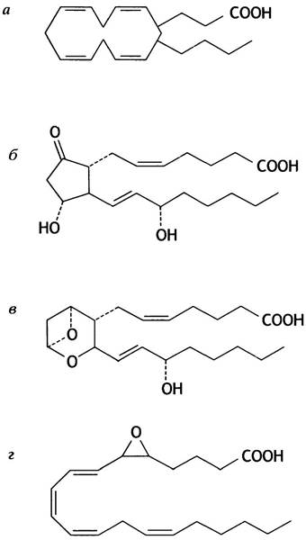
The initial step in prostaglandin synthesis is catalyzed by cyclooxygenase, which converts arachidonic acid into a cyclic compound (see Fig. 10.7, a). This enzyme is inhibited by aspirin (acetylsalicylic acid), which acetylates the hydroxyl group of a serine residue located in the enzyme's Active Site.
Because thromboxanes (Fig. 10.7, c) are formed via the modification of certain prostaglandins, aspirin also blocks their synthesis.
Prostaglandins perform A wide variety of physiological functions. Immediately upon synthesis, they are released From Cells to act as local Hormones by binding to receptors on neighboring cells. They induce pain, inflammation, and fever, stimulate smooth Muscle contraction, participate in Blood Pressure Regulation, and suppress Hydrochloric acid secretion in The Stomach. Thromboxanes promote platelet aggregation, thereby stimulating blood clotting.
By inhibiting cyclooxygenase, aspirin suppresses many of these effects. Specifically, at low doses (100 mg daily), it blocks thromboxane synthesis, thereby reducing platelet aggregation. This preventative therapy helps prevent Heart attacks by lowering the likelihood of clot formation in the coronary Arteries.
Leukotrienes
The structure of a representative leukotriene is shown in Fig. 10.7, d. Leukotrienes are also synthesized from arachidonic acid, but via a different enzyme—lipoxygenase. By inducing sustained smooth muscle contraction, they constrict the Airways, thus playing a key role in the onset of asthma attacks. In addition, leukotrienes are involved in regulating leukocyte activity.
Cholesterol Synthesis and Its Regulation
It is generally believed that most cells in the body are capable of synthesizing cholesterol. Cholesterol is an essential component of Plasma Membranes and serves as a precursor for Bile acids and Steroid Hormones. The liver, and to a lesser extent the Small Intestine, are the primary sites of cholesterol production. Chapter 6 discussed the two mechanisms by which cholesterol in the liver and other tissues forms a single, well-balanced pool.
Remarkably, acetyl-CoA serves as the sole Starting Material for the synthesis of this large, complex cholesterol molecule. This biosynthetic pathway is highly intricate. The initial stages are of particular interest because it is at this level that The rate of cholesterol production is determined. This involves the biosynthesis of mevalonic acid, the first intermediate dedicated exclusively to cholesterol synthesis. The corresponding reactions, illustrated in Fig. 10.8, take place in the cytosol. Hydroxymethylglutaryl-CoA also serves as a precursor for acetoacetate in the Formation of Ketone bodies, though for this purpose it is synthesized within the mitochondria.
Fig. 10.8. Synthesis of mevalonate from acetyl-CoA
At the level of hydroxymethylglutaryl-CoA reductase, cholesterol synthesis is regulated through three mechanisms. Via feedback inhibition, cholesterol: 1) suppresses the Synthesis of the reductase at the Gene level; 2) triggers the degradation of the enzyme; 3) inactivates the enzyme via phosphorylation (for the latter, see p. 161). Special pharmaceutical drugs have been developed to lower blood cholesterol levels for therapeutic purposes. Structurally resembling mevalonic acid, they interact with hydroxymethylglutaryl-CoA reductase to act as transition-state analogs, ensuring their tight binding to the Active Site of the enzyme. Two such drugs are known as lovastatin and simvastatin.
Chapter 10 Questions
1. In the Cytology/cytology/16.html">Early stages of fatty acid synthesis, acetyl-CoA is first carboxylated and then almost immediately decarboxylated. What is the purpose of this?
2. Draw a diagram illustrating the main steps of the fatty acid elongation cycle, omitting the reduction reactions.
3. What is the Structural organization of fatty acid synthase in eukaryotes? How does it differ from the E. coli enzyme? Where is fatty acid synthesis more efficient—in bacteria or in animals?
4. Draw the structures of NAD+ and NADP+. What is the reason for the simultaneous existence of both coenzymes?
5. In which tissues is fatty acid synthesis concentrated in eukaryotes?
6. Palmitate synthesis from acetyl-CoA takes place in the cytoplasm, whereas the primary producer of acetyl-CoA—pyruvate decarboxylase—is located in the mitochondria. Acetyl-CoA cannot cross the membrane. How does it get from the mitochondria into the cytoplasm?
7. What is the source of the NADPH required for fatty acid synthesis?
8. Describe how triglycerides are synthesized from fatty acids.
9. What role does CTP play in Lipid Metabolism?
10. What are eicosanoids? What are their precursors? What is their physiological significance? What is their connection to aspirin?
11. How do drugs that inhibit cholesterol synthesis work?
Last update: 06/08/2026
Editorial and Educational Adaptation: This material has been compiled based on the primary/original source text. The project team performed an editorial review, corrected technical inaccuracies, structured sections, and adapted the content for an educational format.
What was processed:
- elimination of formatting defects (OCR errors, structural breaks, corrupted characters);
- editorial organization of content;
- standardization of terminology in accordance with academic sources;
- verification of factual statements against the original source text.
All mentions of the author, publication year, and origin of the primary text have been preserved in accordance with the source.