BIOCHEMISTRY AND MOLECULAR BIOLOGY - W. ELLIOTT - 2002
CHAPTER 8. GLYCOLYSIS, THE CITRIC ACID CYCLE, AND THE ELECTRON TRANSPORT SYSTEM: REACTIONS OF THESE METABOLIC PATHWAYS
Stage 1. Glycolysis
Glycolysis is defined as The breakdown of a glucose molecule or a Glycogen glucosyl residue into two Pyruvate molecules.
Glucose or glycogen?
Up to this point, our Structure/133.html">Discussion has focused primarily on glucose METABOLISM. However, under normal physiological conditions, most Tissues maintain a glycogen store, which undergoes glycolysis to a greater extent than free glucose. There is a fundamental difference between these two processes. When glycogen is broken down, it yields glucose-1-phosphate, which is subsequently converted into glucose-6-phosphate (see p. 86). In the Liver, the latter is hydrolyzed to release glucose into the bloodstream. In all tissues, however, glucose-6-phosphate also enters The Glycolytic Pathway and is ultimately degraded to pyruvate. It is worth noting that glucose-6-phosphate is generated not only from glycogen but also from glucose via ATP-dependent phosphorylation. This reaction constitutes one of the steps in glycogen synthesis (see p. 86). Whether glucose-6-phosphate is channeled into glycogen storage, routed to pyruvate, or used to maintain Blood glucose levels (in the liver) depends entirely on the physiological needs of the Organism and regulatory mechanisms. The interplay between glycolysis, glucose, and glycogen is illustrated in Fig. 8.1. Phosphorylating a glucose molecule to form glucose-6-phosphate consumes one molecule of ATP. Conversely, no ATP is expended when glucose-6-phosphate is produced directly from glycogen, because the energy of the glucosyl group in glycogen (the ∆G0' of glucosyl group Hydrolysis) is equivalent to that of a phosphoester bond.
Class="center">Fig. 8.1. Formation of glucose-6-phosphate from glycogen or glucose and its subsequent metabolic fates. For details on The regulatory mechanisms governing these processes, see Chapter 12

Why does the initial stage of glycolysis consume ATP?
It may seem paradoxical that a metabolic pathway designed to generate ATP begins by consuming it. What is the purpose of this ATP expenditure? The reason is that glycolysis relies on phosphorylated intermediates, and phosphorylating glucose requires "spending" a high-energy phosphate bond. However, this initial investment is later recovered, as the subsequent steps of glycolysis yield a net surplus of two ATP molecules.
Why is glucose-6-phosphate converted into fructose-6-phosphate?
The next step in glycolysis involves the isomerization of the aldo-sugar derivative, glucose-6-phosphate, into the keto-sugar derivative, fructose-6-phosphate. The ultimate objective of glycolysis is to cleave glucose into two C3 fragments. An aldol Condensation reaction takes place between an aldehyde and a ketone (or between two aldehydes), condensing them into an α-keto alcohol known as an aldol or a β-hydroxycarbonyl compound (Fig. 8.2). Because aldol formation is reversible, the reverse reaction can be harnessed for their Cleavage. Returning to glycolysis: unlike glucose-6-phosphate, fructose-6-phosphate possesses an aldol structure, which is clearly visible in the straight-chain formulas of these compounds shown in Fig. 8.3.
Fig. 8.2. Aldol condensation: reaction of an aldehyde with a ketone or another aldehyde

Fig. 8.3. Linear formulas of monosaccharide isomers: aldoses and ketoses. In solution, glucose-6-phosphate exists in equilibrium with its cyclic pyranose (six-membered) form, whereas fructose-6-phosphate favors the furanose (five-membered) form

Glucose-6-phosphate is isomerized into fructose-6-phosphate in a reaction catalyzed by glucose phosphate isomerase. Subsequently, another enzyme, Phosphofructokinase, utilizes ATP to convert fructose-6-phosphate into fructose-1,6-bisphosphate. Following this, aldolase catalyzes the Cleavage of the hexose bisphosphate into two triose phosphates: glyceraldehyde-3-phosphate and dihydroxyacetone phosphate (Fig. 8.4).
Fig. 8.4. Conversion of glucose-6-phosphate into two C3 compounds. Sugars are depicted as straight chains for clarity

Figure 8.5 illustrates the same reaction using cyclic sugar structures rather than linear ones, with fructose-6-phosphate shown as a five-membered ring (the furanose form). The Standard Free energy change for this aldol cleavage is ∆G°' = +24.3 kJ • mol-1. At first glance, such a reaction should be thermodynamically unfavorable, allowing only the reverse reaction to proceed. However, there is a subtle thermodynamic catch: one molecule of fructose-1,6-bisphosphate is converted into two product molecules. Consequently, the actual (rather than standard) free energy change (∆G) depends heavily on the concentrations of the reactants and products. Under intracellular conditions, the value of ∆G is small, rendering the reaction readily reversible.
Free Energy Changes ∆G°′ and ∆G in aldol cleavage
Although the standard free energy change for aldol cleavage is ∆G°′ = +24.3 kJ • mol-1, the reaction proceeds smoothly under physiological conditions, even though far less endergonic cellular reactions are often blocked. We know that ∆G°′ corresponds to standard conditions where the concentrations of reactants and products are all 1 M. Because intracellular metabolite concentrations typically range from 10-4 to 10-3 M, the actual ∆G always deviates from ∆G°′. While knowing ∆G°′ generally helps assess the feasibility of a given reaction, this approach is less straightforward for aldolase because the intracellular ∆G values for the forward and backward reactions differ very little. This accounts for the unusual thermodynamic behavior of the aldolase reaction.
Fig. 8.5. Conversion of glucose-6-phosphate into two C3 compounds, showing the most prevalent cyclic structures of the sugars

This occurs because the number of molecules doubles during the forward reaction. The actual free energy change, ∆G, is related to the standard free energy change, ∆G°′, by the following equation (see Chapter 1):

or in this case:

Since the product concentration is low, the term RТ • ig {[products]/[reactants]} has a large negative value, which makes the reaction reversible within The Cell. Assuming that the concentration of all reactants in the cell is 10-4 M, then

The value of ∆G at the chosen reactant concentrations allows the reaction to proceed freely in both directions.
Interconversion of dihydroxyacetone phosphate and glyceraldehyde-3-phosphate
Glyceraldehyde-3-phosphate and dihydroxyacetone phosphate are isomer molecules. Their interconversion is catalyzed by the enzyme Triosephosphate isomerase.

The two substances are in equilibrium, but because glyceraldehyde-3-phosphate is continuously consumed in the subsequent step of glycolysis, all the dihydroxyacetone phosphate is gradually converted into glyceraldehyde-3-phosphate.
Glyceraldehyde-3-phosphate dehydrogenase. Generation of a high-energy phosphate bond
The aldehyde group in glyceraldehyde-3-phosphate undergoes oxidation mediated by NAD+. A typical oxidation of a -CHO group to a -COO- group is
characterized by a large negative ∆G value, quite comparable to the Free energy of formation of a high-energy phosphoryl group. Therefore, in this case, the main task of the catalyzing enzyme is to harness the free energy of oxidation to synthesize a high-energy phosphate.
The Active Site of glyceraldehyde-3-phosphate dehydrogenase contains a Cysteine amino acid residue whose side chain features a thiol (sulfhydryl) group that reacts with the substrate's aldehyde group to form a thiohemiacetal:

This compound undergoes oxidation, transferring electrons to the NАD+ molecule. In the process, the thiohemiacetal is converted into a thioester:

Like acetyl-CoA, the thioester (R-СО-S-) is a high-energy compound that, upon phosphorolysis, is converted into a mixed anhydride of phosphoglyceric and phosphoric acids:

The high-energy group R-CO-PO2-3 can transfer its phosphoryl residue to ADP to form ATP. For purely nomenclatural reasons, The enzyme catalyzing this transfer is named after the reverse rather than the forward reaction: phosphoglycerate kinase. Kinases are always named after the compound that shares the reaction equation side with ATP (Fig. 8.6).
Fig. 8.6. Conversion of glyceraldehyde-3-phosphate to 3-phosphoglycerate

The phosphoryl group generated during this process remains attached to the enzyme's true substrate. Consequently, this process is termed substrate-level phosphorylation. 3-Phosphoglycerate is a low-energy phosphate compound incapable of phosphorylating ADP. However, in subsequent steps of glycolysis, intramolecular rearrangements convert this low-energy phosphoester into a high-energy phosphoryl group that can be transferred to ADP to yield ATP.
Final stages of glycolysis
First, the phosphoryl group of 3-phosphoglycerate is transferred from position 3 to position 2 by phosphoglycerate mutase.

In this form, the intramolecular phosphate transfer reaction occurs only in plants. In rabbit Muscle, for instance, the process is more complex. The enzyme contains a phosphate residue that is transferred to the 2-OH group of 3-phosphoglycerate, converting it into 2,3-diphosphoglycerate. Subsequently, the 3-phosphoryl group from 2,3-diphosphoglycerate is transferred back to the enzyme to replenish the donated phosphate, resulting in The formation of 2-phosphoglycerate from 3-phosphoglycerate. The next stage of glycolysis involves the removal of a Water molecule from 2-phosphoglycerate. Although dehydrating Enzymes are typically called dehydratases, in this case the historical name enolase is used, which reflects the Chemical Nature of the reaction—the formation of a substituted enol.

Although the standard free energy change of the reaction catalyzed by enolase is only ∆G°' = +1.8 kJ • mol-1, phosphoenolpyruvate, unlike phosphoglycerate, is a high-energy compound; its hydrolysis corresponds to ∆G°' = -62.2 kJ • mol-1. The reason for this significant difference in ∆G°' values lies in the fact that the reaction product—the enol form of pyruvate—spontaneously converts into the keto form in a reaction characterized by a large negative ∆G°'. Keto-enol Tautomerism is well studied. The keto form (in this case, pyruvate) is usually thermodynamically more stable than the enol form. Therefore, the substantial negative free energy change of the overall reaction is a result of the contribution from this isomerization. The phosphate group from phosphoenolpyruvate is transferred to ADP by the enzyme pyruvate kinase (again, following the convention where kinases are named after the reverse phosphorylation reaction of the substrate, even if that specific reaction does not occur in this direction). The irreversibility of The conversion of phosphoenolpyruvate to pyruvate is of great importance for Gluconeogenesis. Interestingly, in plants and microorganisms, pyruvate is converted into phosphoenolpyruvate, but this is mediated by a different enzyme and, more importantly, consumes twice as much ATP rather than an equimolar amount.
The complete Metabolic Transformations in glycolysis are shown in Fig. 8.7.
Fig. 8.7. The glycolytic pathway. Irreversible reactions are marked with red arrows

ATP Balance in Glycolysis
If glycolysis starts with glucose, 2 molecules of ATP are consumed in the formation of glucose-1-phosphate and fructose-1,6-diphosphate. However, the phosphoglycerate kinase and pyruvate kinase reactions generate 2 molecules of ATP (see Fig. 8.7). Furthermore, considering that 1 molecule of fructose-1,6-diphosphate splits into 2 molecules of glyceraldehyde-3-phosphate, their subsequent conversion to pyruvate is accompanied by The production of 4 ATP molecules. Consequently, the net yield of high-energy compounds resulting from the glycolytic breakdown of 1 glucose molecule is 2 ATP molecules. If glycolysis starts from glycogen, only 1 ATP molecule is consumed to form fructose-1,6-diphosphate while the same 4 molecules are synthesized, resulting in a net yield of 3 ATP molecules.
Reoxidation of Cytoplasmic NADH by Electron Shuttle Systems
Under aerobic conditions, the NADH produced during glycolysis undergoes reoxidation via The transfer of electrons into the Mitochondria. NADH itself cannot cross the mitochondrial membrane. Two systems exist to facilitate this electron transfer.
The first system utilizes dihydroxyacetone phosphate (DHAP), which is formed during the cleavage of fructose-1,6-diphosphate by aldolase. In the Cytoplasm, electrons from NADH are transferred to DHAP, yielding glycerol-3-phosphate. This reaction is catalyzed by the enzyme glycerol-3-phosphate dehydrogenase (here too, the enzyme is named after the reverse reaction). The glycerol phosphate shuttle system is illustrated in Fig. 8.8.
Fig. 8.8. Scheme of the glycerol phosphate shuttle system, transferring Electrons from cytoplasmic NADH to the mitochondrial Respiratory Chain

Glycerol-3-phosphate reaches The inner mitochondrial membrane (recall that the outer membrane is freely permeable to most low-molecular-weight substances), where membrane-bound glycerol-3-phosphate dehydrogenase (which contains FAD as its prosthetic group) transfers electrons from glycerol-3-phosphate into the Mitochondrial Electron Transport chain. The resulting DHAP returns to the cytoplasm, thus completing the shuttle cycle. It should be noted that the electron carrier glycerol-3-phosphate does not enter the mitochondrial matrix. Its sole function is to transfer electrons from cytoplasmic NADH to the mitochondrial Electron Transport Chain.
The other shuttle system—the malate-aspartate shuttle—transfers electrons from cytoplasmic NADH to mitochondrial NAD+. This results in the formation of mitochondrial NADH, which is subsequently oxidized in The electron transport chain. In the cytoplasm, NADH reduces oxaloacetate to malate. Malate then enters the mitochondria via a specific transporter, where it is reoxidized to oxaloacetate with the reduction of NAD+. Since oxaloacetate cannot cross back out of the mitochondrial membrane, it is first converted into aspartate, which is then transported out into the cytoplasm. In the cytoplasm, aspartate undergoes deamination to form oxaloacetate, thereby completing the shuttle cycle (Fig. 8.9). The net outcome of this cycle is The oxidation of cytoplasmic NADH by mitochondrial NAD+.
The glycerol phosphate and malate-aspartate shuttle mechanisms presumably operate with varying intensities across different tissues. They differ significantly. The first system uses cytoplasmic NADH to reduce mitochondrial FAD, which serves as the prosthetic group of the flavoprotein glycerol-3-phosphate dehydrogenase. The Redox Potential of FADH2 is higher than that of NADH. FADH2 passes electrons further down the transport chain. The farther oxygen is located from the carrier in this chain, the more ATP will be synthesized as electrons move toward oxygen. The oxidation of one molecule of cytoplasmic NADH coupled with the reoxidation of mitochondrial FADH2 yields 1.5 molecules of ATP. Starting from a molecule of cytoplasmic NADH, the malate-aspartate shuttle cycle culminates in the reduction of a mitochondrial NAD+ molecule, the reoxidation of which yields 2.5 molecules of ATP.
Fig. 8.9. The malate-aspartate shuttle system for transferring electrons from cytoplasmic NADH to mitochondrial NAD+. Unlike the glycerol phosphate system, the interconversion mechanism of oxaloacetate and aspartate is reversible and supplies NADH to the mitochondria only when the cytoplasmic NADH/NAD+ ratio exceeds that of the mitochondrial matrix

Pyruvate Transport into Mitochondria
Alongside NADH, pyruvate is another major product of glycolysis. Unless it is reduced to lactate, pyruvate enters the mitochondria via a specific transport system that operates as an antiport mechanism exchanging pyruvate for OH- ions.
Stage 2. The Citric Acid Cycle
Pyruvate, the product of glycolysis, enters the metabolic Reactions of the citric acid cycle not directly, but only after being first converted into acetyl-CoA. Therefore, we begin this section by examining the mechanisms of acetyl-CoA formation from pyruvate.
Conversion of Pyruvate to Acetyl-CoA: A Preliminary Step of The Citric Acid Cycle
Within the mitochondrial matrix, pyruvate is converted into acetyl-CoA, after which the acetyl group enters the citric acid cycle. Overall, the reaction catalyzed by pyruvate dehydrogenase can be described by the following equation:
Pyruvate + NAD+ + CoA-SH —> Acetyl-S-CoA + NADH + H+ + CO2.
The conversion of pyruvate to acetyl-CoA is an irreversible reaction (∆G°′ = -33.5 kJ • mol-1). This means that Fatty acids in animal organisms cannot be converted directly into glucose, although Bacteria and plants accomplish this via a specialized mechanism.
Pyruvate dehydrogenase has a rather complex structure. It consists of multiple polypeptide chains and integrates three distinct enzymatic activities within a single molecule, each responsible for a specific reaction stage. The First stage is decarboxylation, during which CO2 is cleaved from pyruvate, while the remaining part of the molecule is converted into a hydroxyethyl group

which attaches to the cofactor thiamine pyrophosphate. This cofactor is synthesized in the body from Vitamin B1 (thiamine), a deficiency of which leads to impaired Carbohydrate Metabolism.

The hydroxyethyl group of pyruvate undergoes several steps to be converted into the acetyl moiety of acetyl-CoA, with this conversion being accompanied by the reduction of NAD+. For obvious reasons, this process is known as oxidative decarboxylation. Another important cofactor participating in these redox reactions is Lipoic Acid, which can exist in two forms: acyclic (reduced) and cyclic (oxidized).

The lipoic acid residue is incorporated into the enzyme as a prosthetic group, attached to the side chain of a Lysine residue via a -CO-NH- amide bond. In Fig. 8.10, which illustrates the involvement of lipoic acid in acetyl-CoA synthesis, the lipoic-lysine residues are depicted as a disulfide moiety, while symbols E1, E2, and E3 designate the three enzymes that make up the single multienzyme complex.
Fig. 8.10. Mechanism of the pyruvate dehydrogenase reaction. TPP — thiamine pyrophosphate; E1, E2, E3 — enzymes within the complex

What is the true magic of the cycle?
At first glance, the citric acid cycle appears to be merely a series of standard Chemical Reactions; however, it exemplifies nature's remarkable ingenuity in generating universal fuel in the form of the reduction equivalents NADH and FADH2, partly produced through the cleavage of water. This fuel is subsequently utilized by the mitochondrial electron transport chain to generate ATP from ADP and Pi. Unlike in Photosynthesis (see Chapter 14), water cleavage here does not serve as the primary energy storage process, and oxygen is released not in free form, but as part of CO2. The energy derived from the Conversion of the acetyl group of acetyl-CoA is harnessed to drive this process. A sequential examination of the cycle's details is essential to fully understand its nature and significance.
Acetyl-CoA enters the cycle by reacting with oxaloacetate, yielding citrate (the anion of citric acid). This reaction consumes a water molecule. As the cycle completes, oxaloacetate is regenerated, and the acetyl group of acetyl-CoA is consumed. A single turn of the cycle (Fig. 8.11) results in the formation of 2 molecules of CO2; the reduction of 3 molecules of NAD+ to NADH; the reduction of 1 molecule of FAD to FADH2; the synthesis of 1 molecule of GTP from GDP and Pi; and the conversion of acetyl-CoA into CoA-SH as it loses its acetyl group.
Fig. 8.11. Entry and exit of substances in the citric acid cycle. Individual reactions of the cycle are not shown

Summing up all the reduction equivalents generated—three in NADH and one in FADH2—yields a total of eight, since reduction is a two-electron process in both cases. To this number, we must add one, as another equivalent is released in the form of the newly formed thiol group in CoA-SH. Thus, a single turn of the cycle releases 9 reduction equivalents, which is equivalent to 9 hydrogen atoms.
Three hydrogen atoms and one oxygen atom are supplied by the CH3CO-S-CoA acetyl group. It remains to determine the Origin of the remaining six hydrogen atoms and three oxygen atoms. Oxygen does not participate in the cycle directly, so we must identify the source of the three oxygen atoms required to form two CO2 molecules (the fourth one originates from the acetyl group). These, much like the hydrogen atoms, can be provided by water molecules. One water molecule is consumed during citrate synthesis, and another during the formation of oxaloacetate. THE ORIGIN OF the remaining two hydrogen atoms and one oxygen atom still needs to be explained, though this will be more conveniently addressed a bit later (see p. 120).
The result is quite striking. On the surface, it appears as though the electrons from water spontaneously ascend the energy scale to reach the level of the reduction equivalents NADH and FADH2 (the same holds true for the electrons of the acetyl group of acetyl-CoA). However, this does not represent a direct transfer of hydrogen atoms from water to NADH and FADH2; rather, it is a chemical balance in which hydrogen derived from water reduces NAD+ and FAD, while oxygen from water is consumed to form CO2. Naturally, these are endergonic processes that require an input of free energy. The source of this energy is the conversion of the pyruvate acetyl group into acetyl-CoA and subsequent derivatives.
Simplified Description of the citric acid cycle
When studying the numerous chemical reactions that make up the cycle one by one, one can easily lose sight of its overall "architecture." Therefore, it makes sense to first examine a simplified version of the cycle.
Oxaloacetate is the substance where everything begins and ends. Acetate is CH3COO-, the oxalyl group (the acyl residue of oxalic acid) is -OOC-CO-, and oxaloacetate has the structure:

The acetyl group of acetyl-CoA attaches to oxaloacetate, converting it into citrate. Examining the formula of the latter reveals the site of attachment:

Citrate, the anion of the optically symmetrical tricarboxylic C6-acid, is isomerized into the anion of another tricarboxylic acid, isocitrate, whose molecule contains a single asymmetric center. Isocitrate is then successively converted into α-ketoglutarate (C5), succinate (C4), fumarate (C4), malate (C4), and finally back to the starting oxaloacetate (Fig. 8.12).
Fig. 8.12. Reactions of the citric acid cycle. The diagram illustrates The structure of the acids participating in the cycle and their interrelationships.

The cycle is now complete, and the acetyl residue has disappeared. Let us move on to a more detailed examination of the Chemical transformations of all substances involved in the cycle.
Mechanisms of the Citric Acid Cycle Reactions
For convenience, these reactions can be divided into three groups: 1) the synthesis of citrate, the reaction through which the acetyl group enters the cycle; 2) the conversion of citrate (C6) into α-ketoglutarate (C5); 3) the transformations of C4 compounds, from succinate to oxaloacetate.
Citrate Synthesis
The enzyme responsible for citrate synthesis is called citrate synthase (note: synthase, not synthetase, since no ATP is involved in the reaction). It catalyzes the condensation of acetyl-CoA with oxaloacetate, yielding citryl-CoA, which is unstable and rapidly hydrolyzes to citrate and free CoA. The overall reaction is irreversible, as ∆G°′ = -32 kJ • mol-1.

Conversion of Citrate to α-Ketoglutarate
The Essence of the Citrate —> Isocitrate reaction lies in the relocation of the hydroxyl group of the symmetrical citrate molecule from position 3 to position 2, forming
an asymmetric isocitrate molecule. The reaction mechanism involves the simultaneous, reversible dehydration of citrate and isocitrate into their common intermediate product, cis-aconitate (aconitic acid was first discovered in plants of the genus Aconitum).

Both reactions are catalyzed by the enzyme aconitase. This name is a nod to tradition, as enzymes of this class are conventionally referred to as dehydratases.

Within the cell, isocitrate is readily converted into α-ketoglutarate; therefore, the reaction equilibrium is shifted toward the formation of isocitrate:
Citrate —> cis-Aconitate —> Isocitrate.
Using Lactate dehydrogenase as an example, we have already encountered NAD+-dependent dehydrogenases. The oxidation reactions of isocitrate and lactate belong to the same reaction type:

In the cycle, isocitrate dehydrogenase catalyzes the reaction leading to the formation of α-ketoglutarate:

The intermediate product of isocitrate oxidation is oxalosuccinate, which, like all β-keto acids (where the keto group is in the β-position relative to the central carboxyl), is unstable and readily loses a carboxyl group as CO2. Decarboxylation occurs directly on The surface of isocitrate dehydrogenase, yielding α-ketoglutarate (C5).
Four-Carbon Acids
α-Ketoglutarate is an analogue of pyruvate, and both of these compounds share a common general formula:

where pyruvate corresponds to R= -СН3, and α-ketoglutarate to R= -СН2СН2СОО-. Earlier, we looked at the conversion of pyruvate into acetyl-CoA and СO2, which is catalyzed by pyruvate dehydrogenase:

Similar to the pyruvate dehydrogenase complex, α-ketoglutarate dehydrogenase (also an enzyme complex) catalyzes the formation of succinyl-CoA from α-ketoglutarate:

However, whereas acetyl-CoA is used in the cycle to synthesize citrate, succinyl-CoA is hydrolyzed to succinate and free CoA-SH. The standard Gibbs free energy change for the hydrolysis of this thioester is ∆G°′ = -35.5 kJ • mol-1. This is sufficient to drive the synthesis of a high-energy phosphate bond, which takes place in the next step.
Coupling of Succinyl-CoA Hydrolysis with GTP Synthesis
The overall equation for the coupled reactions is:
Succinyl-CoA + GDP + Рi <-> Succinate + GTP + CoA-SH
∆G°' = -2.9 kJ • mol-1.
The enzyme that catalyzes this reaction is called succinyl-CoA synthetase (for nomenclature reasons, the enzyme is named after the reverse reaction, which does not actually occur in the cell). Unlike animals, plants utilize an analogous enzyme that synthesizes ATP instead of GTP.
The succinyl-CoA synthetase reaction begins with the phosphorolysis of the thioester group in succinyl-CoA:

Earlier (see p. 118), we drew up a balance sheet for hydrogen and oxygen atoms in the cycle, and balancing it left us two hydrogen atoms and one oxygen atom short. We can now explain the reason for this imbalance: the participation of inorganic phosphate in the cleavage of succinyl-CoA. For clarity, let us express the true reaction as an equivalent combination of two reactions:
GDР + Рi —> GТР + Н2O,
Succinyl-CoA + Н2O —> Succinate + CoA-SН.
It should be emphasized that this combination of two reactions does not reflect the true mechanism of the process, but it does help us understand where the missing water molecule comes from.
Conversion of Succinate to Oxaloacetate
In the first stage of this pathway, the FAD-containing enzyme succinate dehydrogenase oxidizes succinate to fumarate. This oxidation is accompanied by the removal of two hydrogen atoms:

Why does this oxidation use FAD rather than NAD+? The answer lies in the redox potential of the succinate-fumarate pair. For thermodynamic reasons, electrons flow from a pair with a lower redox potential to one with a higher redox potential. Succinate turns out to be too weak a reducing agent to reduce NAD+, but it can reduce FAD. This reaction is described by the equation:

The final step, which completes the cycle, is the conversion of fumarate into oxaloacetate. First, The addition of a water molecule to fumarate yields malate (malate is the anion of malic acid). Although it would be logical to name the enzyme carrying out this reaction fumarate hydratase, it is commonly known by its traditional name, fumarase.

Malate is subsequently oxidized to oxaloacetate by the NAD+-dependent enzyme malate dehydrogenase:

This is an endoergic reaction (∆G°′ = +29.7 kJ • mol-1), and it would not proceed spontaneously on its own. However, the formation of oxaloacetate closes the cycle, and this is followed by the first of the reactions we examined: the conversion of oxaloacetate into citrate. This is a strongly exergonic process, resulting in a very low concentration of oxaloacetate. Consequently, the equilibrium of the Malate <-> Oxaloacetate reaction is shifted toward the formation of oxaloacetate. In other words, the actual value of ∆G is much smaller than the standard ∆G°′, which enables the oxidation of malate.
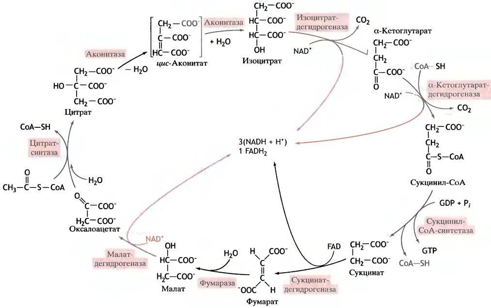
The complete citric acid cycle is shown in Fig. 8.13.
Fig. 8.13. The complete citric acid cycle. The formation of reducing equivalents is highlighted in color. FAD is the prosthetic group of succinate dehydrogenase.
What determines the overall direction of the reactions in the cycle?
As seen from Fig. 8.13, all reactions in the cycle proceed in a coordinated manner in a single direction. This is because three of these reactions have such large negative standard free energy changes (∆G°′) in absolute value that they are practically irreversible. One such reaction is the synthesis of citrate from acetyl-CoA and oxaloacetate (∆G°' = -32.2 kJ • mol-1), the second is the decarboxylation of isocitrate to α-ketoglutarate (∆G°' = -20.9 kJ • mol-1), and the third is the formation of succinyl-CoA from α-ketoglutarate (∆G°' = -33.5 kJ • mol-1).
As a result, the cycle operates in a single direction, even though the equilibrium of the malate dehydrogenase reaction is shifted in the opposite direction (∆G°' = +29.7 kJ • mol-1). The overall change in standard free energy for all reactions in the cycle is negative, which drives its progression in one direction.
Stoichiometry of the Cycle
The overall equation summarizing the operation of the cycle is as follows:
СН3СО-S-СоА + 2Н2О + 3NAD+ + FAD + GDP + Pi—> 2CO2 + 3NADH + 3H+ + FADH2 + CoA-SH + GTP
∆G°' = -40 kJ • mol-1.
If one attempts to balance the hydrogen and oxygen atoms in this equation, a deficit of one mole of H2O becomes apparent.
Substrate Supply for the Citric Acid Cycle
The cycle begins with the condensation reaction of oxaloacetate and acetyl-CoA and concludes with the regeneration of oxaloacetate. The cycle does not operate in isolation from other
metabolic processes, and some of the acids participating in it are utilized for other purposes. However, they are not ingested in sufficient quantities through diet. The metabolism of dietary CARBOHYDRATES is accompanied by the formation of large amounts of C3 compounds in the form of pyruvate, while Lipid Metabolism leads to the appearance of C2 units as acetyl groups. The C4, C5, and C6 acids characteristic of the cycle can be formed from Amino Acids, yet they are also consumed in the synthesis of Certain amino acids (see Chapter 15) and other metabolites, which limits their intracellular concentration. In reality, complex steady-state conditions are established among Various metabolic pathways. The primary function of the cycle is to ensure an uninterrupted supply of "fuel" for energy generation in mitochondria; therefore, of particular importance is the so-called anaplerotic reaction, which involves the synthesis of oxaloacetate from pyruvate and CO2:

The reaction is catalyzed by the enzyme pyruvate carboxylase (not to be confused with Yeast pyruvate decarboxylase!). A distinctive feature of the anaplerotic reaction is that it represents a unique process of converting a C3 acid into a C4 acid. The working moiety of pyruvate carboxylase is biotin. It serves as a cofactor in synthetic carboxylation reactions that utilize so-called active CO2. Biotin is a water-soluble B vitamin. Covalently bound to the enzyme, it reacts with bicarbonate to form carboxybiotin. This reaction is energetically coupled with the hydrolysis of ATP. Carboxybiotin is a reactive yet sufficiently stable compound; it can carboxylate other substances by transferring the carboxyl group to them. The driving force behind such reactions is the large negative free energy change associated with the release of CO2 from carboxybiotin (∆G°' = -19.7 kJ • mol-1). In this case, pyruvate serves as the enzyme's substrate, but other carboxylase enzyme systems also utilize biotin as a cofactor.

Pyruvate carboxylase contains two catalytic sites: one for the carboxylation of biotin, and another for the transfer of the carboxyl group from biotin to pyruvate (in some bacteria, these Functions are performed by two different enzymes). Biotin forms an amide bond with one of the protein's lysine residues. Thus, it is attached to the protein via a long, flexible "arm." It is believed that the flexing of this arm allows biotin to move from one catalytic site of the enzyme to the other (Fig. 8.14).
Fig. 8.14. Biotin in the active site of pyruvate carboxylase. The active moiety of biotin is attached to the protein by a long "arm," which allows biotin to shuttle between different active sites.

Stage 3. Electron Transport Chain from NADH and FADH2 to Oxygen
The oxidation of glucose (or glycogen) comprises three main stages. The first is glycolysis, and the second is the citric acid cycle. We have now arrived at The final stage. From the standpoint of energy generation, the yield from the first two stages is quite modest: only 2 molecules of ATP per molecule of glucose are produced during glycolysis, and the same number is generated in the citric acid cycle (taking into account the energy equivalence of GTP and ATP); one additional molecule of ATP is produced if glycogen serves as the source of glucose. Crucially, the bulk of the energy after the first two stages is stored in the form of 10 molecules of NADH (two from glycolysis, two from the pyruvate dehydrogenase reaction, and six from the citric acid cycle) and 2 molecules of FADH2 (from the citric acid cycle). It should be remembered that 2 molecules of pyruvate are formed from glucose, which drive 2 turns of the cycle.
Thus, the majority of ATP synthesized from ADP and Pi during glucose oxidation is produced as a result of the oxidation of NADH and FADH2.
Electron Transport Chain
Electron carriers are located on the surface or embedded within the inner mitochondrial membrane. This membrane is folded into cristae (see Fig. 7.4), the number and packing density of which correlate with the energy demands of the cell.
What are electron carriers?
Many electron carriers are Proteins containing a heme group as their prosthetic group. These carriers are called Cytochromes because of their red coloration. Different cytochromes are designated by letter subscripts: c1, c, a, and a3, in order of their position along the chain (The Role of two b-type cytochromes will be discussed later). The main Structural Features of heme are shown in Fig. 8.15, and its complete structure is depicted in Fig. 27.3.
Fig. 8.15. Cytology/cytology/92.html">SCHEMATIC STRUCTURE OF heme

The most important thing to know about heme as the prosthetic group of cytochrome carriers is The change in the valence state of its iron atom during their function: it becomes Fe2+ upon receiving an electron from the preceding carrier in the chain, and Fe3+ after transferring the electron to the next one. The properties of the heme molecule itself depend on the protein to which it is attached. Furthermore, the Hemes in different cytochromes may differ in their side chains and the way they attach to the apoprotein. Therefore, there is no contradiction in the fact that cytochromes have different redox potentials even though their prosthetic groups are nearly identical.
Another type of non-heme iron-containing electron carriers consists of proteins in which iron atoms are bound to The sulfhydryl groups of cysteine residues and to sulfide anions, forming iron-sulfur complexes, or centers. The simplest of these is shown in Fig. 8.16.
Fig. 8.16. STRUCTURE OF THE simplest iron-sulfur center. More complex centers contain a greater number of iron and sulfur atoms

As in cytochromes, the iron atoms in these centers can accept and donate electrons, alternating between the ferrous (Fe2+) and ferric (Fe3+) states. Iron-sulfur centers function in concert with flavin-containing enzymes, accepting electrons from succinate dehydrogenase and dehydrogenases involved in Fatty acid oxidation (see Chapter 9). Another type of carrier is the FMN-containing protein. FMN (flavin mononucleotide) is a compound that constitutes the flavin moiety of the FAD molecule (see p. 101). FMN transfers electrons from NADH to the iron-sulfur centers.
The only non-protein electron carrier is ubiquinone (Fig. 8.17), named so because it is both a quinone and is ubiquitous (found everywhere). It is abbreviated as CoQ, UQ, or simply Q. All iron-sulfur centers pass their electrons to ubiquinone.
Upon reduction, ubiquinone acquires not only electrons but protons as well. One-electron reduction converts it into a semiquinone (an organic free radical), while two-electron reduction converts it into a hydroquinone. It is this intermediate formation of a free radical that allows ubiquinone to function as a single-electron as well as a two-electron carrier. A very long hydrophobic "tail" (consisting of 40 carbon atoms in ten sequential isoprenoid units) enables ubiquinone to easily embed itself and move freely within the nonpolar layer of the inner mitochondrial membrane.

Thus, the mitochondrial electron transport chain contains:
✵ an FMN-protein;
✵ proteins featuring non-heme iron-sulfur centers;
✵ unbound ubiquinone, which moves freely within the membrane;
✵ cytochromes—heme-containing proteins.
Fig. 8.17. Ubiquinone, coenzyme Q (a), and its redox transformations (b). R1 is a long hydrophobic tail; R2 = СН3; R3 = ОСН3. The semiquinone may exist in an anionic form (Q-.)

An important point is that one of the cytochromes—cytochrome c (a small, water-soluble protein with a Molecular Weight of ~12.5 kDa, containing just over 100 amino acid residues)—is loosely bound to the outer surface of the inner mitochondrial membrane and readily dissociates from it. All other protein carriers are integral proteins that occupy strictly fixed positions and have a specific orientation within the membrane.
Arrangement of Electron Carriers
Chapter 7 discussed the redox potentials of electron acceptors, specifically noting that the flow of electrons between carriers is directed from a carrier with a higher reducing potential (i.e., a lower redox potential) to one with a lower reducing potential (i.e., a more oxidized one, with a higher redox potential). Within the mitochondrial chain, the carriers possess varying redox potentials.
The values of redox potentials are directly related to changes in free energy (∆G°′). The electron carriers are arranged in the chain such that ∆G°′ progressively decreases, while the redox potential correspondingly increases. Consequently, free energy is released at each step of electron transfer to the next carrier in the chain (Fig. 8.18).
Fig. 8.18. Redox potentials of major Components of the mitochondrial electron transport chain (respiratory chain). Arrows indicate the direction of electron flow

When applied to glucose oxidation, the challenge facing the electron transport chain is to transfer electrons from NADH and FADH2 to oxygen. Although this process involves numerous carriers, they can be grouped into four distinct complexes embedded within the inner mitochondrial membrane (Fig. 8.19). Electrons are shuttled between these complexes by mobile carriers: ubiquinone and cytochrome c. Ubiquinone accepts electrons from complexes I and II and delivers them to complex III, whereas cytochrome c acts as a mediator between complexes III and IV. Complex I transfers electrons from NADH to Q; complex II transfers them from succinate via FADH2 to Q; complex III uses QH2 to reduce cytochrome c; and complex IV transfers electrons from cytochrome c to oxygen. Complexes I, III, and IV are designated, respectively, as NADH-CoQ reductase, CoQH2-cytochrome c reductase, and cytochrome c oxidase.
Fig. 8.19. The four electron carrier complexes of the mitochondrial respiratory chain. Complex I is NADH-CoQ reductase; complex II is succinate-CoQ reductase; complex III is CoQH2-cytochrome c reductase; and complex IV is cytochrome c oxidase. Q denotes ubiquinone, or coenzyme Q. FADH2 is generated in the citric acid cycle from succinate via the succinate dehydrogenase reaction. All complexes are embedded within the inner mitochondrial membrane. Coenzyme Q and cytochrome c function as mobile electron carriers. FADH2 is a component of flavoprotein dehydrogenases that oxidize succinate and fatty acyl-CoA derivatives.

Complex IV, or cytochrome c oxidase, is a multi-subunit protein. It receives electrons from cytochrome c on the outer face of the inner mitochondrial membrane. On their path to oxygen, these electrons pass through cytochromes a and a3, which contain copper atoms that alternate between the Cu+ and Cu2+ oxidation states. Cytochrome c oxidase catalyzes the reduction of molecular oxygen:
O2 + 4е- + 4Н+ —> 2Н2O.
Later (see Chapter 17), we will discuss why it is critical that cytochrome c oxidase transfers all four electrons required to reduce oxygen specifically to water, rather than to partially reduced intermediates such as hydrogen peroxide or reactive free radicals.
How is the free energy released during electron transport utilized for ATP synthesis?
The Mechanism of ATP synthesis from ADP and Pi coupled to Electron transport is fundamentally different from substrate-level phosphorylation during glycolysis (see p. 112). In glycolysis, ATP synthesis is inseparable from the glycolytic reactions themselves; the reaction cannot proceed without generating ATP, which is an integral component of the process. The same holds true for GTP synthesis in the citric acid cycle.
When tracing the movement of electrons along the carrier chain, we did not even mention ATP synthesis. Although ATP production is the ultimate purpose of this chain, electron transport still occurs in damaged mitochondria that are entirely incapable of synthesizing ATP. For decades, this situation baffled biochemists. If electron transport drives ATP generation in Cells and intact mitochondria, why does it fail to produce ATP in damaged mitochondria where electron transfer proceeds successfully? This question could not be answered in terms of familiar substrate-level phosphorylation as seen in glycolysis. The solution was discovered by the British biochemist Peter Mitchell. Working in his home laboratory in 1961, he conceived a radically new concept for coupling electron transport to ATP synthesis. The idea was so original that initially no one took it seriously, and it took considerable time before it gained universal acceptance (Mitchell was eventually awarded the Nobel Prize in 1978). Mitchell's core idea is remarkably simple: gradients serve as the energy source for performing work. For instance, a water level differential drives hydroelectric turbine generators, atmospheric pressure gradients create winds that turn windmills, and so forth. Chemical gradients are no exception. Molecules move down a concentration gradient (from high to low concentration), and if a suitable device is placed in their path, this flow can be harnessed to perform useful work.
Based on his concept, Mitchell concluded that coupling electron transport to ATP synthesis requires three conditions. First, electron transport must generate a gradient; second, the reverse flow against this gradient can occur only through a device that utilizes the gradient's energy to synthesize ATP from ADP and Pi. The third condition of Mitchell's theory is that the biochemical machinery operating on these principles must form a closed vesicle whose intact membrane is essential for maintaining the gradient. A gradient can exist only if the vesicle membrane is impermeable to the substance generating it. This provided a simple and elegant answer to the question of why ATP synthesis ceases when the mitochondrial membrane is damaged and all gradients dissipate, while electron transport remains completely unaffected.
Mitchell discovered that the flow of electrons pumps protons out of the mitochondria into the surrounding medium, thereby establishing a transmembrane proton gradient (lowering the external pH). Because protons are positively charged particles, their outward pumping generates an electrical potential difference across the membrane (negative inside) alongside a pH gradient (higher inside). Together, the electrical and concentration gradients constitute what Mitchell termed the proton-motive force, which acts as the energy source for ATP synthesis. Obviously, the native membrane must remain impermeable to protons for this to work. Conversely, such a membrane must contain specialized channels: as protons flow back through these channels from the surrounding medium into the matrix, they release their energy to drive ATP synthesis.
These specialized proton channels correspond to the mushroom-like protrusions that stud the inner surface of the cristae (Fig. 8.20). These structures are ATP synthase complexes that harness the proton flux to synthesize ATP from ADP and Pi. Each complex consists of two linked components, F0 and F1, each comprising multiple protein subunits. F0 is embedded within the membrane, whereas F1 projects from its surface. ATP is synthesized within F1, while F0 functions as the actual proton channel. Here, a channel refers to a spatially ordered sequence of charged amino acid side chains along which protons hop from one group to the next.
Fig. 8.20. The ATP synthesis system in the inner mitochondrial membrane. F1 consists of nine subunits, and F0 also comprises multiple subunits.

The overall scheme illustrating the chemiosmotic mechanism (as Mitchell's concept is known) is presented in Fig. 8.21.
Fig. 8.21. Mitochondrial ATP synthesis via the chemiosmotic mechanism. FADH2 derived from the oxidation of succinate and fatty acids is utilized in an identical manner.

Two questions naturally arise.
1. How does the reduction of oxygen by NADH and FADH2 drive the pumping of protons from the mitochondrial matrix into the surrounding medium?
2. How does the inward flux of protons from the medium back into the matrix act as the driving force for ATP synthesis from ADP and Pi?
How are protons pumped?
The exact mechanism of proton translocation driven by electron flow through complexes I and IV is not yet fully understood. The situation is different, however, for complex III. The hypothesis originally proposed by Mitchell to couple proton and electron flows is as ingenious as it is simple. It posits that hydrogen atoms are formed on the matrix-facing side of the membrane from protons taken up from the matrix and electrons supplied by the chain. These atoms are generated
on a molecule of ubiquinone Q, which is converted to its reduced form, QH2. The latter then diffuses to the opposite side of the membrane, where the reverse events take place: electrons are split off from the hydrogen atoms, and the resulting protons are released to the outside. In other words, Q is reduced on one side of the membrane, travels as QH2 to the other side, becomes oxidized back to Q, and returns. This occurs because Q participates in two redox couples located on opposite sides of the membrane, capable of transferring hydrogen atoms across the membrane from one surface to the other. Both the reduction of Q and the oxidation of QH2 are catalyzed by specialized proteins. This entire system is known as the Q-cycle (Fig. 8.22).
Fig. 8.22. Coupling of electron transfer through respiratory complex III with transmembrane proton transport. The pathways of mobile electron carriers (ubiquinone and its derivatives) are shown in color; chemical transformations are shown in black; and electron transport is indicated by the dashed black line.

At first glance, its scheme appears complex, but in reality, it is quite straightforward.
The complete scheme of the Q-cycle involves two distinct cyclic processes, each resulting in the extrusion of 2 protons from the membrane. There is a steady-state common pool of Q and QH2 within the membrane. The left side of Fig. 8.22 shows that the reduction of Q to QH2 by complexes I and II is accompanied by the uptake of 2 protons from the mitochondrial matrix. The resulting QH2 migrates to the outer surface of the membrane, where it donates 1 electron to reduce cytochrome c, which transfers it to complex IV (like ubiquinone, cytochrome c is mobile, but in water rather than the membrane, and is located on the outer side of the membrane, as is the electron-accepting region of complex IV). Simultaneously with the loss of an electron, QH2 releases 2 protons into the external environment and is converted into the semiquinone radical anion Q-.. The latter then donates another 1 electron, not to cytochrome c this time, but to cytochrome b. These events conclude the first half of the story of the Q-cycle. The QH2 molecule that left complex II has been oxidized, 2 electrons (one by one) were transferred from it to Cytochromes c and b, and 2 hydrogen atoms in the form of protons emerged into the extramitochondrial space. Ubiquinone enters the common pool of this substance in the membrane. The second half of the story begins with a second QH2 molecule undergoing the exact same oxidation as the first, releasing 2 protons into the environment. Thus, following the oxidation of 2 QH2 molecules, 2 electrons are transferred to complex IV via cytochrome c, and another 2 electrons to cytochrome b. Next, the electrons from one heme belonging to cytochrome b are transferred to another heme bound to the same protein but possessing a higher redox potential (or, equivalently, lower energy). This transfer is equivalent to the movement of electrons to the other side of the membrane facing the matrix. Here, they are consumed in the reduction of Q to QH2, with hydrogen atoms being extracted from the matrix as protons. Subsequently, QH2 returns to the outer side of the membrane, the cycle is completed, and this "carousel" is ready for another turn. (For simplicity and clarity, Fig. 8.22 depicts each of the illustrated molecules sequentially running through the cycle, much like a part moving along an automated production line. In reality, each redox reaction takes place with a molecule randomly chosen from the common pool.) If we consider only the processes occurring within complex III, they can be described by an equation according to which the oxidation of a single QH2 is accompanied by the extrusion of not two, but four protons from the membrane:
QH2 + 2Н+(matrix) + 2 Cyt c (Fe3+) —> Q + 4Н+(Cytosol) + 2 Cyt c (Fe2+).
Fig. 8.23. Approximate arrangement of the main electron carriers relative to the redox potential scale. On the right is a scale indicating how much energy is released when a pair of electrons is transferred from a given carrier to oxygen. Q stands for ubiquinone

Analysis of the Q-cycle clearly demonstrates that the work performed during proton pumping is powered by the free energy released as electrons are transferred down the redox potential gradient.
How can a proton flow drive ATP synthesis?
F1 mushroom-shaped protrusions exhibit ATPase activity in vitro (hydrolyzing ATP to ADP and Pi), but in mitochondria they carry out the reverse reaction—synthesizing ATP.
The question is how the proton flow enables the reaction ADP + Рi —> АТР + Н2O. The most striking feature of ATP synthesis by mitochondrial F1 synthase is that no evidence has been found for any intermediates in which ADP or phosphate is covalently bound to the protein. It appears as though a direct condensation of these two molecules takes place. Apparently, when ADP and Pi are bound to the catalytic site, only a small change in free Energy is required to form ATP (also bound to the catalytic site); however, a substantial energy input is necessary to release ATP from the catalytic site. It is believed that the free energy released during Electron transport along the respiratory chain induces Conformational Changes in the protein molecule. These conformational transitions presumably require an energy input of the same order of magnitude as ATP Synthesis in solution.
Up to this point, we have encountered membrane transport driven by ATP hydrolysis. ATP synthase can be viewed as an ATP-driven proton pump operating in reverse.
There is something magical about Mitchell's concept. For millions of years, all aerobic life forms on Earth have survived thanks to the generation of a pH and charge gradient across The Lipid Bilayer of the membrane. Truly, this is one of the greatest and most remarkable concepts in biology!
Transport of ADP into and ATP out of mitochondria
In most Eukaryotic cells, the bulk of ATP is synthesized inside mitochondria, whereas the primary ATP consumers are located outside them. Consequently, mechanisms must exist to ensure the influx of ADP and phosphate into the mitochondrial matrix and the efflux of ATP from it. These charged molecules cannot cross the lipid bilayer on their own. The so-called ATP-ADP translocase exchanges intramitochondrial ATP for extramitochondrial ADP (Fig. 8.24).
Fig. 8.24. Transmembrane transport in mitochondria associated with ATP synthesis. All fluxes occur via specialized protein translocases

Where does the energy for the ADP-ATP exchange come from? We have already mentioned that the pumping of protons out of mitochondria establishes not only a pH gradient across the inner membrane, but also an electrical potential difference (inside negative). An ATP molecule carries four negative charges, whereas ADP carries only three. It is easy to see that ADP-ATP exchange is equivalent to the net transfer of a single negative charge from the mitochondrial matrix to the exterior—that is, down the potential gradient. In other words, the driving force for this exchange is the Membrane Potential generated by electron transport. Calculations show that about a quarter of the free energy released during electron transport along the chain is consumed by the ADP-ATP exchange. Other transport systems can also be powered by the electrochemical
gradient. These include, for example, the translocase that supplies the mitochondria with the phosphate required for ATP synthesis (see Fig. 8.24).
Balance between ATP synthesis and electron transport
Assuming that ADP and phosphate are already inside the matrix, the synthesis of each ATP molecule corresponds to the passage of 3 protons through the synthase. Net ADP-ATP exchange generates an electrical potential across the membrane because these molecules differ in charge by one unit. For this exchange to become electroneutral, an equimolar amount of protons must enter the mitochondrion, only to be pumped back out again. Therefore, taking these "transport costs" into account, the synthesis of 1 molecule of ATP (available for use in the cytoplasm) is coupled to the extrusion of 4 protons from the matrix.
Each pair of electrons transferred from NADH to oxygen corresponds to 10 protons pumped out of the mitochondrial matrix. Thus, the oxidation of 1 molecule of NADH already residing inside the mitochondrion should yield 2.5 molecules of ATP, whereas the oxidation of 1 molecule of FADH2 (equivalent to a molecule of succinate) yields 1.5 molecules of ATP. Previously, these values were assumed to be three and two molecules of ATP, respectively. These ratios are conventionally termed P/O ratios, since the transfer of 2 electrons is equivalent to the reduction of 1 oxygen atom.
NADH molecules generated during glycolysis are located in the cytosol. They pass their pairs of electrons either to intramitochondrial NAD+ or to mitochondrial FAD, depending on which shuttle mechanism predominates in the given cell (see p. 114). Because of this Variability, a single cytoplasmic NADH molecule yields between 1.5 and 2.5 molecules of ATP.
ATP yield from the oxidation of a glucose molecule to CO2 and H2O
If a glucose molecule rather than glycogen enters the oxidation pathway, the gross yield of ATP per fully oxidized substrate molecule ranges from 30 to 32 molecules, depending on the type of shuttle system used for cytosolic NADH.
Where do these values come from?
A certain amount of ATP is synthesized via substrate-level phosphorylation: glycolysis yields 2 ATP molecules (4 are produced, but 2 are consumed), and another 2 ATP molecules (or GTP in mammals) are generated in the citric acid cycle (since 1 glucose molecule yields 2 acetyl-CoA molecules, which drive two turns of the cycle). Thus, substrate-level phosphorylation produces a total of 4 ATP molecules, while all the rest are synthesized in mitochondria via the electron transport chain.
During glycolysis in the cytoplasm, 2 NADH molecules are also produced per 1 glucose molecule. Their oxidation increases the ATP yield by 3-5 molecules, depending on the type of shuttle system used. In addition, per 1 glucose molecule, pyruvate dehydrogenase produces 2 NADH molecules, and the citric acid cycle yields 6 NADH molecules. Their oxidation leads to the synthesis of 20 ATP molecules. Another 3 ATP molecules are formed through the oxidation of FADH2 during the conversion of succinate to fumarate.
Summing up all these values (with the values for the glycerol-3-phosphate shuttle given in parentheses), we get:
2 + 5(3) + 2 + 20 + 3 = 32 (30).
Note that this figure is approximate. The ATP yield can be accurately estimated only for substrate-level phosphorylation, and the ratio between proton translocation and ATP synthesis is not fixed.
The Escherichia coli bacterium is, in a certain sense, similar to a mitochondrion. Its Plasma Membrane acts as the inner mitochondrial membrane, and its cytoplasm serves as the matrix. Such a cell does not need to rely on shuttle systems to deliver NADH to the respiratory chain. It requires no transport systems for ADP and ATP, which is why the ATP yield per oxidized glucose molecule is noticeably higher in E. coli.
Is potential energy in the form of the proton-motive force used to drive other cellular tasks unrelated to ATP synthesis?
In newborn infants, maintaining a constant body Temperature requires enhanced heat production, which is provided by brown fat cells. They owe their color to an Abundance of mitochondria containing pigmented cytochromes. The rate of ATP synthesis in mitochondria depends on the permeability of the inner membrane to protons; if it is negligible, the return flow of protons is forced to pass through ATP synthase. When membrane permeability increases, a "short circuit" occurs: no ATP is produced, and all the energy is dissipated as heat. Brown fat cell mitochondria contain a special protein, thermogenin, which creates proton channels in the inner membrane. The exact same effect can be induced in normal mitochondria using compounds
that increase the proton permeability of lipid bilayers (the most popular being 2,4-dinitrophenol). All substances acting in this manner are called Oxidative Phosphorylation uncouplers, because they disrupt the transfer of energy from oxidative systems to phosphorylating systems.
Bacteria also use proton pumps to generate a proton gradient across The Plasma Membrane and proton ATP synthases to synthesize ATP (as we noted earlier, the bacterial cell in many ways resembles a mitochondrion). However, bacterial cells have their own specific features: they utilize membrane gradients to pump various substances from the environment into the cytoplasm. An example is the symport of protons and lactose, which operates similarly to the symport of Na+ ions and metabolites in animal cells. Strikingly, the proton gradient also drives the rotation of bacterial flagella.
Questions for Chapter 8
1. Provide a chemical rationale for the glucose-phosphate isomerase reaction.
2. What is meant by substrate-level phosphorylation? Give Examples.
3. The reaction that gave pyruvate kinase its name actually never runs in reverse. Why was the enzyme given this name, and why does this reaction not proceed?
4. How many ATP molecules are formed during the breakdown of 1 molecule of glucose and 1 glucosyl residue of glycogen via the glycolytic pathway?
5. Why is it impossible to give a definitive answer to the question: how many ATP molecules are produced when a molecule of cytosolic NADH is oxidized by Introduction/5.html">Eukaryotic Cell mitochondria?
6. In the citric acid cycle, the conversion of each acetyl group corresponds to the formation of 9 reduction equivalents (or, equivalently, 9 hydrogen atoms) and 2 CO2 molecules (4 oxygen atoms). Where did they come from? 3 hydrogen atoms and 1 oxygen atom come from the acetyl group, while another 4 hydrogens and 2 oxygens belong to the 2 water molecules entering the cycle. Thus, there are 7 hydrogen atoms and 3 oxygen atoms in total. The elemental balance is missing 1 H2O molecule. Where does it come from?
7. Why does the product of isocitrate oxidation undergo decarboxylation, whereas isocitrate itself does not?
8. Explain The Significance of anaplerotic reactions for the citric acid cycle.
9. What cofactor participates in the decarboxylation reaction? Explain how it works.
10. How do respiratory complexes assemble into a unified electron transport chain?
11. What do ubiquinone and cytochrome c have in common, and how do they differ as electron carriers? Where are they localized within the cell?
12. What is the electron transport chain used for in mitochondria?
13. The ATP yield from glucose oxidation in eukaryotic cells is 30 or 32 molecules. In bacterial cells, the yield is higher. What causes this difference?
Last update: 06/08/2026
Editorial and Educational Adaptation: This material has been compiled based on the primary/original source text. The project team performed an editorial review, corrected technical inaccuracies, structured sections, and adapted the content for an educational format.
What was processed:
- elimination of formatting defects (OCR errors, structural breaks, corrupted characters);
- editorial organization of content;
- standardization of terminology in accordance with academic sources;
- verification of factual statements against the original source text.
All mentions of the author, publication year, and origin of the primary text have been preserved in accordance with the source.