BIOCHEMISTRY - L. Stryer - 1984
VOLUME 2
PART II GENERATION AND STORAGE OF METABOLIC ENERGY
CHAPTER 14. OXIDATIVE PHOSPHORYLATION
14.9. Electrons from cytoplasmic NADH enter mitochondria via the glycerol phosphate shuttle
Intact Cell/35.html">Mitochondria are impermeable to NADH and NAD+. How then is cytoplasmic NADH oxidized by the Respiratory Chain? NADH is generated during Glycolysis in The oxidation of glyceraldehyde 3-phosphate. For glycolysis to continue, NAD+ must be regenerated. The solution to this problem is that rather than NADH itself crossing the mitochondrial membrane, the electrons it carries are transferred instead. One of the carriers is glycerol 3-phosphate, which readily passes through the outer mitochondrial membrane. The first step in this shuttle mechanism (Fig. 14.11) is The transfer of electrons from NADH to dihydroxyacetone phosphate to form glycerol 3-phosphate. This reaction, catalyzed by glycerol 3-phosphate dehydrogenase, takes place in the Cytosol. Glycerol 3-phosphate then enters the mitochondria, where it is re-oxidized to dihydroxyacetone phosphate by a dehydrogenase with a bound FAD prosthetic group linked to The inner mitochondrial membrane. The FAD-dependent mitochondrial glycerol dehydrogenase differs from the NAD+-dependent cytosolic glycerol dehydrogenase. The dihydroxyacetone phosphate formed by the oxidation of glycerol 3-phosphate then diffuses out of the mitochondria into the cytosol, completing the shuttle cycle.
Class="center">Fig. 14.11. The glycerol phosphate shuttle mechanism

The overall reaction can be represented as follows:

The reduced flavin inside the mitochondria transfers its electrons to the respiratory chain at the level of coenzyme Q. Thus, the oxidation by the respiratory chain of NADH transported via the glycerol phosphate shuttle yields two molecules of ATP rather than three. At first glance, it might seem that one molecule of ATP is lost in each cycle of this process. This lower yield is due to the preferential use of FAD rather than NAD+ as the electron acceptor in the reaction catalyzed by glycerol 3-phosphate dehydrogenase in mitochondria. The Use of FAD makes it possible to transfer electrons into the mitochondria from cytoplasmic NADH against the cytosolic NADH concentration gradient. The "cost" of this transport is one molecule of ATP per two electrons. The glycerol phosphate shuttle plays a particularly important role in insect flight Muscle.
In The Heart and Liver, electrons are transported into mitochondria from cytoplasmic NADH via the malate-aspartate shuttle mechanism, which is mediated by two membrane carriers and four Enzymes. In the cytosol, electrons are transferred from NADH to malate, which crosses the inner mitochondrial membrane and is then re-oxidized to form NADH in the mitochondrial matrix. Oxaloacetate does not readily pass through the inner
mitochondrial membrane and must be converted by Transamination into aspartate, which can cross this barrier. The overall reaction of the malate-aspartate shuttle is described by the following equation:
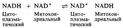
Unlike the glycerol phosphate shuttle, this mechanism is readily reversible. Consequently, NADH can enter the mitochondria via the malate-aspartate shuttle only if the [NADH]/[NAD+] ratio is higher in the cytosol than in the mitochondrial matrix. When electrons are transferred from NADH to the mitochondrial respiratory chain using this mechanism, no energy is consumed, and three molecules of ATP are synthesized for each transported molecule of NADH.
14.10. The influx of ADP into mitochondria requires the efflux of ATP
ATP and ADP cannot diffuse freely across the inner mitochondrial membrane. The translocation of these highly charged molecules across this barrier is made possible by a specific carrier. An interesting feature of this transport is the coupling of ATP and ADP fluxes. ADP enters the mitochondrial matrix only if ATP leaves, and vice versa. This coupled efflux of ATP and influx of ADP is an example of facilitated exchange diffusion. It is mediated by the ATP-ADP translocase, a dimer composed of identical subunits with a Molecular Weight of 29 kDa each. The translocase is abundant in the inner mitochondrial membrane, accounting for about 6% of the total protein. ADP is preferentially transported to the cytoplasmic side of the membrane. This explains in part why the [ATP]/[ADP][Pi] ratio on the cytoplasmic side of the membrane is 10 times higher than on the matrix side. The coupled Transport of ATP and ADP by the translocase is likely driven by the proton gradient across the inner mitochondrial membrane. The ATP-ADP translocase is specifically inhibited by very low concentrations of atractyloside, a plant glycoside, or bongkrekic acid, a mold antibiotic. Shortly after The addition of these inhibitors, Oxidative Phosphorylation ceases because ADP can no longer enter the mitochondria.
14.11. Mitochondria contain numerous transport systems for ions and metabolites
The ATP-ADP translocase is just one of many Mitochondrial Transport Systems. The inner mitochondrial membrane contains a variety of carriers for ions and charged metabolites. For example, the dicarboxylate carrier mediates the facilitated exchange diffusion of malate, succinate, fumarate, and Pi. Tricarboxylate carriers provide for the exchange of OH- for Pi. Cytosolic Pyruvate enters the mitochondrial matrix in exchange for OH- via the pyruvate carrier. The reciprocal exchange of glutamate and aspartate is carried out by the glutamate carrier, which can also transport OH-. Mitochondria also possess a calcium ion transport system. The immediate source of Free energy for Ca2+ accumulation in the mitochondrial matrix is not ATP, but the proton-motive force generated by electron transport.
Last update: 06/08/2026
Editorial and Educational Adaptation: This material has been compiled based on the primary/original source text. The project team performed an editorial review, corrected technical inaccuracies, structured sections, and adapted the content for an educational format.
What was processed:
- elimination of formatting defects (OCR errors, structural breaks, corrupted characters);
- editorial organization of content;
- standardization of terminology in accordance with academic sources;
- verification of factual statements against the original source text.
All mentions of the author, publication year, and origin of the primary text have been preserved in accordance with the source.