BIOCHEMISTRY - L. Stryer - 1984
VOLUME 2
PART II GENERATION AND STORAGE OF METABOLIC ENERGY
CHAPTER 14. OXIDATIVE PHOSPHORYLATION
14.7. Protons are Ejected by Asymmetrically Oriented Transmembrane Complexes
The generation of a proton gradient during electron flow through the three energy-conserving sites of the Respiratory Chain requires their asymmetrical orientation. Furthermore, the three corresponding enzyme complexes must be embedded in the membrane in such a way that protons can be pumped from the matrix side to the cytoplasmic side. The Use of submitochondrial particles, generated by sonication of Cell/35.html">Mitochondria (Fig. 14.10), greatly facilitated The Study of these important aspects of the Electron Transport Chain. The outer surface of submitochondrial particles corresponds to the matrix-facing surface of the inner membrane of an intact mitochondrion. Thus, both surfaces of The inner mitochondrial membrane—the cytoplasmic-facing surface in the intact mitochondrion and the matrix-facing surface of the submitochondrial particles—are accessible to experimental investigation. The arrangement of the protein Components of the respiratory chain in these preparations has been studied using Proteolytic Enzymes, specific Antibodies, Lectins, and membrane-impermeable labeling Reagents. For example, subunits II, V, and VI of cytochrome c oxidase can be labeled only from the matrix side. Cytochrome c oxidase binds cytochrome c exclusively on the cytoplasmic surface and pumps protons in only one direction. These experiments, as well as analogous studies conducted with NADH-Q reductase and QH2-cytochrome c reductase (Table 14.2), have shown that all three energy-conserving systems span the inner mitochondrial membrane and are oriented asymmetrically.
Class="center">Table 14.2. Components of the Mitochondrial Electron Transport chain

Fig. 14.10. Electron micrographs. A - submitochondrial particle with F1 knobs on its surface; B - submitochondrial particle treated with urea, which removes the F1 knobs; C - isolated F1; and D - reconstituted submitochondrial particle formed by adding F1 (coupling factor) to "stripped" membranes. The particle shown in Fig. B can transfer electrons to O2, but is incapable of ATP synthesis. The reconstituted particle shown in Fig. D carries out Oxidative Phosphorylation

Where do the translocated protons come from? One possibility is that they are protons directly involved in the chemical Reactions Catalyzed by the three systems. For instance, when NADH transfers two electrons to the NADH-Q reductase complex, two H+ ions are involved in the process—one from NADH itself and the second from the solvent. According to another hypothesis, translocated protons may arise indirectly via conformational interactions between the catalytic and protein-binding sites at various locations within the enzyme complex. The stoichiometry of Electron transfer reactions suggests that only one proton per electron, or two protons per energy-conserving site (phosphorylation site), can be "pumped" via a direct mechanism. At the same time, the H+/energy-conserving site ratio has been found to be in the range of 3 to 4, which is inconsistent with a direct mechanism. It seems likely that electron flow through each energy-conserving site induces alternating conformational changes that facilitate the movement of protons from the matrix side to the cytoplasmic side of the membrane. Recall that the protons involved in the Bohr effect in Hemoglobin originate from a site on the molecule distant from the heme group (Section 4.16).
14.8. ATP is Synthesized by the Reverse Flow of Protons into the Matrix Through Proton Channels
Let us now turn to the utilization of the proton gradient for ATP synthesis. The enzyme catalyzing this process appears in Cytology/cytology/93.html">ELECTRON MICROGRAPHS OF submitochondrial particles as spherical projections On the surface (Fig. 14.10). In intact mitochondria, such projections are located on the side of the inner mitochondrial membrane that faces the matrix. In 1960, Efraim Racker discovered that these rounded projections (85 Å in diameter) could be removed by mechanical agitation. "Stripped" submitochondrial particles retain The ability to transfer electrons through their electron transport chain, but ATP synthesis no longer occurs. Conversely, the detached 85-Å projections catalyze the Hydrolysis of ATP. However, the most striking finding in Racker's observations was that The addition of these ATPase projections to "stripped" submitochondrial particles restored their ability to synthesize ATP. These projections are designated as coupling factor 1, or F1. The Physiological Role of F1 is to catalyze the synthesis of ATP. The ATPase activity exhibited by solubilized F1 (in the absence of a proton gradient) is a result of the reversal of its inherent physiological reaction.
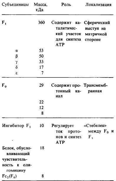
The F1 component, which has a mass of 360 kDa and contains five types of polypeptide chains (Table 14.3), represents only a part of the mitochondrial ATP-synthesizing machinery. Another essential component of this complex is F0, a hydrophobic segment consisting of four polypeptide chains anchored in the inner mitochondrial membrane. F0 acts as the proton channel of the complex. The "stalk" between F0 and F1 includes several other Proteins (Table 14.3). One of them confers sensitivity to oligomycin, an antibiotic that blocks ATP synthesis by disrupting the utilization of the proton gradient.
Table 14.3. Components of the mitochondrial ATP-synthesizing complex
The flow of protons through the F0 channel from the cytoplasmic side of the membrane to the matrix drives the synthesis of ATP carried out by F1. How is proton flow coupled to ATP synthesis? As in the case of proton pumping, both Direct and Indirect mechanisms are conceivable. It has been suggested that proton flow directly drives the ATP synthesis reaction. According to this scheme, Pi is activated and simultaneously interacts with ADP to form ATP. Another hypothesis is that the coupling of proton flow to ATP synthesis is mediated by conformational changes transmitted through the enzyme complex.
Last update: 06/08/2026
Editorial and Educational Adaptation: This material has been compiled based on the primary/original source text. The project team performed an editorial review, corrected technical inaccuracies, structured sections, and adapted the content for an educational format.
What was processed:
- elimination of formatting defects (OCR errors, structural breaks, corrupted characters);
- editorial organization of content;
- standardization of terminology in accordance with academic sources;
- verification of factual statements against the original source text.
All mentions of the author, publication year, and origin of the primary text have been preserved in accordance with the source.