BIOCHEMISTRY - L. Stryer - 1984
VOLUME 2
PART II GENERATION AND STORAGE OF METABOLIC ENERGY
CHAPTER 14. OXIDATIVE PHOSPHORYLATION
14.5. Coupling of Oxidation and Phosphorylation is Mediated by a Proton Gradient
Let us now consider the flow of electrons from NADH to O2, which is an exergonic process:
NADH + 1/2O2 + Н+⇄ Н2O + NАD+
∆G0' = — 52.6 kcal/mol.
This Free energy of oxidation is utilized for ATP synthesis:
ADP + Рi + Н+⇄ АТР + Н+2O
∆G0' = + 7.3 kcal/mol.
Class="center">Fig. 14.7. Electron micrograph of the two-dimensional crystal Structure of cytochrome c oxidase

The synthesis of ATP is carried out by molecular assemblies in The inner mitochondrial membrane. The corresponding enzyme complex (Sec. 14.8) is termed mitochondrial ATPase because it was discovered through its ability to catalyze hydrolytic reactions.
Fig. 14.8. Electron transport along the Respiratory Chain generates a proton gradient and a Membrane Potential across the inner mitochondrial membrane

How is the coupling between NADH oxidation and ADP phosphorylation achieved? It was initially postulated that electron transfer leads to The formation of a covalent high-energy intermediate, which serves as a precursor to ATP. This chemical coupling hypothesis was based on The Mechanism of substrate-level phosphorylation, exemplified by the glyceraldehyde-3-phosphate dehydrogenase reaction, which yields 1,3-BPG as a high-energy intermediate (Sec. 12.14). Another suggestion was put forward, namely that the free energy of oxidation is captured by a protein in an activated conformation, which then stimulates ATP synthesis. Researchers in numerous laboratories spent decades trying to isolate these putative high-energy intermediates, but all such attempts proved unsuccessful.
A radically different mechanism, the chemiosmotic hypothesis, was postulated by Peter Mitchell in 1961. He proposed that the coupling of electron transfer and ATP synthesis is driven by a proton gradient rather than a high-energy covalent intermediate or an activated protein. According to this model, electron transport through the respiratory chain results in the extrusion of protons from the matrix to the cytoplasmic side of the inner mitochondrial membrane, thereby increasing the concentration of H+ ions. Consequently, a membrane potential is generated, with a positive charge on the cytoplasmic side of the membrane (Fig. 14.8). This proton-motive force, the author postulates, drives ATP synthesis via the ATPase complex. In this model, the interaction between the electron-transfer chain and the ATP-synthesizing complex is mediated solely by the membrane potential. The model requires that the electron carriers of the Cell/36.html">Respiratory Chain and the ATPase possess a vectorial Organization; that is, they must be oriented in a specific manner relative to the two surfaces of the inner mitochondrial membrane. Furthermore, the inner mitochondrial membrane must be completely impermeable to protons, as the existence of a proton gradient requires a sealed compartment. The fundamental premise of the proposed mechanism is that the primary energy-storing act is the translocation of protons across the inner mitochondrial membrane.
Mitchell's hypothesis of the coupling of oxidation and phosphorylation by a proton gradient has since received overwhelming experimental support.
1. A proton gradient across the inner mitochondrial membrane is established during electron transport. The external pH is 1.4 units lower than the internal pH, and the membrane potential is 0.14 V, with the outer surface carrying a positive charge. The total Electrochemical Potential ∆p (in volts) is composed of the membrane potential (∆ ) and the concentration gradient of H+ ions (∆pH). In the equation below, R is the gas constant, T is the absolute Temperature, and F is the Faraday constant.

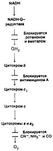
Fig. 14.9. Sites of action of various ELECTRON TRANSPORT INHIBITORS
This total proton-motive force of 0.224 V corresponds to a free energy of 5.2 kcal per mole of protons.
2. When a pH gradient is artificially established across the membrane of Mitochondria or METABOLISM/14.html">Chloroplasts (Sec. 19.13), ATP synthesis takes place in the absence of electron transport.
3. Upon illumination, the purple membrane protein of halobacteria pumps protons (Section 19.21). Synthetic vesicles containing this bacterial protein and purified ATPase from bovine Heart mitochondria synthesize ATP upon illumination. In this experiment, the purple membrane protein replaces the respiratory chain; consequently, the respiratory chain and ATPase are biochemically distinct systems linked solely by a proton gradient.
4. Both the respiratory chain (Section 14.7) and ATPase (Section 14.8) exhibit a vectorial organization within the inner mitochondrial membrane.
5. Compartment closure is essential for Oxidative Phosphorylation. Soluble preparations or membrane fragments lacking well-defined inner and outer compartments fail to catalyze electron transport-coupled ATP synthesis.
6. Protonophores—substances that transport protons across the inner mitochondrial membrane—dissipate the proton gradient and thereby uncouple oxidation from phosphorylation (Section 14.14).
14.6. The Proton Gradient Is Generated at Three Sites
Protons are extruded at three distinct sites along the electron flow pathway of the respiratory chain from NADH to O2 (Fig. 14.9): site 1 is the NADH-Q reductase complex; site 2 is the QH2-cytochrome c reductase complex; and site 3 is the cytochrome c oxidase complex. The proton gradient generated at each of these sites during The transfer of a pair of electrons from NADH is utilized to synthesize one molecule of ATP. Various experimental approaches have been employed to identify these sites.
1. Comparison of ATP yields upon The oxidation of various substrates. The oxidation of NADH yields three ATP molecules, whereas the oxidation of succinate yields two. Electrons from FADH2 enter the Electron Transport Chain at the level of coenzyme Q, which operates at a lower energy level than phosphorylation site 1. The oxidation of the non-physiological substrate ascorbate yields only a single ATP molecule because ascorbate-derived electrons enter at the level of cytochrome c, which is at a lower energy level than phosphorylation site 2. Oxidative phosphorylation is frequently characterized by the P:O ratio, defined as the number of moles of inorganic phosphate incorporated into an organic form per atom of oxygen consumed. The P:O ratios for the oxidation of NADH, succinate, and ascorbate are 3, 2, and 1, respectively.
2. Thermodynamic measurements. The standard free-energy change, $\Delta G^{0\prime}$, for Electron transfer from NADH to the lower-energy Fe-S center in NADH-Q reductase is — 12 kcal/mol; for electron transfer from cytochrome b to c1 in QH2-cytochrome c reductase, it is 10 kcal/mol; and for electron transfer from cytochrome a to O2 in cytochrome c oxidase, it is — 24 kcal/mol. These redox reactions are sufficiently exergonic to drive ATP synthesis under standard conditions ($\Delta G^{0\prime}$ = — 7.3 kcal/mol). The $\Delta G^{0\prime}$ values for other electron transfer steps mediated by coenzyme Q and cytochrome c are too small to support ATP synthesis.
3. Specific inhibition of electron flow. Rotenone and amytal specifically inhibit electron transfer within the NADH-Q reductase complex, thereby preventing proton gradient generation at site 1 (see Fig. 14.9). However, these inhibitors do not impair succinate oxidation, as electrons from this substrate enter the transport chain downstream of the coenzyme Q block. Antimycin A halts electron flow between Cytochromes b and c1, preventing the ATP Synthesis Coupled to proton gradient generation at site 2. This block can be bypassed by adding ascorbate, which directly reduces cytochrome c. Electrons then flow from cytochrome c to O2, concomitantly driving ATP synthesis coupled to the proton gradient at site 3. Finally, electron flow can be blocked between the cytochrome oxidase complex and O2 by CN-, N-3, and CO. Cyanide and azide react with the ferric form of this carrier, whereas carbon monoxide inhibits the ferrous form. In the presence of these inhibitors, the blocked electron flow abolishes phosphorylation coupled to proton gradient generation at site 3.
The sites of action of these inhibitors were elucidated using the crossover theorem. Britton Chance introduced an elegant spectroscopic method to determine the oxidation-reduction state of each electron carrier. This technique relies on the distinct absorption spectra characteristic of the oxidized and reduced forms of each carrier. The addition of an electron transport inhibitor alters The ratio of these forms. For instance, the addition of antimycin A causes carriers located in The electron transport chain between NADH and cytochrome b to shift toward a more reduced state, whereas those between cytochrome c and O2 shift toward a more oxidized state. From this, one can conclude that antimycin A inhibits The conversion of cytochrome b to cytochrome c1, because this step constitutes a crossover point.
4. Reconstitution of vesicle systems containing a single proton-extrusion site. Each of the three proton-extrusion sites has been successfully reconstituted into synthetic phospholipid vesicles containing ATPase. The Addition of an oxidizable substrate to such vesicles generates a proton-motive force sufficient to synthesize one molecule of ATP per electron pair.
Last update: 06/08/2026
Editorial and Educational Adaptation: This material has been compiled based on the primary/original source text. The project team performed an editorial review, corrected technical inaccuracies, structured sections, and adapted the content for an educational format.
What was processed:
- elimination of formatting defects (OCR errors, structural breaks, corrupted characters);
- editorial organization of content;
- standardization of terminology in accordance with academic sources;
- verification of factual statements against the original source text.
All mentions of the author, publication year, and origin of the primary text have been preserved in accordance with the source.