General Microbiology - Schlegel H. 1987
Basic mechanisms of metabolism and energy conversion
Respiratory chain and electron transport-coupled phosphorylation
While most anaerobic organisms can synthesize ATP only via substrate-level phosphorylation, aerobes are capable of incomparably more efficient ATP regeneration. They possess a specialized machinery: a respiratory (electron transport) chain and the enzyme ATP synthase. In prokaryotes, both systems are located in Cell/30.html">The Plasma Membrane, whereas in eukaryotes, they reside in The inner mitochondrial membrane. Derived from substrates, reduction equivalents (H or electrons) enter the Respiratory Chain within these membranes, and electrons are transferred to O2 (or other TERMINAL ELECTRON ACCEPTORS). The reactions taking place in the respiratory chain represent a biochemical analogue of hydrogen combustion. They differ from the chemical burning of molecular hydrogen in that a significant portion of the Free energy is converted into a biologically available form—namely, ATP—with only a small fraction dissipated as heat.
Class="center">
Fig. 7.8. Scheme of Oxidative Phosphorylation in the bacterial plasma membrane and the inner mitochondrial membrane. A. Oxidation of NADH2 and proton extrusion. B. Electrochemical gradient between the inner and outer sides. C. ATP regeneration resulting from the reverse proton flux.
Mechanism of Oxidative phosphorylation. Reduction equivalents (protons and electrons) released by substrates are transferred to the plasma membrane or the inner mitochondrial membrane. They are transported across the membrane in such a way that an electrochemical gradient is established between its inner and outer sides, with a positive potential on the outside and a negative one on the inside (Fig. 7.8). This potential difference arises from the specific arrangement of the respiratory chain components within the membrane.
Some of these components transfer electrons, while others transfer hydrogen. The spatial arrangement of the carriers within the membrane is such that during The transport of electrons from the substrate to oxygen, protons (H+) are bound on the inner side of the membrane and released on the outer side. One can imagine that electrons follow a zigzag pathway through the membrane while simultaneously ferrying protons from the inside out. This electron- and proton-transporting system is known as the respiratory or Electron Transport Chain. It is sometimes figuratively referred to as a "proton pump," since The primary function of this system is proton translocation.
The nonequilibrium distribution of charges—that is, the electrochemical gradient—serves as the driving force for ATP regeneration (and other energy-requiring processes). The membrane contains a specialized enzyme, ATP synthase, which synthesizes ATP from ADP and Pi. This enzyme protrudes from the membrane on its inner side. During ATP synthesis, protons flow back from the outer side of the membrane to the inner side. The synthesis of ATP driven by the energy of transmembrane Electron transport is called oxidative phosphorylation or respiratory chain phosphorylation.
To understand The Mechanism of Respiration, it is essential to know
1) the Components of the respiratory chain, 2) their redox potentials, and 3) their spatial arrangement within the membrane.
Membranes as the site of respiration. The components of the respiratory chain are enzyme Proteins with relatively tightly bound low-molecular-weight prosthetic groups. The long-standing concept that "respiration is catalysis effected by iron at surfaces" (O. Warburg) holds true. Indeed, the Enzymes of the respiratory chain are structurally linked. In eukaryotes, they are localized in the inner mitochondrial membrane, whereas in prokaryotes, they reside in the plasma membrane. The mode of action and localization of respiratory chain components in both types of membranes are largely similar. The respiratory chain of *Alcaligenes eutrophus* and *Paracoccus denitrificans* is virtually identical to that of Mitochondria.
The components of the respiratory chain are embedded in The Lipid Bilayer. These comprise A large number of enzymes, Coenzymes, and prosthetic groups, various dehydrogenases, and transport systems involved in electron and hydrogen transfer. The protein components can be isolated from the membrane. The most important components participating in hydrogen oxidation are Flavoproteins, iron-sulfur proteins, Quinones, and Cytochromes.
Flvoproteins are enzymes containing either flavin mononucleotide (FMN) or flavin adenine dinucleotide (FAD) as prosthetic groups. These enzymes function as hydrogen carriers. Their active moiety is the isoalloxazine ring system (Fig. 7.9, A), which acts as a reversible redox system. The reactive centers are two nitrogen atoms, each of which can bind one [H]. Binding can occur in two steps via a semiquinone intermediate. Due to their ability to transfer either two hydrogen atoms or one at a time, flavoproteins can act as intermediaries between two Different types of hydrogen transfer processes.
Iron-sulfur proteins are electron-transporting redox systems. They contain iron atoms linked, on the one hand, to the sulfur of Cysteine residues and, on the other hand, to inorganic sulfide sulfur (Fig. 7.9, B). The latter is very easily split off as hydrogen sulfide upon acidification. Cysteine residues are part of polypeptide chains; the Fe–S centers can be regarded as prosthetic groups of the polypeptide. The [2Fe + 2S] centers involved in the respiratory chain are capable of transferring only a single electron. Iron-sulfur Proteins of the [2Fe + 2S] type, containing two labile sulfur and iron atoms each, are found in several enzyme complexes of the respiratory chain. Of the total iron present in the plasma membrane, 80% is bound in Fe–S proteins and only 20% in cytochromes.
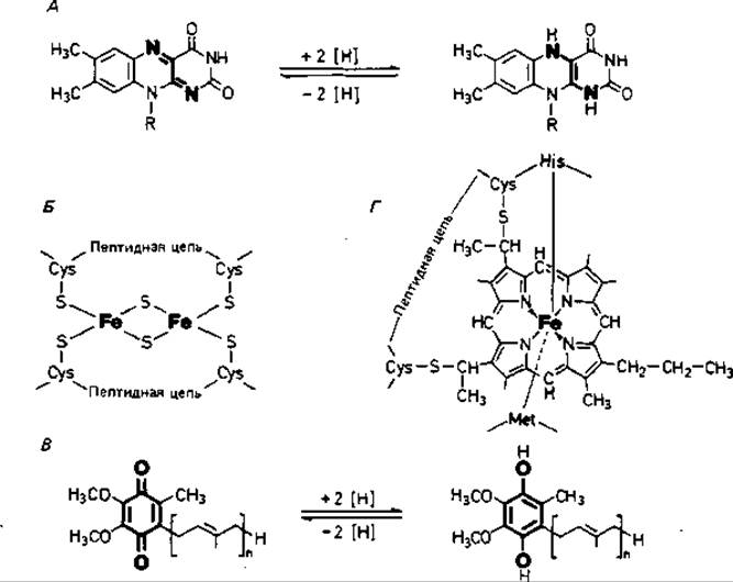
Fig. 7.9. Structural formulas of some Key Components of the respiratory chain. A. Isoalloxazine ring system of FMN or FAD in oxidized and reduced forms. B. [2Fe + 2S] center of an iron-sulfur protein. C. Reduction of ubiquinone to ubihydroquinone. D. Active Site of cytochrome c.
In addition to electron transport in membranes, Fe–S proteins participate in MOLECULAR Nitrogen Fixation, sulfite and nitrite reduction, Photosynthesis, the release and activation of molecular hydrogen, and alkane oxidation. Fe–S proteins have a relatively low molecular weight and a strongly negative redox potential; their $E'_0$ values range from –0.2 to –0.6 V. Besides iron-sulfur proteins with [2Fe + 2S] centers (found in METABOLISM/14.html">Chloroplasts and aerobic Bacteria), there are others with a [4Fe + 4S] center (in *Clostridium*, *Chromatium*), as well as those containing two [4Fe + 4S] centers (in *Clostridium*, *Azotobacter*). Some iron-sulfur proteins are named after their origin or function: ferredoxin, putidaredoxin, rubredoxin, adrenodoxin.

Another group of redox systems in the respiratory chain comprises quinones. The inner mitochondrial membrane and Gram-negative bacteria contain ubiquinone (coenzyme Q; Fig. 7.9, C); Gram-positive bacteria contain naphthoquinones, while chloroplasts contain plastoquinones. Quinones, particularly ubiquinone, are lipophilic and therefore localized in the lipid phase of the membrane. They are capable of transferring hydrogen or electrons. This transfer can take place in two steps, with the semiquinone serving as an intermediate form. Compared to other respiratory chain components, quinones are present in a 10- to 15-fold excess. They act as "collectors" of hydrogen supplied by various coenzymes and prosthetic groups in the Respiratory Chain and pass it on to the cytochromes.
Cytochromes are redox systems that transport electrons only; they do not transport hydrogen. Electrons are delivered to cytochromes from the quinone pool. Upon electron transfer, an equivalent number of protons is released into solution. As a prosthetic group, cytochromes contain a heme moiety (Fig. 7.9, D). The central iron atom of the heme ring participates in electron transfer by changing its valence state. Cytochromes are colored proteins characterized by distinct Absorption Spectra and redox potentials. They include cytochromes a, a3, b, c, o, and A number of others. In cytochrome c, the heme groups are covalently linked to cysteine residues of the apoprotein; thanks to this stable bond, it is Water-soluble and can be extracted from the membrane using salt solutions. Cytochrome c is found in almost all organisms possessing a respiratory chain, whereas the distribution of other cytochromes exhibits notable variations.
Cytochromes are also involved in The transfer of electrons to oxygen. Cytochrome c oxidase (cytochrome aa3) is the terminal oxidase that reacts with oxygen, transferring four electrons to it:

Cytochrome o, which frequently occurs in bacteria, can also react with molecular oxygen. This terminal oxidase can be inhibited by cyanide or carbon monoxide.
For a long time, the presence of cytochromes was considered a hallmark of aerobic or phototrophic organisms. The discovery of cytochrome c3 in *Desulfovibrio* initially seemed surprising, but it subsequently became clear that sulfate reduction by these sulfate-reducing bacteria enables them to carry out oxidative phosphorylation under anaerobic conditions, thus formally corresponding to respiration. Recently, it has been revealed that the aerotolerant lactic acid bacteria *Streptococcus lactis* and *Leuconostoc mesenteroides*, as well as the anaerobe *Bifidobacterium*, also synthesize cytochromes when grown on media containing hemin or Blood. Cytochromes have likewise been detected in the strict anaerobes *Selenomonas ruminantium*, *Veillonella alcalescens*, *Vibrio succinogenes*, *Clostridium formicoaceticum*, and *C. thermoaceticum*. It is quite possible that cytochromes and the capacity for some degree of electron transport-coupled phosphorylation will also be discovered in other strictly anaerobic bacteria.
Redox potential. Hydrogen transport and electron transport are equivalent processes. The respiratory chain can be viewed as an electron transfer chain. The components of the respiratory chain alternate between oxidized and reduced states—that is, they behave as typical redox catalysts. They possess a redox potential that can be measured directly (in cytochromes) or indirectly (for NAD, FAD).
The redox potential serves as a quantitative measure of the tendency of given compounds or elements to donate electrons. This potential is measured relative to the potential of molecular hydrogen. By definition, a hydrogen half-cell—a platinized or platinum electrode immersed in an acid solution and flushed with gaseous H2 at a pressure of 1.013 bar and pH 0—has a potential equal to zero:

Similar to chemical elements, biological substances can also be arranged in a series According to the magnitude of their redox potential at a 1/2 degree of reduction (equal concentrations of the oxidized and reduced forms); this value is designated as E0. In biochemistry, the value E0' adjusted to pH 7 is used. At this pH, the hydrogen electrode has a potential of — 0,42 V. Fig. 7.10, A shows the dependence of the potential, referred to the potential of the hydrogen half-element, on the pH value. From the Nernst equation
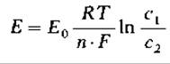
it follows that the dependence of the measured potential of the redox system E' on the concentrations of the oxidized and reduced forms is expressed by the formula


Fig. 7.10. Redox potentials. A. Dependence of the redox potential (referred to the potential of the hydrogen half-element (n-H2 electrode) on the pH value; normal potentials E'0 are indicated for certain compounds. B. Dependence of the measured potential E'0 on the concentrations of the oxidized and reduced forms of the compound for two redox systems.
The value of E' is more negative the smaller The ratio of the concentration of the oxidized form to the concentration of the reduced form (Fig. 7.10, B).
The redox potential serves as a measure of the maximum useful work that a system can perform, i.e., a measure of The change in free energy (AG0) in a given reaction. From the difference in redox potentials of two reacting systems, AE0, the change in free energy for a given reaction can be calculated:
∆G0= - n ∙ F∙ ∆Е0 = - n ∙ 96,5 ∙ ∆E0 (kJ/mol)
The E'0 values of individual components of the respiratory chain range from — 0.32 V for NADH2/NAD [— 0.08 V for flavoprotein (FADH2/FAD); — 0.04 V for cytochrome b (Fe2 + /Fe3 +)] to + 0.81 V for O2-/1/2O2. Values for a number of substrates can also be given: lactate/Pyruvate — 0.186 V; malate/oxaloacetate — 0.166 V; succinate/fumarate — 0.03 V.
Table 7.4. Redox potentials of the respiratory chain components. Potential differences between individual components and equivalent Free Energy Changes
|
Respiratory chain components |
E'0, V |
Difference in E'0 values, V |
-∆ G'0 |
|
|
kJ/mol |
kcal/mol |
|||
|
Hydrogen |
- 0,42 |
0,10 |
19,3 |
4,61 |
|
NAD |
-0,32 |
0,24 |
46,4 |
11,1 |
|
Flavoprotein |
-0,08 |
0,04 |
7,7 |
1,84 |
|
Cytochrome b |
-0,04 |
0,31 |
59,8 |
14,30 |
|
Cytochrome c |
+ 0,27 |
0,02 |
3,8 |
0,92 |
|
Cytochrome a |
+ 0,29 |
0,52 |
100,4 |
24,0 |
|
Oxygen |
+ 0,81 |
Since the difference between the E'0 values for H2 and O2 is — 0.42-0.81 = — 1.23 V, the free energy change in the oxyhydrogen gas reaction AG0' should be equal to — 2∙96.5∙1.23 = — 237.4 kJ/mol. In The Cell, during hydrogen transfer from NADH2, the potential difference is only [(+ 0.81 V) - (- 0.32 V)] = 1.13 V, i.e., ∆G0 = - 218 kJ/mol. Similarly, the corresponding energy yield for any two electron carriers in the respiratory chain can be calculated from the potential difference (Table 7.4).
Arrangement and Functions of redox systems in the respiratory chain. According to their redox potential, the components of the respiratory chain can be arranged in a series starting with NAD (most negative potential) and ending with cytochrome oxidase and oxygen (Fig. 7.11). Quinones and cytochromes serve as auxiliary substrates. These components are reduced by hydrogen supplied by various Donors. Hydrogen obtained via NAD is transferred to quinone with the help of NADH dehydrogenase; similarly, hydrogen from succinate is transferred to quinone (via succinate dehydrogenase) and hydrogen obtained by dehydrogenation of Fatty acids (via other specific dehydrogenases). Quinone serves as a pooled reservoir of hydrogen from substrates in the respiratory chain. Some of the enzymes involved in hydrogen transfer contain FMN or FAD, as well as iron-sulfur proteins.
Reduced quinones are reoxidized by the cytochrome system. Cytochromes accept electrons and transfer them to oxygen; hydrogen is ionized and leaves the membrane (Fig. 7.8 and 7.12, D). The sequence of redox systems deduced from the respective potentials was confirmed experimentally using spectrometry and specific inhibitors.

Fig. 7.11. General scheme of the respiratory chain in eukaryotic mitochondria and in the membranes of many bacteria. DH - dehydrogenase; UQ - ubiquinone; FP - flavoprotein.
Respiratory Chain Inhibitors. The respiratory chain is inhibited or blocked by various cellular poisons. Amytal, rotenone, and piericidin A suppress The activity of NADH dehydrogenase; antimycin A blocks electron transfer between cytochromes b and c. Cyanide and carbon monoxide inhibit only cytochrome oxidase; in cytochrome c, the iron ion is apparently embedded so deep within the protein that it does not interact with either CN- or CO. The specific action of these poisons and Changes in the characteristic absorption spectra of the respiratory chain components served as indicators used to study this chain.
The P/O ratio and Energy balance. Consideration of the redox potentials (Table 7.4) shows that there are only three oxidation steps in the respiratory chain that release at least as much energy as is contained in one "high-energy" bond. When 2 [H] are transferred from NADH2 to oxygen, only three electron transitions can be coupled with the phosphorylation of ADP to ATP, so that at best only three phosphate molecules can be incorporated into an organic compound. This Coupling of oxidation with phosphorylation is usually expressed as the P/O ratio (the number of ATP molecules formed per 1 atom of consumed oxygen). For animal mitochondria, using isocitrate or malate as hydrogen donors (which transfer their hydrogen to NAD), an experimental P/O ratio of 3 can be obtained; for succinate, from which hydrogen can be incorporated into the respiratory chain only at the flavoprotein level, the P/O ratio is only 2.
Knowing the fact that three ATP molecules are formed upon the transfer of two hydrogen equivalents along the respiratory chain makes it possible to compile the energy balance for the case of glucose oxidation. If Glucose Catabolism proceeds via the fructose bisphosphate pathway and through The Tricarboxylic Acid Cycle, and all the hydrogen is burned to water in the respiratory chain, then for 1 mole of glucose consumed, the following are formed: a) via the fructose bisphosphate pathway — 2 moles of NADH2; b) during pyruvate dehydrogenation — 2 moles of NADH2; c) in the tricarboxylic acid cycle — 2∙3 moles of NADH2 and 2 moles of FADH2; total — 10 moles of NADH2 and 2 moles of FADH2. With P/O ratios equal to 3 and 2, the number of synthesized moles of ATP will be 10∙3 + 2∙2 = 34. If we add to this the two moles of ATP synthesized in the fructose bisphosphate pathway and the two moles formed during The oxidation of 2-oxoglutarate, we obtain a grand total of 38 moles of ATP.
This calculation holds true for mitochondria and many bacteria. However, bacteria often have only two phosphorylation sites, i.e., even for hydrogen supplied by NADH2, the P/O ratio is only two. This is the case, for example, in Escherichia coli Cells growing under aerobic conditions, in which the oxidation of 1 mole of glucose leads to The formation of only 26 moles of ATP.
Phosphorylation in the respiratory chain. The regeneration of ATP during phosphorylation in the respiratory chain and photosynthetic phosphorylation takes place in membranes. ATP synthase, just like the components of the respiratory chain, is an integral part of the membrane. How the hydrogen and electron transfer occurring in the respiratory chain is coupled with ATP synthesis is not yet fully understood. However, numerous experiments have shown that ATP regeneration occurs only in spaces completely surrounded by membranes — in vesicles. The processes of hydrogen and electron transfer are closely coupled with the movement of protons, and this process in turn is necessary for ATP regeneration.
Proton transport. By adding molecular oxygen to a suspension of aerobic bacteria or mitochondria that were previously under anaerobic conditions, a decrease in the pH of the medium can be observed. This leads to the Conclusion that protons are extruded from Bacterial cells and mitochondria during respiration (Fig. 7.12, A and B). If vesicles are prepared from bacterial or mitochondrial membranes in which the former inner side faces outward (inverted vesicles), then during respiration a "reversed" proton transfer will be observed, leading to alkalinization of the suspension medium (Fig. 7.12, C). As a result of proton translocation, an Electrochemical Potential gradient is created. The internal space of intact mitochondria or bacteria is electronegative relative to the suspension medium and is characterized by a higher pH. Both transmembrane gradients — the pH and electrical potential gradients — tend to drive the translocated proton back inside. This difference in electrochemical proton potentials (also called proton motive force) is composed of the electrical Membrane Potential (∆ψ) and the pH difference between the outer and inner sides of the membrane (∆pH) in accordance with the equation

where Z = 2.3∙R∙T/F, i.e., 59 mV at 25°C. The proton potential can be due solely to the pH difference, solely to the electrical membrane potential, or to both simultaneously.

Fig. 7.12. Proton transport during substrate respiration. Protons are released into the suspension medium from a bacterial cell (A) or a mitochondrion (B). In "submitochondrial particles" (C), the membranes are inverted (inside out), so protons are transported inward. D. Pathway of proton and electron transfer during NADH2 oxidation according to the chemiosmotic hypothesis. KCт - Cell wall; ПМ - plasma membrane; ВМ and НМ - inner and outer mitochondrial membranes; Q - coenzyme Q; Z - hypothetical hydrogen carrier; FeS - iron-sulfur proteins; b, c, a, a3 - cytochromes.
The following concepts are consistent with experimental data. The plasma membrane of bacteria and the inner membrane of mitochondria are impermeable to ions, including H+ and OH-; the electrical conductivity of the membranes is low. The membrane is asymmetrical; although the lipid bilayer appears symmetrical, the topography of functional proteins (electron transport components, ATP synthase, permeases, etc.) imparts an asymmetrical character to the membrane. The spatial orientation of enzyme molecules determines directed mass transfer. According to Mitchell's hypothesis, the respiratory chain consists of alternating hydrogen carriers and electron carriers arranged in the membrane (Fig. 7.12, D) in such a way that substrate oxidation leads to the consumption of protons on the inner side of the membrane and their release on the outer side. If the chain forms three loops, then the oxidation of NADH2 drives the extrusion of six protons. Such proton translocation driven by respiratory energy leads to the generation of an electrochemical gradient across the inner and outer sides of the membrane. The proton motive force is precisely the driving force that ultimately performs phosphorylation, i.e., ensures ATP synthesis. Biochemical energy conversion via ATP regeneration is thus the result of proton gradient generation and is accompanied by a decrease in membrane potential. Such is The Essence of Mitchell's chemiosmotic theory.
Regeneration of ATP from ADP and Pi. The synthesis of ATP from ADP and inorganic phosphate (Pi) is catalyzed by ATP synthase. This enzyme converts the energy delivered by the electron flux into the energy of the phospho-ester bonds of ATP. The enzyme is found in all energy-transducing membranes, namely in the membranes of mitochondria, chloroplasts, and bacteria. It is quite large (mol. mass 350∙103) and has a complex Structure (Fig. 7.12, D)—it consists of a HEAD composed of several subunits, a stalk, and a base; the latter is embedded in the lipid bilayer of the plasma membrane. ATP synthase catalyzes The addition of phosphate to ADP with the Cleavage of a water molecule, resulting in the formation of ATP. Exactly how the proton flux or proton gradient drives this phosphorylation reaction is still unknown; it is possible that protons flow back into the interior of the mitochondrion or bacterium through some channel or pore in the enzyme molecule, and The energy released in the process is utilized for phosphorylation.
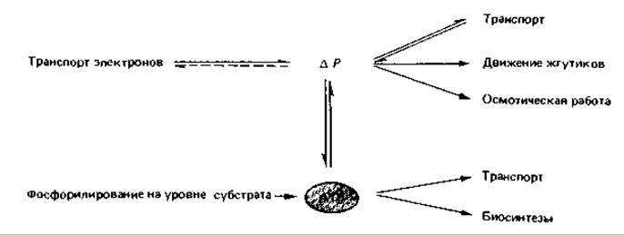
ATP synthase is identical to F1-ATPase, so its activity can be detected by ATP Hydrolysis: ATP + H2O → ADP + Pi + H+. The reversibility of the ATP synthase reaction is of paramount importance for the cell. For instance, ATP generated via substrate-level phosphorylation can be utilized by ATP synthase to build up a proton gradient. Thus, this enzyme can also function as a "proton pump." Due to the reversibility of the processes occurring within the plasma membrane, the proton gradient and ATP can, as it were, be interconverted. This is of great significance for a number of other processes, such as Active Transport, flagellar motility, and biosynthetic pathways, which can be illustrated by the following scheme:
Reverse electron transfer driven by ATP energy. Particular problems arise for bacteria that utilize an electron donor whose redox potential is more positive than that of pyridine NUCLEOTIDES. Reduced pyridine nucleotides are essential for biosynthetic processes, specifically for the reduction of 3-phosphoglycerate during autotrophic CO2 fixation. Therefore, the regeneration of reduced pyridine nucleotides is also required when sulfide, thiosulfate, sulfur, nitrate, or Fe2+ serve as electron donors. Since direct reduction of NAD by such donors is thermodynamically unfavorable, one must assume that NAD is reduced here via Reverse Electron Transport driven by ATP energy, and that ATP regeneration occurs exclusively at the terminal (oxygen) segment of the respiratory chain. Such ATP-dependent reverse electron transport coupled with NAD reduction has already been discovered in Nitrobacter and Thiobacillus.
Toxic Effect of molecular oxygen on aerobic and anaerobic organisms. Oxygen acts as the terminal electron acceptor in aerobic respiration and is therefore required by all aerobic organisms. Ever since Pasteur studied Butyric acid Fermentation by bacteria, it has been known that oxygen is toxic to strictly anaerobic species. Unexpectedly, oxygen can also exert toxic effects on aerobic organisms. Consequently, most organisms possess enzymes capable of protecting the cell against toxic products derived from O2.
In biological environments, Three types of oxygen activation should be distinguished, depending on the number of electrons simultaneously transferred to the O2 molecule:

Reaction (1) is catalyzed by cytochrome c oxidase, the terminal enzyme of The electron transport chain. Here, the transfer of
four electrons occurs simultaneously, resulting in two O2- ions, each of which reacts with two protons to form water. Of all enzymes, only cytochrome c oxidase and certain copper-containing "blue enzymes" (tyrosinase, laccase) are capable of transferring four electrons to O2 at once.
Reaction (2) is characteristic of certain flavin-containing enzymes (glucose oxidase, amino acid oxidases, xanthine oxidase). These enzymes simultaneously transfer two electrons and reduce O2 to the peroxide ion O22-, which reacts with protons to form H2O2. Hydrogen peroxide is toxic to the cell because it oxidizes, for example, SH-groups; catalase and peroxidase exert a protective effect in this regard:

Since flavin-containing enzymes are present in many anaerobic and aerobic bacteria, it becomes clear why most aerobic organisms possess catalase. In reaction (3), catalyzed by many oxidases (xanthine oxidase, aldehyde oxidase, NADPH oxidase, etc.), only a single electron is transferred. This yields the superoxide radical ion O2-, which, being a radical, is highly reactive. We are dealing here merely with a side reaction of the aforementioned enzymes; however, the superoxide radical and the product of its reaction with H2O2 (O2- + H2O2 + H+ → O2 + H2O + OH•)—the hydroxyl radical—are highly reactive and trigger the formation of highly reactive compounds within the cell. Superoxide dismutase exerts a protective effect:

Thus, superoxide dismutase, in conjunction with catalase, converts superoxide radicals into harmless oxygen (in its ground state).
It is believed that oxygen can be tolerated only by those organisms that possess superoxide dismutase. This enzyme has been found in almost all aerotolerant bacteria studied to date. However, it is premature to draw broad generalizations, as intensive research is still ongoing.
Electron transport processes in anaerobic bacteria. Under anaerobic conditions, i.e., in the absence of oxygen, chemoorganotrophic organisms can obtain biochemical energy (in the form of ATP) in two ways: via fermentation and via electron transport-coupled phosphorylation. Organisms performing fermentation have access to few reactions dedicated to ATP synthesis. These are Substrate-Level Phosphorylation Reactions (Sec. 7.21).
Many bacteria, however, utilize oxidative (electron transport) phosphorylation even under anaerobic conditions; in this process, electrons derived from substrate cleavage are transferred along a (shortened) electron transport chain to exogenous (supplied in the nutrient medium) or endogenous (generated during substrate degradation) acceptors. Electron acceptors can include nitrate, sulfate, carbonate, and fumarate ions, as well as elemental sulfur; the corresponding bacterial species are grouped into the physiological categories of nitrate-reducing, denitrifying, sulfate-reducing, methanogenic, and acetogenic bacteria, as well as sulfur-reducing bacteria. All these bacteria play a crucial role in the natural balance. Since electron transport-coupled phosphorylation was long considered a hallmark of aerobic respiration, the term "Anaerobic respiration" is nowadays also used when referring to energy conversion via oxidative phosphorylation under anaerobic conditions (see Chapter 9).
Electron transport phosphorylation with fumaric acid as the electron acceptor occurs not only in bacteria, but also in worms and even mammals. The reaction catalyzed by fumarate reductase can be detected by the accumulation or excretion of succinic acid.
Last update: 13/08/2026
Editorial and Educational Adaptation: This material has been compiled based on the primary/original source text. The project team performed an editorial review, corrected technical inaccuracies, structured sections, and adapted the content for an educational format.
What was processed:
- elimination of formatting defects (OCR errors, structural breaks, corrupted characters);
- editorial organization of content;
- standardization of terminology in accordance with academic sources;
- verification of factual statements against the original source text.
All mentions of the author, publication year, and origin of the primary text have been preserved in accordance with the source.