Human Biochemistry, Volume 1 - Murray R. 1993
Bioenergetics and Carbohydrate and Lipid Metabolism
Oxidative Phosphorylation and Mitochondrial Transport Systems
Mechanism of Oxidative Phosphorylation
To explain the mechanism coupling oxidation and phosphorylation, two principal hypotheses have been put forward. The chemical hypothesis postulates direct chemical coupling at all Stages of the process, much like ATP formation during Glycolysis. It is assumed that there exists an energy-rich intermediate (I — X) that links the processes of oxidation and phosphorylation. Since such a compound has not yet been discovered, this hypothesis has been largely discredited and will not be considered further (for a detailed Structure/133.html">Discussion, see the review by Harper, Rodwell, and Mayes, 1979). The chemiosmotic theory postulates that The oxidation of Respiratory Chain components generates hydrogen ions, which are translocated to the outer surface of the coupling mitochondrial membrane. The resulting Electrochemical Potential difference, caused by the asymmetrical distribution of hydrogen ions (protons, H+), is utilized to drive the Mechanism of ATP formation.
Other hypotheses have also been proposed; according to one of them, the energy of oxidation is stored in the form of Conformational Changes in molecules and subsequently used to generate energy-rich phosphate bonds.
The Chemiosmotic Theory
According to Mitchell, the primary event in Oxidative Phosphorylation is the translocation of protons (H+) to the outer side of the coupling membrane (The inner mitochondrial membrane), driven by the oxidation process within the respiratory chain. It is assumed that the membrane is generally impermeable to ions, especially to protons, which accumulate on the outer side of the membrane, creating an electrochemical potential difference (∆μн) across it. This difference is composed of a chemical potential (pH gradient) and an electrical potential. The electrochemical potential difference powers the membrane-bound ATP synthase (or the reverse process catalyzed by the membrane-bound ATP hydrolase), which synthesizes ATP in the presence of Pi + ADP (Fig. 13.9). Thus, there is no need for a high-energy intermediate common to both oxidation and phosphorylation processes, as postulated by the chemical hypothesis.
It is assumed that the respiratory chain in the membrane is arranged as three oxidation-reduction (redox) loops formed by complexes I, III, and IV, respectively. An idealized loop consisting of hydrogen carriers and an electron carrier is shown in Fig. 13.10. A possible configuration of the respiratory chain arranged into three functional redox loops is shown in Fig. 13.11.
According to this scheme, The transfer of each pair of electrons from NADH to oxygen is accompanied by the translocation of 6 protons from the inner to the outer side of the mitochondrial membrane. First, NADH releases one proton and two electrons, which, together with another proton from the mitochondrial matrix, reduce FMN to FMNH2. FMN is part of a large protein complex spanning the entire thickness of the membrane, allowing it to release two protons on the outer side of the membrane and then return two electrons to the inner side via FeS Proteins, which become reduced in the process. Each reduced FeS complex donates one electron to a ubiquinone (Q) molecule, which, upon accepting protons from the inner side of the membrane, is converted into QH2. As a small lipid-soluble molecule, QH2 readily moves to the outer side of the membrane, where it releases a pair of protons and transfers two electrons to the next carrier in the respiratory chain—cytochrome b. This carrier (in the form of a complex of Cytochromes b566 and b562) is also believed to span the mitochondrial membrane, enabling it to transfer electrons to another ubiquinone molecule, which simultaneously picks up two more protons from the matrix. QH2 makes another shuttle run to the outer surface, where two protons are released and two electrons are transferred to two molecules of cytochrome c. The recently postulated Q-cycle, supported by compelling evidence (Fig. 13.11), suggests that the semiquinone QH2 serves as the hydrogen-carrying component of the two redox loops. Next, the electrons pass through the remainder of the cytochrome chain along the membrane to cytochrome a3, located on the inner side of the membrane. Here, two electrons combine with two protons (H+) from the matrix and an oxygen atom to form Water.
Class="center">
Fig. 13.9. Principles underlying the chemiosmotic theory of oxidative phosphorylation. F1 and F0 are protein subunits responsible for phosphorylation. The primary proton flow is generated by coupling oxidation with the translocation of protons transferred from the inner to the outer side of the membrane; this translocation is carried out by respiratory chain complexes I, III, and IV, each acting as a proton pump. Uncouplers, such as dinitrophenol, cause H+ leakage across the membrane, drastically reducing the electrochemical proton gradient. Oligomycin specifically blocks the proton flow through F0.

Fig. 13.10. Oxidation-reduction (redox) proton translocation loop (chemiosmotic theory); Car — carrier.
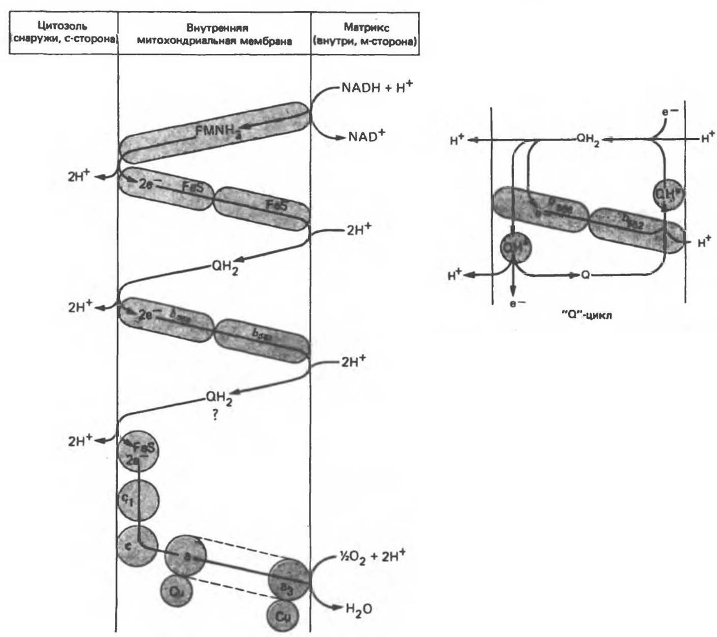
Fig. 13.11. Possible configuration of redox loops in the respiratory chain (chemiosmotic theory). This scheme is largely speculative, particularly regarding the region where Q interacts with cytochrome b, as the exact Nature of the intermediates and their relative positions are not precisely known. It is possible that a "Q cycle" operates in this case, ensuring proton translocation with the participation of the semiquinone (marked with an asterisk), as shown in the right-hand figure. On both sides of the membrane, the semiquinone is anchored by Q-binding proteins, whereas QH2 and Q are mobile. Cytochromes are arranged in the sequence b, c1, c, a, a3 (the latter being part of cytochrome aa3, which spans the membrane). FeS — iron-sulfur protein. Recent data indicate that cytochrome c oxidoreductase Functions as a proton pump (see Fig. 13.9).
The inner membrane contains A number of proteins—respiratory chain Enzymes—arranged adjacently to one another within the membrane, as shown in Fig. 13.11. Phosphorylating subunits responsible for ATP formation are located On the surface of the inner membrane (Fig. 13.12). They consist of several proteins that collectively form the F1 subunit; the latter protrudes into the matrix and functions as ATP synthase (Fig. 13.9). The F1 subunits are connected via a "stalk" to the membrane protein subunit F0, which presumably spans the entire membrane (Fig. 13.9). The passage of a pair of protons through the (F0—F1) complex results in The formation of one ATP molecule from ADP and Pi. Interestingly, similar phosphorylating subunits are found on the inner side of the bacterial Cell/33.html">Plasma Membrane and on the outer side of the chloroplast thylakoid membrane. It is important to note that in Mitochondria and Bacteria, the proton gradient is directed from the outside in, whereas in METABOLISM/14.html">Chloroplasts it has the opposite orientation.
Concepts regarding the mechanism coupling proton translocation with ATP synthesis by the anisotropic (vectorful) ATP synthase system are largely speculative. The model proposed by Mitchell is shown in Fig. 13.13. A pair of protons attacks one of the oxygen atoms of the Pi molecule, producing H2O and an active form of Pi, which immediately combines with ADP to form ATP. According to these views, ATP synthesis is not the primary energy-consuming step; rather, such a step is the release of ATP from the Active Site, which is likely associated with conformational Changes in the F1 subunit.
The chemiosmotic theory is supported by the following experimental evidence:
1. The addition of protons to the medium surrounding mitochondria leads to ATP formation.
2. Oxidative phosphorylation does not occur in soluble systems where the vectorful ATP synthase cannot function. A closed membrane system is required for oxidative phosphorylation to proceed (Fig. 13.9).
3. The Components of the respiratory chain are arranged in the membrane in an orderly, side-by-side fashion across the membrane, as predicted by the chemiosmotic theory (Fig. 13.11).
4. The P:H+ ratio for ATP synthase is 1:2, while the H+:O ratio for the oxidation of succinate and 3-hydroxybutyrate is 4 and 6, respectively; these values are in approximate agreement with the expected ratios. These data correlate with the presence of three redox loops in the respiratory chain.

Fig. 13.12. Structure of mitochondrial membranes. Submitochondrial particles are "verted inside out" and make it possible to study closed membrane systems in which the phosphorylating subunits face outward and the proton gradient has a "reversed" direction.
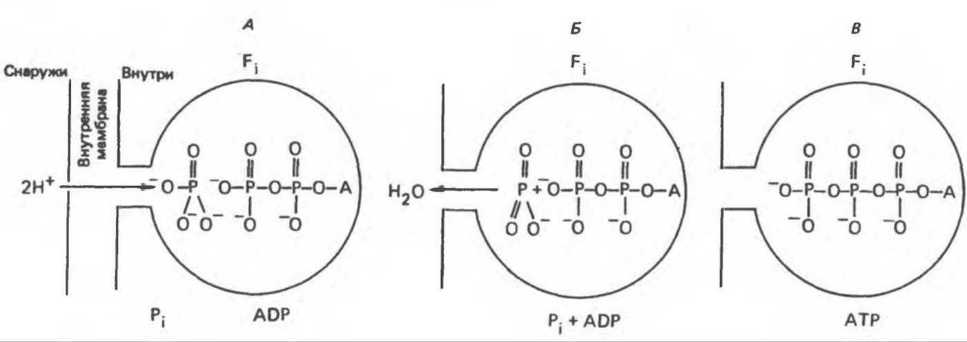
Fig. 13.13. Proton translocation mediated by the ATP synthase system (according to Mitchell).
The chemiosmotic theory explains the following phenomena:
1. The phenomenon of Respiratory Control. The electrochemical potential difference across the membrane, generated by proton translocation, inhibits the further transport of reducing equivalents along the respiratory chain until protons are translocated back across the membrane via the membrane-bound vectorful ATP synthase. This process, in turn, depends on the availability of ADP and Pi.
2. Action of uncouplers. These compounds (e.g., dinitrophenol) are amphipathic (see p. 164) and increase membrane permeability to protons (Fig. 13.9), thereby decreasing the electrochemical potential and short-circuiting the ATP synthase. In this case, oxidation can proceed without phosphorylation.
3. Functioning of Mitochondrial Transport Systems (see below). This phenomenon can be considered a prerequisite for the operation of the coupling membrane, which must remain impermeable to protons and other ions to maintain the electrochemical gradient. The membrane also incorporates transport systems that mediate the exchange of anions for OH- ions and cations for H+ ions. Such systems are essential for the import and export of ionized metabolites while preserving electrical and osmotic neutrality.
Last update: 06/08/2026
Editorial and Educational Adaptation: This material has been compiled based on the primary/original source text. The project team performed an editorial review, corrected technical inaccuracies, structured sections, and adapted the content for an educational format.
What was processed:
- elimination of formatting defects (OCR errors, structural breaks, corrupted characters);
- editorial organization of content;
- standardization of terminology in accordance with academic sources;
- verification of factual statements against the original source text.
All mentions of the author, publication year, and origin of the primary text have been preserved in accordance with the source.