Harper's Illustrated Biochemistry, Volume 1 - Murray R. 1993
Bioenergetics and Metabolism of Carbohydrates and Lipids
Oxidative Phosphorylation and Mitochondrial Transport Systems
Mitochondrial Transport Systems
Mitochondrial membranes and the localization of Key Enzymes within Cell/35.html">Mitochondria
Mitochondria possess an outer membrane permeable to most metabolites and a selectively permeable inner membrane characterized by numerous folds (cristae) projecting into the matrix (the internal space of the mitochondrion) (Fig. 13.12). The outer membrane can be removed by digitonin Treatment; it is distinguished by the presence of monoamine oxidase and several Other Enzymes (such as acyl-CoA synthetase, glycerophosphate acyltransferase, monoacylglycerophosphate acyltransferase, and phospholipase A2). Adenylate kinase and creatine kinase reside in the intermembrane space. The phospholipid cardiolipin is localized within the inner membrane.
The matrix houses the soluble enzymes of The Citric Acid Cycle as well as the enzymes responsible for the β-Oxidation of Fatty acids, which necessitates specialized transport mechanisms for metabolites and NUCLEOTIDES across the inner membrane. Succinate dehydrogenase is localized on the inner surface of The inner mitochondrial membrane, where it transfers reducing equivalents to the Respiratory Chain at the ubiquinone level (bypassing the first redox loop). 3-Hydroxybutyrate dehydrogenase is likewise situated on the matrix-facing side of the inner mitochondrial membrane. Glycerol-3-phosphate dehydrogenase is located on the outer surface of the inner membrane, where it participates in the functioning of the glycerophosphate shuttle mechanism.
Class="center">
Fig. 13.14. Glycerophosphate shuttle mechanism for The transport of reducing equivalents from the Cytosol into the mitochondrion.
Oxidation of extra-mitochondrial NADH via substrate shuttle mechanisms
NADH cannot cross the mitochondrial membrane; it is continuously generated in the cytosol by glyceraldehyde-3-phosphate dehydrogenase, a key glycolytic enzyme (see Fig. 18.2). However, under aerobic conditions, extramitochondrial NADH does not accumulate, as it is oxidized by the mitochondrial respiratory chain. Several mechanisms have been proposed to account for this phenomenon. They involve the transport of reducing equivalents across the mitochondrial membrane via substrate pairs coupled with their respective dehydrogenases. This requires the presence of a specific dehydrogenase on both sides of the mitochondrial membrane. The transfer of reducing equivalents via the glycerophosphate shuttle is illustrated in Fig. 13.14. It should be noted, however, that because the mitochondrial enzyme is linked to the respiratory chain via a flavoprotein rather than NAD, only two, rather than three, ATP molecules are formed per atom of oxygen consumed. In some animal species, The activity of FAD-dependent enzymes decreases following thyroidectomy and increases upon thyroxine administration. While this shuttle operates in insect flight Muscle and white muscle and plays a crucial role in the Liver, other Tissues (such as cardiac muscle) lack mitochondrial glycerol-3-phosphate dehydrogenase. A more ubiquitous transport system is believed to rely on malate, utilizing both cytosolic and mitochondrial malate dehydrogenases. The malate shuttle system is depicted in Fig. 13.15. The complexity of this system stems from the impermeability of the mitochondrial membrane to oxaloacetate; consequently, $\alpha$-ketoglutarate and aspartate—formed via the Transamination of oxaloacetate with glutamate—are transported across the mitochondrial membrane into the cytosol, where oxaloacetate is regenerated from $\alpha$-ketoglutarate.
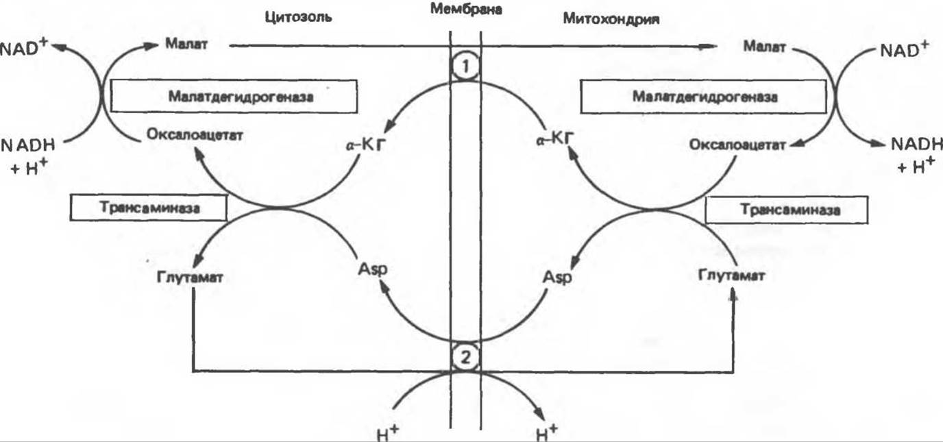
Fig. 13.15. Malate-aspartate shuttle mechanism for the transport of reducing equivalents from the cytosol into the mitochondria. 1 — ketoglutarate carrier, 2 — aspartate and glutamate carrier (note the coupled proton transport).
Energy-Dependent Ion Transport in Mitochondria
In actively respiring mitochondria engaged in Oxidative Phosphorylation, K+, Na+, Ca2+, and Mg2+ cations, as well as Pi, accumulate and are maintained at specific levels. The Uncoupling of Respiration and phosphorylation by dinitrophenol leads to the loss of ions by mitochondria; at the same time, oligomycin does not inhibit ion accumulation—indicating that this process does not rely on the energy stored via ADP phosphorylation. It can be assumed that cation transport is driven by the proton pump.
Transport Systems (Fig. 13.16)
The inner mitochondrial membrane is freely permeable to uncharged small molecules such as oxygen, Water, CO2, and NH3, as well as to monocarboxylic acids such as 3-hydroxybutyric, acetoacetic, and acetic acids. Long-chain Fatty acids are transported into mitochondria via the carnitine system (see Fig. 23.1); there is also a specific Pyruvate carrier that Functions via a symport mechanism utilizing the proton gradient from the outer to the inner surface of the mitochondrial membrane. The transport of dicarboxylate and tricarboxylate anions, as well as Amino Acids, is mediated by specialized carrier systems that facilitate their passage across the membrane. Monocarboxylic acids cross the membrane more readily due to their lower degree of dissociation; the undissociated form of the acid has a higher lipid solubility, and it is widely believed that monocarboxylic acids traverse the lipid membrane in this very form.
The transport of di- and tricarboxylate anions is closely coupled to the transport of inorganic phosphate, which readily crosses the membrane as H2PO-4 ions in exchange for OH-. Malate is carried by the dicarboxylate transport system in exchange for the inward transport of inorganic phosphate. The transport of citrate, isocitrate, and cis-aconitate by the tricarboxylate transport system occurs in exchange for the inward transport of malate. α-Ketoglutarate is also taken up in exchange for malate. Thus, these exchange mechanisms serve to maintain osmotic balance. It should be noted that the transport of citrate across the mitochondrial membrane depends not only on malate transport but also on inorganic phosphate transport. The adenine nucleotide carrier exchanges ATP for ADP, but not for AMP. A vital requirement is ensuring the export of ATP from the mitochondria for extramitochondrial utilization alongside the simultaneous influx of ADP for intramitochondrial ATP synthesis (Fig. 13.17). Na+ ions can be exchanged for H+ ions driven by the proton gradient. It is hypothesized that during The Active Transport of Ca2+ ions into mitochondria, a single positive charge is transferred per ion, which is likely associated with a Ca2+/H+ exchange. Calcium efflux from the mitochondrion is facilitated by its exchange for Na+.
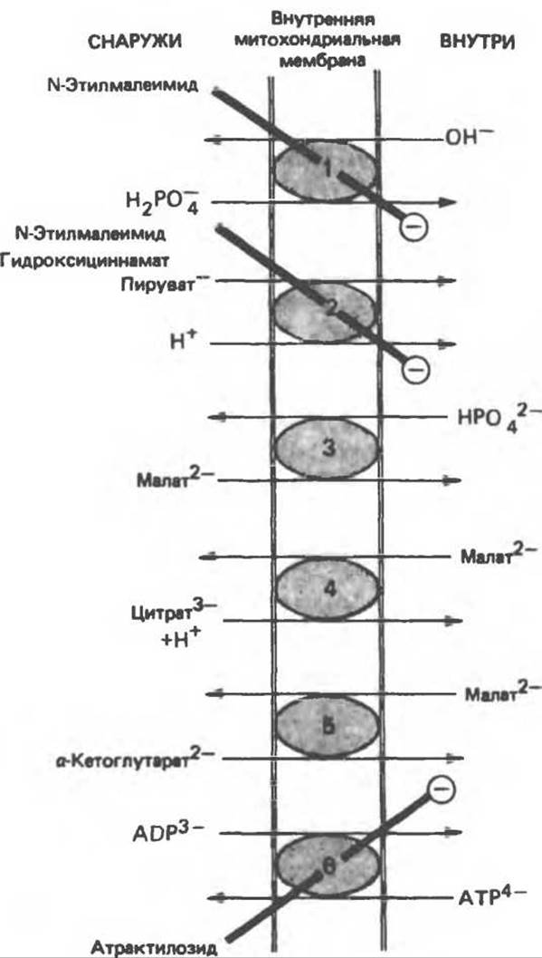
Fig. 13.16. Transport systems of the mitochondrial membrane. 1 — phosphate carrier, 2 — pyruvate symport, 3 — dicarboxylate carrier, 4 — tricarboxylate carrier, 5 — α-ketoglutarate carrier, 6 — adenine nucleotide carrier. N-Ethylmaleimide, hydroxycinnamate, and atractyloside inhibit (⊝) these systems. Also present (not shown in the figure) are transport systems for aspartate and glutamate (see Fig. 13.15), glutamine, Ornithine, and carnitine (see Fig. 23.1).

Fig. 13.17. Combined action of the phosphate carrier (1) and the adenine nucleotide carrier (2) in the ATP synthesis system. The H4/Pi symport is equivalent to the Pi/OH- antiport shown in Fig. 13.16: For every molecule of ATP exported from the mitochondrion, three protons enter the mitochondrion. If, however, ATP is utilized intramitochondrially, only two protons enter. This model accounts for the stoichiometric ratio of 3 translocated protons per electron pair at each coupling site (in contrast to Cross's hypothesis, which assumes the translocation of only two protons); it does not contradict Mitchell's original hypothesis (Fig. 13.11) (2 protons per electron pair at each coupling site) given a P/O ratio of 2 for NADH-dependent oxidation and 1.3 for succinate oxidation (according to Hinkle).
Action of Ionophores
The compounds in question derive their name from their ability to specifically bind certain cations and facilitate their transport across Introduction/36.html">Biological Membranes. These properties of ionophores stem from their lipophilic nature, which enables them to cross lipid membranes, particularly the mitochondrial membrane. A prime example is the antibiotic valinomycin, which carries K+ across the mitochondrial membrane and thereby decreases the Membrane Potential between its inner and outer surfaces. Nigericin also acts as an ionophore for K+ ions, but in exchange for H+; in this case, the pH gradient across the membrane is diminished. In the simultaneous presence of valinomycin and nigericin, both the membrane potential and the pH gradient are abolished, resulting in the complete inhibition of phosphorylation. Classical uncouplers, such as dinitrophenol, are essentially proton ionophores.
Impairments of the Respiratory Chain
Fatal infantile mitochondrial myopathy and Renal Dysfunction are associated with a reduced content or complete absence of most respiratory chain oxidoreductases.
Cross R. L. The mechanism and Regulation of ATP synthesis by F1-ATPases, Annu. Rev. Biochem., 1981, 50, 681.
Harper H. A., Rodwell V. W., Mayes P. A. Page 276. In: Review of Physiological Chemistry, 17th ed., Lange, 1979.
Hatefi Y. The Mitochondrial Electron Transport and oxidative phosphorylation system, Annu. Rev. Biochem., 1985, 54, 1015.
Hinkle P. C., McCarty R. E. How Cells make ATP, Sсi. Am. (March), 1978, 238, 104.
Hinkle P. C., Yu M. L. The phosphorus/oxygen ratio of mitochondrial oxidative phosphorylation, J. Biol. Chem., 1979, 254, 2450.
Mitchell P. Keilin’s respiratory chain concept and its chemiosmotic consequences, Science, 1979, 206, 1148.
Nicholls D. G. Bioenergetics: An Introduction to the Chemiosmotic Theory, Academic Press, 1982.
Tyler D. D. The mitochondrial ATP synthase, Page 117. In: Membrane Structure and function, Vol. 5, Bittar E. E. (ed.), Wiley, 1984.
Tyler D. D., Sutton С. M. Mitochondrial transporting systems, Page 181. In: Membrane Structure and Function, Vol. 5, Bittar E. E. (ed.), Wiley, 1984.
Last update: 06/08/2026
Editorial and Educational Adaptation: This material has been compiled based on the primary/original source text. The project team performed an editorial review, corrected technical inaccuracies, structured sections, and adapted the content for an educational format.
What was processed:
- elimination of formatting defects (OCR errors, structural breaks, corrupted characters);
- editorial organization of content;
- standardization of terminology in accordance with academic sources;
- verification of factual statements against the original source text.
All mentions of the author, publication year, and origin of the primary text have been preserved in accordance with the source.