Human Physiology - William F. Ganong 2002
Urine Formation and Excretion
Renal Function and Urination
Renal Dysfunction
Various Kidney diseases are characterized by a wide range of functional impairments. Renal renin secretion and its relationship with The Development of arterial Hypertension are discussed in Chapters 24 and 33. In various renal disorders, urine often contains protein, leukocytes, erythrocytes, and casts—compacted fragments of protein material that accumulate in the tubules and are subsequently flushed out into the bladder with urine. Other major consequences of kidney disease include the loss of renal concentration or dilution capacity, uremia, acidosis, and excessive Na+ retention.
Proteinuria
As a result of many kidney diseases, the permeability of glomerular capillaries increases, allowing protein to enter the urine in amounts exceeding normal trace levels. This phenomenon is termed proteinuria. If the protein in question is albumin, it is referred to as albuminuria. The relationship between albuminuria and the charge of the glomerular capillary membrane was discussed above. The amount of protein in the urine can be substantial, particularly in various nephroses. In such cases, protein loss may occur faster than the body can synthesize Plasma Proteins. Consequently, hypoproteinemia develops, leading to a decrease in plasma oncotic pressure and circulating Blood volume, while edema fluid accumulates intensively in the Tissues.
Class="center">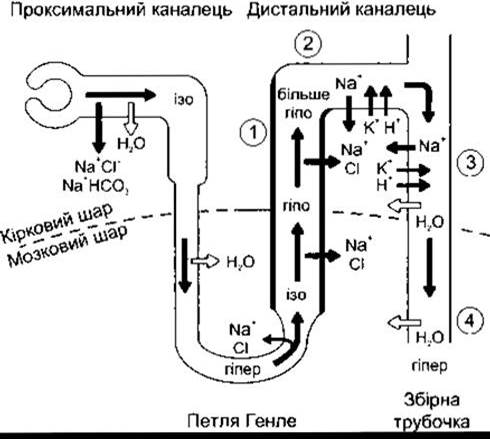
Fig. 38-25. Sites of action of various Diuretics. 1 - furosemide and other loop diuretics act on the thick ascending limb of the Loop of Henle; 2 - thiazide diuretics act on the early Distal convoluted tubule; 3 - aldosterone antagonists, triamterene, and amiloride act primarily on the collecting ducts; 4 - vasopressin V2-receptor antagonists act on the collecting ducts.
Proteinuria is also observed in a benign condition that is not typically considered a disease. In some completely healthy individuals, albumin appears in the urine while in an upright posture. This condition is known as orthostatic albuminuria. Urine produced by these individuals while lying down is protein-free.
Loss of Renal Concentration and Dilution Capacity
In kidney disease, urine becomes less concentrated, and its daily volume often increases, manifesting as symptoms such as polyuria (excessive urine output) and nocturia (the need to wake up at night to empty the bladder). While The ability to produce dilute urine is preserved, as the disease progresses, the urinary osmotic pressure becomes fixed at a level roughly approximating that of Blood Plasma. This indicates the loss of not only the concentration capacity but also the dilution ability of the Kidneys. One reason for this is disruption of the countercurrent multiplier mechanism; however, these impairments are more largely driven by the loss of a significant proportion of functional nephrons. If one kidney is surgically removed, the number of functional nephrons is halved. The nephrons of the remaining single kidney must filter and excrete a larger load of osmotically active solutes, effectively establishing an osmotic diuresis. In osmotic diuresis, urinary concentration approaches that of blood plasma (see above). The same process occurs when the number of functional nephrons decreases due to various diseases. The remaining nephrons bear a heavier workload, which ultimately leads to their damage. It is believed that the morphological substrate of nephron injury resulting from sustained hyperfiltration is fibrosis of the proximal tubular Cells, although this remains to be definitively established. Through this positive feedback loop, There is a progressive loss of functionally active renal parenchyma and a deepening of renal failure, culminating in oliguria or even anuria.
Uremia
When protein breakdown products accumulate in the blood in significant amounts, a syndrome known as uremia develops. Its manifestations include lethargy, somnolence, apathy, loss of appetite, nausea and vomiting, pruritus, mental disorders, Muscle twitching, convulsions, and ultimately coma. Serum urea and creatinine levels are markedly elevated. The concentrations of these substances in the blood are used as indicators of uremic severity. The symptoms of uremia are caused not so much by urea and creatinine themselves, but by the progressive accumulation of other toxic compounds, most likely organic acids and phenols.
The toxic compounds responsible for uremic symptoms can be removed from the blood using an artificial kidney machine (hemodialysis). Another effective technique is hemofiltration, in which blood is filtered through a membrane under pressure, and the lost fluids are subsequently replenished. These Methods can sustain life and maintain a satisfactory condition in patients for many months, even in the presence of complete anuria or bilateral nephrectomy.
Other hallmarks of chronic renal failure include anemia, primarily caused by the loss of renal Erythropoietin production (see Chapter 24), and secondary hyperparathyroidism resulting from 1,25-dihydroxycholecalciferol deficiency (see Chapter 21).
Acidosis
Acidosis is frequently observed in Chronic Kidney Disease due to the inability of the kidneys to adequately secrete acidic metabolic products (see Chapter 39). In a rare syndrome—Renal Tubular Acidosis—there is a specific impairment in the kidneys' ability to acidify urine, while other renal Functions remain intact. However, in most kidney diseases, the urine is maximally acidic, and acidosis develops due to limited overall H+ secretion resulting from impaired tubular NH4+ production.
Disorders of Na+ METABOLISM
Many patients with kidney disease retain excessive amounts of Na+ and develop pronounced edema. There are at least three reasons for enhanced renal Na+ retention. In acute Glomerulonephritis—a disease affecting predominantly the glomeruli—Na+ filtration is markedly reduced. In Nephrotic Syndrome, increased aldosterone secretion leads to salt retention. This condition is accompanied by low plasma protein levels; consequently, fluid shifts from the plasma into the interstitial spaces, reducing total plasma volume. This activates the Renin-Angiotensin System, which in turn stimulates aldosterone secretion. The third cause of Na+ retention and edema in renal disease is Heart Failure (see Chapter 33). Renal pathology contributes to the development of heart failure partly through the arterial hypertension it frequently induces.
Differential Responses to Aldosterone
As noted in Chapter 20, normal individuals do not collapse upon administration of excessive mineralocorticoids. They cease retaining Na+ and excrete large amounts of K+. Conversely, patients with marked edema due to nephrotic syndrome, cirrhosis, or heart failure continue to effectively retain Na+ and do not waste K+. One reason for this is the reduction in the amount of Na+ reaching the distal nephron; Na+ in the tubular fluid helps maintain the potential difference between the tubular fluid and tubular cells, which promotes enhanced K+ secretion. The decreased Na+ content in the distal tubule is achieved through enhanced proximal Na+ reabsorption and reduced Na+ filtration.
Last update: 10/08/2026
Editorial and Educational Adaptation: This material has been compiled based on the primary/original source text. The project team performed an editorial review, corrected technical inaccuracies, structured sections, and adapted the content for an educational format.
What was processed:
- elimination of formatting defects (OCR errors, structural breaks, corrupted characters);
- editorial organization of content;
- standardization of terminology in accordance with academic sources;
- verification of factual statements against the original source text.
All mentions of the author, publication year, and origin of the primary text have been preserved in accordance with the source.