BIOCHEMISTRY AND MOLECULAR BIOLOGY - W. ELLIOTT - 2002
CHAPTER 7. GENERATING ENERGY FROM FOOD. INTRODUCTION
Energy generation in the form of ATP is accomplished through complex and intertwined pathways, which are best understood by first examining The breakdown of glucose.
Energy production from glucose
MAIN STAGES OF glucose oxidation
The overall oxidation of glucose can be described by the following summary equation:
С6Н12О6 + 6О2 —> 6СО2 + 6Н2О.
The standard Gibbs Free energy change, ∆G°′, for this reaction is -2820 kJ • mol-1. Within Cells, The oxidation of a glucose molecule is coupled with the synthesis of more than 30 ATP molecules from ADP and Pi. The oxidation of glucose to CO2 and H2O can be divided into three stages.
1. Glycolysis — The process of breaking down glucose into two three-carbon fragments (Pyruvate molecules), coupled with the reduction of an electron carrier; it takes place in the Cell Cytoplasm.
2. The Krebs cycle (also known as The Citric Acid Cycle or Tricarboxylic Acid Cycle) — a series of reactions in which the second and third carbon atoms of pyruvate are converted into CO2, accompanied by the reduction of electron carriers. Molecular oxygen is not involved in this process, which occurs inside the Mitochondria.
3. The Electron Transport Chain — a pathway that transfers electrons to O2, which then combines with protons from the surrounding medium to form H2O. In eukaryotes, this stage takes place in The inner mitochondrial membrane and is responsible for The production of the vast majority of ATP. Before examining glucose oxidation in greater detail, we must first discuss The Nature of Biological Oxidation.
Biological oxidation and hydrogen-transfer systems
Oxidation does not necessarily require the involvement of oxygen. In general terms, the term simply reflects the loss of electrons. Oxidation can occur either as a direct transfer of electrons, such as The conversion of a ferric ion to a ferrous ion: Fe2+ —> Fe3+ + е-, or as an electron transfer coupled with the removal of hydrogen (a proton) from the oxidized molecule: АН2 —> А + 2е- + 2Н+.
In chemical systems, oxidation can be viewed as The transfer of electrons from a donor molecule to an acceptor molecule. Protons may be released into the surrounding solution or transferred along with electrons to the acceptor, which is equivalent to the transfer of hydrogen atoms.
In aerobic cells, oxygen serves as the ultimate oxidizing agent. Oxygen is electrophilic, meaning it readily accepts electrons; this process yields Water, with the required protons being drawn directly from the solution:
O2 + 4e- + 4Н+ —> 2Н2O.
Situated between oxygen (the terminal oxidant) and the metabolite being oxidized are intermediate electron acceptors. By binding electrons and passing them on to the next acceptor, they form a chain that shuttles electrons from the initial metabolite to oxygen. Each of these carriers alternately acts as an electron acceptor and an electron donor. It is this property that allows every member of the chain to function as an electron carrier. The Synthesis of the bulk of cellular ATP is linked to this electron transport chain. The general Organization of such a chain is illustrated in Fig. 7.1. Two participants in this chain play a particularly crucial role, and their properties are essential to understand thoroughly.
Class="center">Fig. 7.1. General concept of electron transfer to oxygen and ATP generation

NAD+ - an important electron carrier
The primary electron carrier that directly interacts with most oxidized metabolites is nicotinamide adenine dinucleotide, or NAD+. We have already encountered NUCLEOTIDES—compounds consisting of a nitrogenous base, a sugar, and a phosphate group—when studying AMP (see Fig. 1.6).
In the AMP molecule, the nucleic base is adenine (the structures of adenine and other bases are covered in Chapter 18). In the case of NAD+, we are dealing with two nucleotides linked together via their phosphate groups, which is why the compound is termed a dinucleotide. Structurally, NAD+ differs from the dinucleotide Building Blocks of Nucleic Acids, in which mononucleotides are also joined by phosphodiester bonds. The Structure of NAD+ can be represented as follows: base-sugar-phosphate-phosphate-sugar-base. One of these bases is adenine, just as in ATP, while the other is nicotinamide (niacinamide); both sugars are identical molecules of ribose. Thus, the structure of NAD+ can be depicted as follows:

NAD+ Functions as a coenzyme: a term used for low-molecular-weight Organic compounds that participate in enzymatic reactions. Unlike a typical substrate, NAD+ differs in that, once reduced, it dissociates from one enzyme and is immediately oxidized by another. Both Enzymes act "merely" as catalysts; in reality, NAD+ is reduced by the substrate of the first enzyme and oxidized by the substrate of the second, thereby acting as an electron shuttle that undergoes continuous cycles of Oxidation and reduction.
The "Active Site" of this coenzyme is its nicotinamide moiety; nicotinic acid (also known as niacin) is a vitamin. While it is an essential dietary component for certain animals, The Human Body is capable of synthesizing it independently from The amino acid Tryptophan. The rest of the NAD+ molecule serves as a binding domain for enzymes and does not undergo chemical alteration during the catalytic cycle.
Nicotinamide has the following structure:
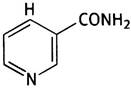
The structure of NAD+ can be represented as follows:

where R is the remaining part of the NAD+ molecule.
The reduction of NAD+ can be viewed as The addition of two electrons and a proton or, equivalently, a so-called hydride ion (H-). The reduced product (NADH) has the following structure:

NAD+ serves as a coenzyme for various dehydrogenases that catalyze Reactions of the type:
АН2 + NAD+ «-» А + NADH + Н+
The reduced NAD+ then diffuses to another enzyme, where it undergoes oxidation:
В + NADH + Н+ «-» ВН2 + NAD+
Thus, NAD+ acts as a carrier of two hydrogen atoms from substrate A to substrate B:
АН2 + В —> А + ВН2.
In biochemistry, such reactions are often depicted as a cycle:

Although formally the process can be represented as the transfer of two hydrogen atoms, in reality, one of them "travels" with the reduced NAD+, whereas the movement of the other is accomplished through two sequentially occurring and spatially separated events: the release of a proton into the surrounding medium and the binding of a proton from the surrounding medium. Therefore, using the abbreviation NADH always implies the combination of NADH + Н+.
FAD is an important electron carrier
FAD - flavin adenine dinucleotide - serves as an important electron (hydrogen) carrier. It is a derivative of vitamin B2 (riboflavin). Upon reduction, FAD attaches two hydrogen atoms and is converted into FADH2. Unlike NAD+, which moves from one dehydrogenase to another, FAD is bound to the apoenzyme, serving as its prosthetic group.
FAD has the following structure: isoalloxazine ring system-ribityl-phosphate-phosphate-ribose-adenine. Isoalloxazine is the chemical name of the ring system in riboflavin; ribityl is a pentahydric alcohol whose residue is also part of the riboflavin structure. The term "dinucleotide" is even less applicable to FAD than to NAD, since ribityl is not a sugar. The structural changes during the reduction of FAD, specifically of the isoalloxazine residue, are shown below:

Another electron carrier is flavin mononucleotide, or FMN. It has the following structure: isoalloxazine ring system-ribityl-phosphate, and differs from vitamin B2 (riboflavin) only by the presence of a phosphate group.
Both the oxidized and fully reduced forms of FMN and FAD have identical structures of the isoalloxazine residue.
Both Flavin Coenzymes can also exist in the form of so-called semiquinones. Two electrons are required for the complete reduction of flavin, and one for partial reduction. This allows flavins to participate in both single-electron and two-Electron transfer reactions.
Glycolysis is The First stage of glucose oxidation
Glucose oxidation occurs in three stages, the first of which is glycolysis. This is an anaerobic process during which only two ATP molecules are synthesized per glucose molecule. As shown in Fig. 7.2, the End products of glycolysis are pyruvate (i.e., the anion of pyruvic acid) and NADH. If glycolysis proceeds under aerobic conditions (in the presence of oxygen), both of these substances enter the mitochondria, where pyruvate is oxidized to CO2 and H2O, and NADH is oxidized to NAD+.
Fig. 7.2. Aerobic glycolysis of glucose (General Overview)
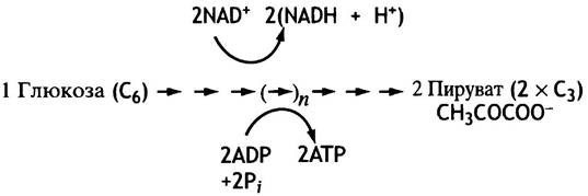
However, the body's oxygen supply does not always match its metabolic demands. For instance, during the initial Phases of the alarm reaction, when The Heart rate has not yet increased significantly, The rate of glycolysis spikes to provide contracting Muscles with the necessary ATP. Because NAD+ acts as a catalyst present in cells in limited amounts, it is crucial that NADH is rapidly reoxidized back to NAD+—since without NAD+, glycolysis grinds to a halt. We cannot afford to let glycolysis stall, as it remains the sole energy source for muscles when mitochondria fall short of the demand. To prevent this, a backup system is in place to regenerate NAD+ from NADH through the pyruvate-driven reduction of NADH. This reaction is catalyzed by Lactate dehydrogenase, which is abundant in Muscle cells.

The production of lactate (the conjugate base of lactic acid) from glucose is termed anaerobic glycolysis, distinguishing it from aerobic glycolysis, which yields pyruvate and an NADH that is subsequently oxidized in the mitochondria. It should be emphasized that the physiological purpose of anaerobic glycolysis is not to produce lactate. Rather, it is simply a mechanism to sustain ATP synthesis when the normal pathway of NADH reoxidation is blocked (Fig. 7.3). Anaerobic glycolysis is an extremely inefficient way to generate ATP, yielding a net gain of only two ATP molecules per consumed glucose molecule. Once the heart shifts into a faster rhythm and oxygen delivery to the muscles matches their workload, NADH reoxidation resumes, and glycolysis becomes aerobic. The lactate accumulated in the muscles is not metabolic waste; it is released into the bloodstream and cleared by the Liver (see p. 151).
Fig. 7.3. Anaerobic glycolysis of glucose (general overview)

Notably, using a similar strategy, Yeast can survive indefinitely in the absence of oxygen relying solely on anaerobic glycolysis. In yeast cells, pyruvate is decarboxylated (i.e., loses CO2) to yield acetaldehyde. The latter is then consumed to oxidize NADH in a reaction catalyzed by Alcohol dehydrogenase. Because of the low ATP yield, massive amounts of glucose must be broken down, producing alcohol and CO2.
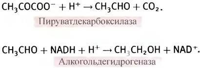
The description of glycolysis above has been simplified to highlight its core features. In reality, most of the glucose within muscles is stored as Glycogen. If ATP production is calculated relative to a glucose residue within glycogen rather than free glucose, glycolysis yields three molecules of ATP per glucose unit instead of two.
The Citric Acid cycle: the Second Stage of glucose oxidation
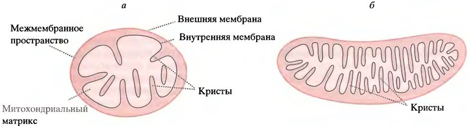
Mitochondria are small, membrane-bound intracellular Organelles (see Fig. 3.15). They can be considered the power plants of The Cell, as this is where the vast majority of ATP is synthesized. Mitochondria are enclosed by a double membrane. The outer membrane is permeable to many compounds and plays no direct role in energy production. In contrast, the inner membrane has extremely low permeability to most substances, except those with dedicated transport systems. This inner membrane folds into structures called cristae, which dramatically increase its surface area. The higher the energy demand of a tissue, the more cristae its mitochondria possess and the more densely packed they are (Fig. 7.4). The interior of the mitochondrion is filled with a concentrated solution of enzymes known as the matrix. The second stage of glucose METABOLISM—the citric acid cycle—takes place within the matrix, with only a single reaction of the cycle occurring in the inner membrane.
Fig. 7.4. Liver (a) and cardiac muscle (b) mitochondria. The density of cristae reflects the cellular demand for ATP
As noted earlier, aerobic glycolysis taking place in the cytoplasm yields pyruvate and NADH. Pyruvate enters the mitochondrial matrix via a specialized transport system, whereas NADH cannot cross the mitochondrial membrane. Instead, its "reducing equivalents" are shuttled across the inner membrane via specialized shuttle mechanisms and delivered to intra-mitochondrial NAD+- and FAD-linked enzymes. Ultimately, the NADH generated during glycolysis is reoxidized by the mitochondria, while NAD+ remains in the cytoplasm to support ongoing glycolysis. Inside the mitochondria, both NADH and FADH2 are oxidized by The electron transport chain.
It is worth noting that certain cells, such as erythrocytes (red Blood Cells), lack mitochondria, making glycolysis their sole source of ATP. Naturally, the survival of such cells depends entirely on a continuous supply of glucose.
Let us examine the enzymatic reaction that converts pyruvate—once inside the mitochondria—into a molecule that plays a central role in numerous metabolic pathways: acetyl-coenzyme A, or acetyl-CoA. In biochemical reactions, coenzyme A is often abbreviated as CoA-SH because the thiol group serves as the reactive center of the molecule. Unlike NAD+ and FAD, which function as electron carriers, coenzyme A acts as a carrier of acyl groups. It is a dinucleotide containing a moiety derived from the water-soluble vitamin pantothenic acid.

Interestingly, pantothenic acid plays a different role in coenzyme A compared to other Vitamins, such as niacin in NAD+ and riboflavin in FAD, which participate directly in Chemical Reactions. The pantothenate moiety appears to be required for protein recognition of coenzyme A, although it remains entirely unclear why evolution selected such a structurally unique architecture for this purpose in biochemistry.
The structure of coenzyme A can be represented schematically as follows:

β-Mercaptoethylamine is the primary functional ("working") group of the molecule:

Coenzyme A transports acyl groups in the form of thioesters, such as acetyl-CoA, which has the structure CH3CO-S-CoA. Unlike conventional oxygen esters, thioesters are high-energy (macroergic) compounds. The Standard Free Energy of Hydrolysis, ∆G°′, is -31 kJ • mol-1, whereas the hydrolysis of standard carboxylic acid esters yields a ∆G°′ of -20 kJ • mol-1. This difference in ∆G°′ can be explained by the fact that ordinary carboxylic acid esters are Resonance-stabilized and therefore possess a lower free energy content than non-resonance-stabilized thiol esters. This unique property of thioesters is harnessed across various biochemical systems.
Inside the mitochondria, pyruvate undergoes oxidative decarboxylation, a process involving the removal of CO2 (decarboxylation), the transfer of two electrons to NAD+ (oxidation), and the attachment of the resulting acetyl group to coenzyme A. Yeast also perform pyruvate decarboxylation catalyzed by pyruvate decarboxylase; however, this reaction is not oxidative (NAD+ is not involved) and yields acetaldehyde rather than an acetyl group. The large negative change in standard free energy indicates that The oxidative decarboxylation of pyruvate is irreversible:
Pyruvate + CoA - SH + NAD+ —> Acetyl-S-CoA + NADH + H+ + CO2
∆G°′ = - 33.5 kJ • mol-1.
The acetyl moiety in acetyl-CoA is the form in which pyruvate enters the citric acid cycle. This cycle is alternatively known as the Krebs cycle (named after its discoverer) or the tricarboxylic acid cycle (because A number of the metabolites involved contain three carboxyl groups). The acetyl carbon atoms in acetyl-CoA, previously part of pyruvate, are converted into CO2, while concurrently 3 molecules of NAD+ are reduced to NADH, and 1 molecule of FAD is reduced to FADH2. Furthermore, the overall consequence of these transformations is the synthesis of a single "high-energy" phosphoryl group from Pi for each consumed acetyl group (Fig. 7.5).
Fig. 7.5. Simplified diagram illustrating The Fate of NADH produced during glycolysis and the generation of NADH and FADH2 in mitochondria

Thus, a pyruvate molecule transported from the cytoplasm into the mitochondrion is converted into 3 molecules of CO2; in the process, 3 molecules of NAD+ and 1 molecule of FAD are reduced, and one molecule of ATP is generated. Given that 1 glucose molecule is split into 2 pyruvate molecules, glycolysis combined with the citric acid cycle yields 4 moles of ATP per mole of glucose. However, an additional 30 ATP molecules will be produced subsequently.
Electron transfer to oxygen: the Third Stage of glucose oxidation
This involves the oxidation of NADH and FADH2, which takes place in the inner mitochondrial membrane through the sequential transfer of electrons by specialized carriers.
Hierarchy of electron carriers in the electron transport chain
Recall that the objective is to transfer electrons from NADH and FADH2 to oxygen, yielding water:
NADH + H+ + 1/2O2 —> NAD+ + H2O.
The standard Gibbs free energy change for this process, ∆G°′, is -220 kJ • mol-1. To understand how this process occurs, one must utilize data on the standard reduction potentials (often referred to simply as redox potentials) of the participating compounds. Any oxidation-reduction reaction involves an electron acceptor (oxidizing agent) and an electron donor (reducing agent). The overall reaction:
X- + Y <-> X + Y-
can be conceptually divided into two half-reactions:
X- <-> X + e (1)
Y + e <-> Y- (2)
Each of these involves the oxidized and reduced forms of a single compound, termed a conjugate redox pair or redox pair. Here, the pairs are X and X-, as well as Y and Y-. Clearly, both redox pairs actually participate in the reaction, with one donating an electron and the other accepting it.
Different redox pairs exhibit varying affinities for electrons. Those with a lower affinity tend to transfer electrons to those with a higher affinity. The electron affinity of a redox pair is measured by its reduction potential (or redox potential) E’0. The value of this parameter allows one to predict the direction of electron flow between reactants. Equally important is the direct thermodynamic relationship between E'0 and changes in free energy.
The value of E0 is expressed in volts. The lower (more negative) it is, the lower the affinity of the substance for electrons, the greater its tendency to donate them, the higher its reducing power, and the higher the energy of the electrons.
The reason why a seemingly purely chemical property is expressed in volts lies in the method used to measure redox potentials. A redox reaction requires two redox pairs between which electron transfer takes place. These can be separated into distinct vessels (half-cells). Connecting such half-cells with a copper wire (an electron conductor) allows the flow of electrons to be directed. When measuring E'0, the reaction 2H+ + 2e- <-> H2 catalyzed by platinum black serves as one redox pair (the so-called hydrogen electrode), while the other conjugate pair is a mixture of the oxidized and reduced forms of the compound under investigation. In addition to positive ions, anions are also present in each half-cell. Electrons flow through the wire in a direction determined by the relative potentials of the systems (their electron affinities). Suppose the hydrogen electrode is connected to a half-cell containing Fe2+ and Fe3+ ions. This redistribution of electrons results in A change in cation concentration in both vessels. To maintain current flow, the electroneutrality of each solution must be preserved by allowing a flow of anions to balance the charge. For this purpose, the vessels are connected by a tube filled with an Agar-agar gel (to permit ionic conductivity while preventing bulk fluid flow). This device is known as an agar-salt bridge or agar bridge (Fig. 7.6). The electrical potential difference between the half-cells is measured with a voltmeter wired into the connecting circuit. By convention, the potential of the standard hydrogen electrode is set to zero. Thus, all experimental values of E'0 are calculated relative to this standard and are therefore relative quantities. In physics, electric current is conventionally defined as flowing in the direction opposite to electron movement. Consequently, the half-cell that donates electrons is at a more negative potential.
Fig. 7.6. Schematic diagram of an apparatus for measuring redox potentials. The reference hydrogen electrode (A) contains the H2/2H+ redox pair, the redox reaction of which is catalyzed by platinum black deposited on the electrode. The second half-cell (B) contains the redox pair whose potential is to be measured. If half-cell B is more reduced (i.e., E'0 is more negative than that of the hydrogen electrode) than A, electrons will flow from B to A, reducing 2H+ to H2 (when the half-cells are connected by a copper wire), while anions will migrate from A to B through the agar-salt bridge to neutralize the charge. If B is less reduced than A (i.e., E'0 is more positive than that of the hydrogen electrode), the entire process proceeds in the reverse direction

The symbol E0 (without a prime) denotes the standard reduction potential determined under standard conditions, where the concentrations of all solutes are 1 M and the hydrogen pressure is 1 atmosphere. In biochemistry, it is customary to use E'0 values corresponding to pH 7 (rather than pH 0). Under these conditions, the potential of the reference electrode (the hydrogen electrode) is -0.42 V. For the reaction NAD+ + 2H+ + 2e- —> NADH + H+, E'0 = -0.32 V, whereas for 1/2O2 + 2H+ + 2e- —> H2O, E'0 = +0.82 V. Such a large difference in redox potentials indicates that NADH is capable of reducing oxygen to water, whereas the reverse reaction is thermodynamically impossible.
Redox potentials E'0 are related to the standard Gibbs free energy change ∆G°′ by the Nernst equation:
∆G°′ = - nF∆E0',
where n is the number of electrons transferred in the reaction; F is the Faraday constant (96.5 kJ • V-1 • mol-1); ∆Е'0 is the difference in redox potentials between the electron donor and electron acceptor pairs.
The oxidation reaction of NADH:
NADH + Н+ + 1/2O2 —> NAD+ + Н2O
can be represented as a combination of two half-reactions:
NADH + Н+ —> NAD+ + 2Н+ + 2е- Е'0 = -0.320 V;
1/2O2 + 2Н+ + 2е- —> Н2O Е'0 = -0.816 V.
For the overall reaction: Е'0 = - 0.320 - 0.816 = 1.136 V,
therefore:
∆G°′ = -2(96.5 kJ • V-1 • mol-1)(-1.136 V) = -219.25 kJ • mol-1.
Fig. 7.7. The electron transport chain (schematic diagram). Some carriers accept only electrons, while protons are released into the water. Other carriers transfer both electrons and protons. The final stage is the transfer of electrons to an oxygen molecule. FADH is positioned lower in the transport chain than NADH. (The number of carriers is chosen arbitrarily)

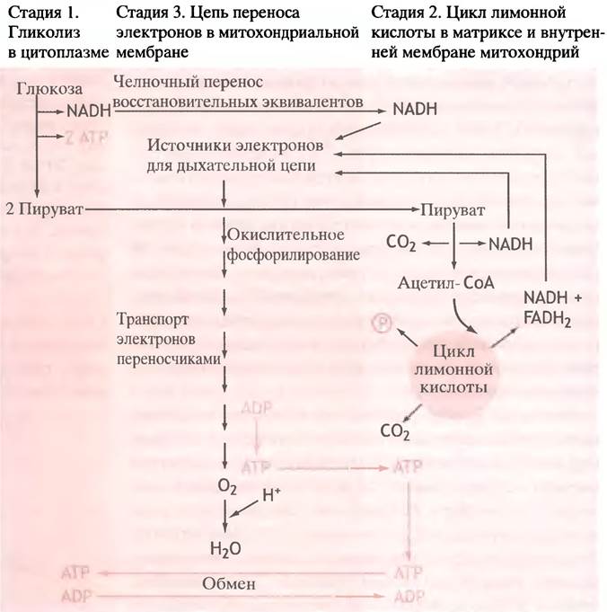
The transport of electrons to oxygen does not occur in a single step. The electron transport system in mitochondria is a chain of electron carriers whose redox potential increases (and, accordingly, the reduction potential decreases) as they approach oxygen. The movement of electrons from NADH and FADH2 to oxygen can be likened to descending a staircase, where the electron carriers serve as the steps. With each jump from step to step, a portion of free energy is released (Fig. 7.7). Instead of being wasted and dissipated as heat, as occurs during glucose combustion, this energy is used via specific mechanisms to synthesize ATP from ADP and Pi. This is why this complex process is called Oxidative Phosphorylation. The oxidation of 1 mole of glucose yields more than 30 moles of ATP. The pathways of all three stages of glucose oxidation discussed above are integrated in Fig. 7.8.
Fig. 7.8. Glucose oxidation. For simplicity, only the transformation products are shown (obviously, reduced NADH is formed from NAD+). During glycogen oxidation, three ATP molecules are produced for each glucose residue during glycolysis. In the citric acid cycle, GTP rather than ATP is produced as a high-energy compound. ATP is exported from mitochondria into the cytoplasm in exchange for ADP. NADH and FADH2 are generated not only during glucose oxidation, but also during the Oxidation of Fatty acids and other molecules. NADH and FADH2 donate electrons to the Respiratory Chain at different sites
Energy generation through the oxidation of fats and Amino Acids
To meet its energy demands in the form of ATP, the body oxidizes not only glucose and glycogen, but also fats and excess amino acids. In the case of triglycerides, from an energy-provision standpoint, the oxidation of the fatty acid is of primary importance rather than the glycerol moiety. The Chemical Structure of Fatty acids has nothing in common with that of glucose, so one would naturally expect their oxidation mechanism to be completely different. This makes the magical simplicity with which the Metabolic pathways of very different substances are interconnected all the more striking. Glucose transformations yield acetyl-CoA, which enters the citric acid cycle. Fatty acids undergo sequential breakdown, during which each step converts a pair of carbon atoms into the acetyl group of acetyl-CoA, which then enters the citric acid cycle. Therefore, the metabolism of Fatty Acids and glucose differs only up to the point of acetyl-CoA formation. Moreover, the Initial Stages of both glucose and fatty acid breakdown produce reduced forms of NAD (NADH) and FAD (FADH2), which are subsequently oxidized in the Mitochondrial Electron Transport chain (Fig. 7.9).
Fig. 7.9. Oxidation of fats and glucose. The diagram illustrates how the electron flow entering the respiratory chain is formed

The oxidation of amino acids is similar to that of glucose and fatty acids, although more complex in detail. All 20 Amino acids can be used for energy generation if their quantities exceed the body's immediate requirements for Protein Synthesis. Despite structural differences, their metabolic pathway is universal: they undergo deamination, and the nitrogen-free hydrocarbon Skeleton is ultimately utilized to form pyruvate and/or acetyl-CoA, which enter the citric acid cycle (Fig. 7.10). Thus, this cycle plays a central role in metabolism.
Fig. 7.10. Oxidation of glucose, fats, and amino acids for energy generation

Interchangeability of different fuel types
Excess glucose can be converted into fats. This occurs because Fatty acids are synthesized from acetyl-CoA:
Glucose —> pyruvate —> acetyl-CoA —> fatty acids.
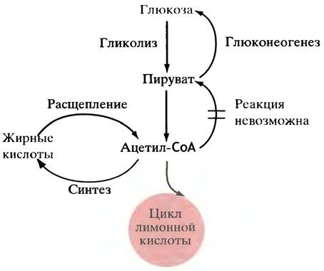
However, in animals, fatty acids cannot be converted into glucose because its synthesis strictly requires pyruvate, which cannot be obtained from acetyl-CoA due to the irreversibility of the reaction catalyzed by pyruvate dehydrogenase. Therefore, fatty acids cannot be converted into glucose (Fig. 7.11), but glucose can be converted into triglycerides.
Fig. 7.11. Simplified diagram illustrating why animals can convert glucose into fats, but fats cannot be converted into glucose. The reverse pathways do not entirely mirror the forward ones. Plants and Bacteria can convert fats into glucose without reversing the pyruvate dehydrogenase reaction.
Those amino acids that yield pyruvate or carboxylic acids for the citric acid cycle upon degradation can be converted into glucose by animals (they are termed glucogenic). During starvation, Muscle Proteins are broken down, and the resulting Amino acids are utilized by the liver to produce glucose. Plants and bacteria are able to convert fat and two-carbon compounds such as
acetate into glucose, but they employ a special pathway for this purpose, known as the Glyoxylate cycle (a variation of the citric acid cycle), which is absent in animals.
Questions for Chapter 7
1. What are the three stages of glucose oxidation, and where do they take place?
2. Describe the structure of NAD+. Draw its electron-accepting group in both the oxidized and reduced forms. How does NAD+ transfer hydrogen between substrates?
3. Explain the Structure and function of FAD.
4. What is the difference between aerobic and anaerobic glycolysis in muscle? When is anaerobic glycolysis utilized?
5. Describe the structure of coenzyme A. Which part of its molecule serves as the acceptor for acyl groups? What is the ∆G°′ of thiol ester hydrolysis? Compare this value with the ∆G°′ of carboxylic ester hydrolysis.
6. Describe the reaction catalyzed by pyruvate dehydrogenase and provide the ∆G°′ values.
7. What is the normal metabolic fate of the acetyl-CoA produced in the pyruvate dehydrogenase reaction?
8. Glycolysis and the citric acid cycle generate NADH and FADH2. What is the subsequent fate of these compounds?
9. The FAD + 2H+ + 2e- —> FADH2 redox pair has an E′0 of -0.219 V, whereas the 1/2 O2 + 2Н+ + 2е- —> Н2O redox pair has an E′0 of +0.816 V. Calculate ∆G0’ for the oxidation of FADH2 by oxygen to water.
10. Apart from the pyruvate dehydrogenase reaction, how else can acetyl-CoA be synthesized in the body?
11. A. Can glucose be converted into fats? Explain your answer. B. Can fatty acids in animals be converted into glucose? Explain your answer. C. Can fatty acids in E. coli be converted into glucose? Explain your answer.
Last update: 06/08/2026
Editorial and Educational Adaptation: This material has been compiled based on the primary/original source text. The project team performed an editorial review, corrected technical inaccuracies, structured sections, and adapted the content for an educational format.
What was processed:
- elimination of formatting defects (OCR errors, structural breaks, corrupted characters);
- editorial organization of content;
- standardization of terminology in accordance with academic sources;
- verification of factual statements against the original source text.
All mentions of the author, publication year, and origin of the primary text have been preserved in accordance with the source.