BIOCHEMISTRY AND MOLECULAR BIOLOGY - W. ELLIOTT - 2002
CHAPTER 6. BIOCHEMICAL MECHANISMS OF NUTRIENT TRANSPORT, STORAGE, AND MOBILIZATION
Transformation of glucose in the body
Mechanism of Glycogen synthesis
First of all, it should be noted that the synthesis of glycogen from glucose is an endergonic process, meaning it requires an energy input. The ∆G°′ of glycosidic bond Hydrolysis is -16 kJ • mol-1, so The equilibrium state favors virtually complete hydrolytic breakdown of glycogen to glucose (glycogen + H2O —> glucose). The formation of the glycosidic bond in the body proceeds coupled with the hydrolysis of a high-energy phosphate derivative.
Glycogen synthesis occurs by elongating an existing glycogen molecule seed (referred to as a primer) through the sequential addition of individual glucose molecules. This primer is the protein glycogenin, to one of whose Tyrosine residues an oligosaccharide chain of 8 glucose residues is attached via an O-glycosidic bond. This protein remains permanently incorporated into the glycogen granule. Glucose molecules are always added to the non-reducing end of the polysaccharide. Glucose belongs to the Class of sugars called aldoses. In aqueous solutions, its cyclic (predominant) form exists in equilibrium with an open-chain form containing an aldehyde group.

The aldehyde group acts as a reducing agent, which is why the C-1 end of the ring form is designated as the reducing end, and the C-4 end as the non-reducing end. Consequently, the glycogen chain always possesses a non-reducing end.
Thus, a glycogen molecule can be schematically represented as follows:

and its Biosynthesis involving ATP can be described by the following scheme:
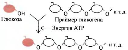
This process is repeated many times, leading to the formation of a long polysaccharide chain.
How is glycogen synthesis supplied with the necessary energy?
When glucose enters The Cell, it undergoes phosphorylation mediated by ATP. This reaction is catalyzed by the enzyme hexokinase in the Brain and Muscles.
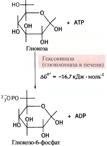
Kinases are a family of Enzymes that transfer a phosphoryl group from ATP to various molecules, and glucose is a representative of the Monosaccharides known as hexoses; hence the name hexokinase. In the Liver, this same reaction is catalyzed by a different enzyme, glucokinase.
Phosphorylation is an exergonic and therefore virtually irreversible reaction. Unlike free glucose, the resulting glucose-6-phosphate is a heavily charged molecule, which prevents it from diffusing across The Plasma Membrane. At the same time, phosphorylation leads to a decrease in the concentration of free glucose within the Cytoplasm. Both of these factors promote the diffusion of glucose into Cells from the surrounding environment along its concentration gradient. Under the Influence of the enzyme phosphoglucomutase, the phosphoryl group in the glucose phosphate molecule can reversibly migrate between the hydroxyl groups at the C-6 and C-1 carbon atoms.

The Standard Free energy values for the hydrolysis of glucose-1-phosphate and the hydrolysis of the α-(1 —> 4)-glycosidic bond of glycogen are identical at -21.0 kJ • mol-1. The Free energy of hydrolysis for the α-(1 —> 4)-glycosidic bond in the disaccharide maltose is -15.5 kJ • mol-1. Therefore, it is natural to assume that glycogen chain elongation occurs via a reaction between glucose-1-phosphate and the glycogen primer. However, this assumption is incorrect. Biochemical reactions are combined in such a way that the overall process becomes thermodynamically irreversible. Meanwhile, a direct synthesis of glycogen from glucose-1-phosphate would be a reversible and, consequently, uncontrolled reaction.
To render glycogen synthesis thermodynamically irreversible, an additional step is introduced. To understand this, let us recall The Structure of ATP (see Fig. 1.6). Its analogue is uridine triphosphate (UTP), in which uracil takes THE PLACE OF adenine. The cell synthesizes UTP using ATP as an energy source. It turns out that the intermediate product in glycogen synthesis is uridine diphosphate glucose (UDP-glucose), formed from UTP and glucose-1-phosphate.
For a purely formal reason (dictated by nomenclature rules), The enzyme catalyzing this reaction is named after the reverse reaction, which does not actually take place in the cell: UDP-glucose pyrophosphorylase. The irreversibility of this reaction is achieved because inorganic pyrophosphate (the product of the forward reaction) is extremely rapidly hydrolyzed by pyrophosphatase within the cell.
It is UDP-glucose, rather than glucose-1-phosphate, that serves as the glucose residue donor during glycogen synthesis. It can be viewed as an activated sugar, and sugar UDP-derivatives participate in biosynthetic reactions in animals in this capacity. Why specifically these compounds are used remains an enigma yet to be solved. In any case, starch synthesis in plants proceeds with the participation of adenine rather than uracil derivatives. The scheme of glycogen synthesis involving UDP-glucose as the glucose residue donor is shown in Fig. 6.2. This reaction is catalyzed by the enzyme glycogen synthase (synthases are enzymes that catalyze a synthetic reaction without utilizing ATP; otherwise, the term synthetases is used). Let us return once more to the issue of the irreversibility of UDP-glucose formation from UTP and glucose-1-phosphate. During this reaction (see Fig. 6.1), inorganic pyrophosphate is released, the hydrolysis of which proceeds irreversibly (∆G°′ = -33.5 kJ • mol-1). This particular circumstance imparts irreversibility to the entire set of reactions involved in glycogen biosynthesis (Fig. 6.3). This also explains why glycogen breakdown cannot occur simply by the reversal of its synthetic pathways.
Fig. 6.2. Synthesis of glycogen by primer elongation
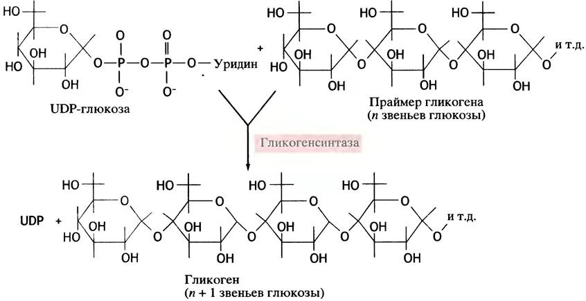
Fig. 6.3. Synthesis of glycogen from glucose

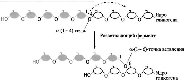
This diagram does not fully capture The process of glycogen synthesis, as glycogen should theoretically be an indefinitely long oligosaccharide chain, similar to amylose, the linear component of plant starch. Meanwhile, glycogen, much like another starch component—amylopectin, features a branched structure. This architecture is conferred by a specialized enzyme known as the branching enzyme. Once the synthase has built a linear chain segment of approximately 11 glucose residues, the branching enzyme transfers its terminal fragment, containing an average of 7 glucose residues, to the hydroxyl group of the C-6 carbon atom of a glucose unit within the same or another chain (Fig. 6.4). The energy required to form the α-(1 —> 6) bond is roughly equivalent to that of the α-(1 —> 4) bond. Consequently, this is a simple transfer reaction that does not require an energy input. As synthesis proceeds, the number of chain ends multiplies exponentially, serving both as the growing tips of the molecule and as sites for its subsequent fragmentation.
Thus, following a meal, glucose from the bloodstream enters Tissues and is converted into glycogen via the reactions discussed above.
Fig. 6.4. Action of the branching enzyme during glycogen synthesis
How is glucose formed in the liver?
The liver stores glucose as glycogen not so much for its own metabolic needs, but to ensure a continuous supply of glucose to other tissues, particularly the brain and erythrocytes. Between meals, the liver breaks down its stored glycogen at a rate sufficient to maintain a relatively constant Blood glucose concentration. This rate is governed by external signals determined by the Glucagon/Insulin ratio.
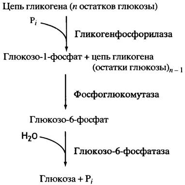
Much like its synthesis, the breakdown of glycogen proceeds from the non-reducing ends of the polysaccharide chains. In this process, the glycosidic bond is attacked not by Water, but by a phosphate anion; by analogy with hydrolysis, reactions of this type are termed phosphorolysis. Accordingly, the enzyme catalyzing this reaction is called Glycogen phosphorylase.

The glucose-1-phosphate produced via phosphorolysis is subsequently isomerized by phosphoglucomutase into glucose-6-phosphate (the reverse of the reaction described above),
which is then hydrolyzed to glucose, and the resulting free glucose is released into the blood (this hydrolysis, catalyzed by glucose-6-phosphatase, occurs exclusively in The Liver and Kidneys):
Glucose-1-phosphate <-> Glucose-6-phosphate
Glucose-6-phosphate + Н2O —> Glucose + Рi.
The general pathway of glucose production from glycogen is illustrated in Fig. 6.5.
Fig. 6.5. Breakdown of glycogen via phosphorolysis, culminating in the release of glucose into the blood
Glucose-6-phosphatase is localized on the membrane of The Endoplasmic reticulum. A notable feature of this enzyme is that its Active Site faces the lumen of the reticulum rather than the cytoplasm. To interact with it, glucose-6-phosphate crosses the membrane via a specialized transport protein and is hydrolyzed only thereafter. In turn, the hydrolysis products—glucose and phosphate—are shuttled back into the cytoplasm by dedicated transport systems.
Branch points in glycogen impede the action of phosphorylase, likely due to the enzyme's size and the spatial constraints of its active site. It cannot proceed with phosphorolysis if a chain end and a branch point are separated by four or fewer glycosidic residues. This obstacle is overcome in a two-stage process. In The First stage, three terminal residues are transferred by the debranching enzyme to the C-4 hydroxyl group of the terminal residue of another short chain, thereby lengthening it and making it accessible to phosphorylase. The Second Stage involves the hydrolytic Cleavage of the remaining side-chain α-(1 —> 6)-glycosidic residue (Fig. 6.6). Notably, both of these processes are catalyzed by a single protein—the debranching enzyme—which consequently possesses two distinct catalytic activities.
Fig. 6.6. Removal of branches in the glycogen molecule

Fig. 6.7. Uptake, storage, and release of glucose in the liver. Insulin and glucagon do not directly affect the enzymes of Glycogen METABOLISM

The overall scheme of glucose uptake, storage, and release by the liver is shown in Fig. 6.7.
Let us highlight several crucial Features of the processes depicted in this diagram.
1. The Synthesis and degradation of glycogen proceed via different pathways, allowing both processes to be regulated independently of each other.
2. Only in the liver and kidneys is glucose-6-phosphate, derived from glycogen, converted into free glucose, which is then released into the bloodstream to become available to other tissues.
3. In both the liver and other tissues, glucose-6-phosphate serves as a substrate for oxidation (see Chapter 8).
4. Insulin stimulates cells to store glycogen while simultaneously blocking its breakdown. Glucagon acts in the opposite manner.
Thus, insulin promotes glycogen synthesis, whereas glucagon stimulates the release of glucose from the liver. The MECHANISM OF ACTION of these Hormones will be discussed in subsequent chapters devoted to the molecular signals that regulate cellular life.
Why does glucokinase operate in the liver while hexokinase Functions in other tissues?
As soon as glucose enters a cell, it is immediately phosphorylated to glucose-6-phosphate. In the liver, this reaction is catalyzed by glucokinase, whereas in the brain and other tissues, it is catalyzed by hexokinase. Under starvation conditions, when supplying glucose to the bloodstream becomes a top priority, its utilization by Muscle and other cells is restricted by insulin levels, whereas glucose uptake by the brain, erythrocytes, and liver is insulin-independent. This may seem illogical. It would imply that muscles sacrifice their own Proteins so that the liver can synthesize glucose from Amino Acids to maintain the viability of brain cells, while at the same time the liver itself competes with the brain for this glucose. However, this does not happen! The reason is that liver glucokinase has a significantly lower affinity for glucose compared to brain hexokinase (Fig. 6.8). This means that during starvation, the liver takes up much less glucose than the brain, because passive Glucose Transport into cells is determined by The rate of its intracellular phosphorylation.
Fig. 6.8. Effect of glucose concentration on The activity of hexokinase (1) and glucokinase (2)

After a meal, when blood glucose levels are high and insulin prompts the liver to synthesize glycogen, glucokinase operates at its maximum possible rate. There is another difference between hexokinase and glucokinase: the latter is not inhibited by its reaction product, glucose-6-phosphate. This allows glycogen synthesis in the liver to proceed even when intracellular glucose-6-phosphate levels are high. Furthermore, the transport protein responsible for glucose uptake into liver cells has a higher Michaelis constant (Km) than those in other cells. A similar situation is observed in pancreatic cells that secrete insulin and coordinate its secretion with blood glucose levels.
What happens to other sugars absorbed in the Small Intestine?
During animal Digestion, along with glucose, many other sugars enter the portal vein from the intestine. These include galactose, derived from the milk sugar lactose, and fructose, the product of the hydrolysis of another dietary disaccharide, sucrose. To provide energy for the Organism, galactose and fructose must be converted either into glucose or into its metabolic intermediates within the liver. Ultimately, these sugars, along with glucose, are converted into glycogen and fat or undergo oxidation to CO2 and H2O.
To convert galactose into glucose, it is necessary to alter the optical configuration of the C-4 atom of the sugar residue (a process known in chemistry as epimerization).

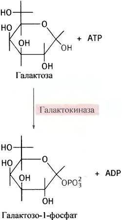
Cells cannot directly epimerize galactose, but they contain an enzyme called epimerase, which converts UDP-galactose into UDP-glucose. The latter is an intermediate in glycogen synthesis. First, galactose is phosphorylated by galactokinase to form galactose-1-phosphate:
Now, one might expect galactose-1-phosphate to react with UTP by analogy with glucose-1-phosphate. Instead, however, galactose-1-phosphate displaces glucose-1-phosphate in UDP-glucose, thereby forming UDP-galactose:

Since this reaction can be viewed as The transfer of a uridine phosphate (uridylyl) moiety from UDP-glucose, the enzyme catalyzing it is called uridylyltransferase. The epimerization reaction takes place only after UDP-galactose has been formed.

Combining all these reactions gives the following pathway for galactose transformation, culminating in The production of glucose-1-phosphate (Fig. 6.9).
Fig. 6.9. Entry of galactose into Major Metabolic Pathways via its conversion into glucose-1-phosphate

A well-known genetic defect manifests in infants as a condition called galactosemia. Affected individuals lack uridylyltransferase, resulting in the accumulation of galactose and its metabolic derivatives within the body. This leads to impaired brain development and blindness. To prevent such severe consequences, it is sufficient to eliminate galactose from the diet. The body's requirement for galactose to synthesize its own Glycolipids and Glycoproteins is met because the epimerization of UDP-galactose to UDP-glucose is reversible, and these synthetic processes specifically require UDP-galactose as a donor of galactosyl residues.
PATHWAYS OF AMINO acid Metabolism in the Organism (in the context of energy supply)
Although amino acids do not have specialized storage mechanisms like glucose and fat, they nevertheless contribute to energy supply. Their role is particularly crucial during starvation: under these conditions, Muscle Proteins are broken down, and the resulting Amino acids are transported to the liver, where they serve as substrates for glucose synthesis.
Metabolic pathways of fats and Cholesterol in the body
It is worth explaining why we discuss cholesterol when analyzing energy supply, even though it does not contribute to it directly. The fact is that after digestion, cholesterol is transported together with fats as part of Lipoproteins. Hence the rationale for considering The Fate of these substances jointly.
Cellular uptake of fat from chylomicrons
After a fatty meal, the blood becomes saturated with chylomicrons—particles whose shell is formed by Phospholipids, cholesterol, and proteins, while the core consists of neutral fats (triglycerides) and cholesterol esters (see Fig. 4.5).
Triglycerides are stored as reserves in adipose tissue, in the Mammary Glands of mammals (secreted with milk), as well as in muscles and other tissues, where they are used to generate energy.
Unlike triglycerides, free Fatty acids readily cross cell membranes. This is presumably why triglycerides in chylomicrons are hydrolyzed in the bloodstream by lipase located On the surface of cells lining the capillaries. The products of this hydrolysis are Fatty Acids and glycerol, which are immediately taken up by nearby cells.

The amount of fatty acids delivered from the blood into the tissue is determined by the total lipase activity in the capillaries permeating the tissue. This activity is particularly high in adipose tissue and in mammary glands during Lactation. The level of lipase depends on the physiological needs of the body; for instance, it increases as the insulin/glucagon ratio rises. Because a chylomicron can be viewed as a lipoprotein (a lipid-protein complex), the capillary lipase is called lipoprotein lipase to distinguish it from other lipases. Presumably, this lipase may also serve as a kind of bridge temporarily anchoring the chylomicron to the cell surface.
As triglycerides are depleted from chylomicrons, the latter shrink in size and are converted into so-called remnants containing the original amounts of cholesterol and its esters, as well as about 10% of the initial triglyceride content. These remnants are taken up by the liver via receptor-mediated endocytosis (Fig. 6.10) and end their lifecycle inside liver cells, delivering triglycerides and cholesterol thither. The complete fate of chylomicrons is illustrated in Fig. 6.11.
Fig. 6.10. Uptake of a chylomicron remnant by a liver cell via receptor-mediated endocytosis, where the receptor is specific for apolipoprotein E in the chylomicron

Fig. 6.11. Transport of fat and cholesterol via chylomicrons (CM): a - General view; b - detailed view

The fate of fats and cholesterol
In addition to chylomicrons, which transport fat and cholesterol directly from the intestine, a substantial contribution to The transport of these substances is made by lipoproteins, in the form of which fat and cholesterol enter the blood from the liver. The liver is capable of synthesizing triglycerides itself from glucose and other metabolites, and it also receives some triglycerides from the outside along with chylomicron remnants. Nevertheless, the liver should by no means be considered a lipid storage site (fatty liver is a pathological condition). Triglycerides are exported from the liver to other tissues as very low-density lipoproteins (VLDL), which structurally resemble chylomicrons. As a result, the liver acts as the primary fat supplier, provisioning the main consumers: muscles, where fats are oxidized, and adipose cells, where they are stored.
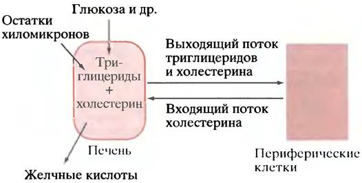
The liver not only collects dietary cholesterol delivered within chylomicron remnants, but is also the main site of Cholesterol synthesis in the body. From the liver, cholesterol is distributed throughout the body via VLDL along with triglycerides. At the same time, There is a counterflow of cholesterol from peripheral tissues back to the liver (Fig. 6.12). The purpose of this reverse movement is not yet entirely clear, especially since most cells can synthesize cholesterol themselves. One possible explanation is that this allows for a more flexible Regulation of the body's overall cholesterol content, as the liver can eliminate excess cholesterol in the form of Bile acids. Another hypothesis suggests that the release of cholesterol from the liver is a necessary consequence of triglyceride "secretion," since cholesterol is an essential component of the VLDL that carry them. According to this hypothesis, the return flow of cholesterol to the liver is simply a way to complete the transport cycle by bringing the carrier (cholesterol) back. Solving this problem is of great medical importance, as it is closely tied to understanding The Nature of atherosclerosis.
Fig. 6.12. Movement of triglycerides and cholesterol to and from the liver (schematic). In the liver, these substances are synthesized from metabolites and incorporated into chylomicron remnants. Here, cholesterol is also converted into bile acids
Utilization of cholesterol in the body
Cholesterol is an essential component of the plasma membrane in animal cells. At the same time, its excess in the body causes cardiovascular disease. This is why mechanisms for eliminating cholesterol from the body are of paramount importance.
The primary mechanism for this is The conversion of cholesterol into bile acids in the liver. In humans, approximately 0.5 g of cholesterol is excreted daily in this manner. From the liver, bile acids are secreted as part of bile into the small intestine and are subsequently reabsorbed by its walls along with fat. Therefore, one therapeutic objective in lowering cholesterol levels is to block the reabsorption of bile acids. This is achieved by administering substances that form stable complexes with bile acids in the intestine, preventing them from being absorbed by epithelial cells. Another, more modern approach involves regulating the synthesis of bile acids. In the Adrenal Glands and reproductive Organs, cholesterol is used to synthesize not only bile acids but also Steroid Hormones. Cholesterol is stored in cells in the form of esters and transported throughout the body within lipoproteins.
Lipoproteins involved in fat and cholesterol transport
The liver produces Two Types of lipoproteins: VLDL and HDL (high-density lipoproteins). This process involves the Endoplasmic reticulum and the Golgi apparatus, where lipoproteins are packaged into membrane-bound vesicles and released via exocytosis. In rats, about 80% of lipoproteins (excluding chylomicrons) are produced in the liver, with the remainder synthesized in intestinal cells. Next, we will discuss the conversion of VLDL into IDL (intermediate-density lipoproteins) and LDL (low-density lipoproteins). Thus, including chylomicrons, we are dealing with five types of lipoproteins. Cytology/cytology/93.html">ELECTRON MICROGRAPHS OF some of these are shown in Fig. 6.13.
Fig. 6.13. Electron micrographs of chylomicrons and lipoproteins: a - Chylomicrons; b - very low-density lipoproteins; c - low-density lipoproteins; d - high-density lipoproteins. The scale bar on each micrograph represents 100 nm. Photographs kindly provided by Dr. T. Forte (Lawrence Berkeley Laboratory, University of California)

Apolipoproteins
Each of the five lipoprotein classes has a characteristic set of apolipoproteins—that is, the purely protein components integrated into their structure. More than a dozen such proteins are currently known. While the precise function of every individual apolipoprotein is not yet fully defined, their collective roles can be summarized as follows.
1. Some apolipoproteins are essential for lipoprotein assembly. Examples include apoB48 in chylomicrons and apoB100 in VLDL.
2. Other apolipoproteins are responsible for recognition by specific receptors on The surface of target cells, which in turn triggers receptor-mediated endocytosis (see Fig. 6.10). This selective recognition ensures that individual lipoproteins bind exclusively to their designated cell types. Notable examples of such targeting apolipoproteins include apoB100 in LDL, which binds to LDL receptors, and apoE, which enables liver cells to recognize chylomicron remnants.
3. Certain apolipoproteins act as enzyme activators. For instance, apoCII in chylomicrons activates lipoprotein lipase, an enzyme that cleaves fatty acids from triglycerides. Not all apolipoproteins are present in lipoproteins from the outset; some are incorporated during extracellular transport. Notably, apoCII is synthesized in the liver but integrates into chylomicrons only after their formation.
Mechanism of Triglyceride and Cholesterol Transport from the Liver to Other Tissues, and Reverse Cholesterol Transport
Let us begin with the export of triglycerides and cholesterol from the liver in the form of VLDL. Patients with impaired VLDL synthesis experience an accumulation of triglycerides within the liver.
As soon as VLDL particles enter the bloodstream, they gradually lose triglycerides through the action of lipoprotein lipase. Concurrently, their relative content of cholesterol and cholesterol esters increases (Table 6.1), their density rises, and their overall size decreases. Consequently, VLDL particles are converted first into IDL and subsequently into LDL. It is primarily LDL that is recognized by receptors on peripheral tissue cells and internalized via endocytosis, thereby supplying these cells with residual triglycerides and cholesterol. The rate of this uptake is regulated by the number of receptors expressed on the surface of the target cells.
Table 6.1. Content of triglycerides and cholesterol in various lipoproteins (as % of dry mass)
Parameter |
Chylomicrons |
VLDL |
IDL |
LDL |
HDL |
Triglycerides |
80 |
50 |
30 |
10 |
8 |
Cholesterol and its esters |
8 |
20 |
30 |
50 |
30 |
The export of cholesterol from the liver is counterbalanced by a reverse flux from peripheral tissues, mediated by HDL. These particles are synthesized primarily in the liver and form a disc-like structure composed of apolipoproteins, phospholipids, and relatively small amounts of triglycerides and cholesterol. In peripheral tissues, HDL particles become enriched with cholesterol, presumably via passive diffusion from Plasma Membranes into the docked lipoprotein. Initially, the hydroxyl groups of the cholesterol molecules are exposed to the aqueous environment surrounding the HDL and are accessible for Esterification. The conversion of amphipathic cholesterol into entirely hydrophobic esters triggers a conformational change, transforming the HDL from a disc into a sphere.
Unlike extracellular esterification, intracellular esterification of cholesterol (CHOL) requires ATP energy; therefore, a different strategy is employed for HDL cholesterol. A specialized enzyme, lecithin-cholesterol acyltransferase (LCAT), catalyzes the transfer of fatty acid residues from lecithin (present in HDL) to cholesterol.

This reaction does not require an external energy source because the initial and resulting esters are energetically nearly equivalent.
Cholesterol esters sequestered within HDL are transferred to chylomicrons, VLDL, IDL, and LDL via a specialized carrier protein known as cholesteryl ester transfer protein (CETP). Although The primary function of IDL and LDL is to deliver their cargo to peripheral tissues, a fraction of these particles returns to the liver cells. Thus, The Mechanism of reverse cholesterol transport can be broken down into several key stages.
1. Cholesterol is transferred from the membranes of peripheral cells to HDL.
2. HDL cholesterol is esterified, and the resulting esters migrate from the periphery of the lipoprotein toward its core.
3. Cholesterol esters are transferred from HDL to chylomicrons, VLDL, IDL, and LDL.
4. A fraction of IDL and LDL (as well as postprandial chylomicrons) delivers cholesterol esters to liver cells, thereby effecting the net transport of cholesterol from peripheral cells back to the liver (Fig. 6.14).
Fig. 6.14. The Role of lipoproteins in cholesterol transport to and from the liver. Bold lines indicate the transfer of cholesterol esters among lipoproteins. All stages are reversible, and the diagram is simplified.

In reality, the process is far more complex, as simultaneous exchanges occur between various lipoproteins, involving triglycerides and cholesterol esters, the exchange of triglycerides for cholesterol esters, and so forth. Many of these phenomena have yet to be fully elucidated in detail.
Medical professionals often refer to LDL as "bad cholesterol" because an excess of cholesterol in the body—which increases the likelihood of atherosclerosis—correlates strongly with the proportion of this lipoprotein type. Atherosclerosis manifests as the formation of specialized plaques within Blood Vessels, largely composed of cholesterol. These plaques narrow and obstruct the vessels. In coronary vessels, this can precipitate Heart attacks. A well-known genetic defect causes cells to express very few LDL receptors. As a result, the clearance of cholesterol-rich LDL from the bloodstream is impaired, leading to elevated levels of LDL-bound cholesterol. This condition, known as familial hypercholesterolemia, is associated with severe cardiovascular complications. Conversely, HDL is termed "good cholesterol" by physicians, as high HDL levels are associated with a reduced risk of atherosclerosis. HDL accelerates the efflux of cholesterol toward the liver, though the exact mechanism of its protective action remains incompletely understood. It is hypothesized (though not yet definitively proven) that HDL can help halt the progression of atherosclerosis by extracting cholesterol directly from atherosclerotic plaques within blood vessels.
How Do Fats Leave Fat Cells?
Lipase releases free fatty acids stored in the form of neutral fats. However, the lipase functioning within fat cells is fundamentally different from the lipase found in blood capillaries. This difference lies in the fact that intracellular lipase is regulated by hormones: it is activated by glucagon and inhibited by insulin (see Chapter 12). After a meal, when insulin levels are high and glucagon levels are low, fat cells absorb glucose from Blood Plasma and fatty acids from chylomicrons, converting them into triglycerides, while the hydrolysis of the latter is inhibited. During starvation, the situation is reversed: Fatty acids are produced, which then leave the fat cells into the bloodstream and are delivered to other tissues. In juvenile diabetes, when patients have a very low insulin-to-glucagon ratio, hormone-sensitive lipase causes a large influx of fatty acids into the blood, which are taken up by the liver and converted into Ketone Bodies, leading to severe ketonemia (an excess of ketone bodies in the blood)—an additional severe complication of Diabetes Mellitus.
Hormone-sensitive lipase is activated not only by glucagon, but also by adrenaline. Following an "adrenaline" signal, fat cells release fatty acids that are delivered to muscles, enabling them to achieve maximum activity (see Chapter 12). Fat cells are innervated by sympathetic nerve
fibers, whose endings secrete noradrenaline upon excitation. This neurotransmitter also activates hormone-sensitive lipase.
How are fatty acids transported by the blood?
If fatty acids were present in the blood in a free state, it would resemble soapy water, because in a neutral environment these acids are completely dissociated. In reality, fatty acids in the blood are adsorbed onto the surface of a protein—serum albumin (SA). Its molecule has hydrophobic regions to which various hydrophobic molecules adhere. The resulting non-covalent complexes are not overly strong, which makes the binding reversible.
Questions for Chapter 6
1. Explain how the thermodynamic irreversibility of glycogen synthesis from glucose-1-phosphate is achieved.
2. How would you interpret the name UDP-glucose pyrophosphorylase?
3. Which tissues are capable of releasing glucose produced during glycogen breakdown into the blood, and why?
4. What reaction is catalyzed by the enzymes glucokinase and hexokinase? Why does the liver contain glucokinase, while the brain and other tissues contain hexokinase?
5. Childhood galactosemia is prevented by a galactose-free diet. Why is this possible thanks to UDP-galactose epimerase?
6. How do triglycerides leave chylomicrons and become assimilated by tissues?
7. What are VLDLs and what is their function?
8. What is meant by the reverse transport of cholesterol?
9. In what way is most of the cholesterol excreted from the body?
10. Cholesterol esterification is an endergonic reaction. How then is cholesterol esterified in HDLs without the participation of ATP?
11. How and under what conditions do fat cells release fat?
Last update: 06/08/2026
Editorial and Educational Adaptation: This material has been compiled based on the primary/original source text. The project team performed an editorial review, corrected technical inaccuracies, structured sections, and adapted the content for an educational format.
What was processed:
- elimination of formatting defects (OCR errors, structural breaks, corrupted characters);
- editorial organization of content;
- standardization of terminology in accordance with academic sources;
- verification of factual statements against the original source text.
All mentions of the author, publication year, and origin of the primary text have been preserved in accordance with the source.