LEHNINGER PRINCIPLES OF BIOCHEMISTRY - VOL 2. BIOENERGETICS AND METABOLISM - 2014
PART II. BIOENERGETICS AND METABOLISM
18. AMINO ACID OXIDATION AND THE PRODUCTION OF UREA
18.3. Catabolism of Amino Acid Carbon Skeletons
Under normal conditions, Amino Acid Catabolism accounts for only 10% to 15% of The Human Body's energy production; it is not as intensive as Glycolysis or Fatty acid oxidation. The various pathways of this catabolic route also vary widely, depending on the balance between biosynthetic requirements and the availability of a given amino acid. The 20 PATHWAYS OF AMINO acid degradation converge to form just six major products, all of which enter The Citric Acid Cycle (Fig. 18-15). From here, the carbon skeletons either enter Gluconeogenesis or ketogenesis, or are completely oxidized to CO2 and H2O.
Class="center">Fig. 18-15. Overview of amino acid catabolism. Amino Acids are grouped According to the major product of their degradation. Some amino acids appear more than once because different parts of their carbon skeletons are broken down into different end products. The most important Catabolic pathways in vertebrates are shown, although minor variations exist among vertebrate species. Threonine, for example, is degraded via at least two distinct pathways (see Figs. 18-19, 18-27), and the physiological significance of a particular pathway can vary depending on metabolic conditions. Glucogenic and ketogenic amino acids are highlighted in different colors. Note that Five amino acids are both glucogenic and ketogenic. Amino acids that degrade to Pyruvate are also potentially ketogenic. Only Two amino acids, leucine and Lysine, are strictly ketogenic.

All or part of the carbon Skeleton of Seven amino acids is ultimately degraded to acetyl-CoA. Five amino acids are converted to α-ketoglutarate, four to succinyl-CoA, two to fumarate, and two to oxaloacetate. Six amino acids are converted, entirely or in part, to pyruvate, which is then transformed into either acetyl-CoA or oxaloacetate. Later, we will summarize the individual Metabolic pathways of the 20 Amino acids as flowcharts, with each pathway leading to a specific entry point in The Citric Acid cycle. In these diagrams, the carbon atoms that enter the citric acid cycle are color-coded. Note that some amino acids appear more than once, reflecting the different metabolic fates of various PARTS OF THE carbon skeleton. Rather than examining every single step of each amino acid catabolic pathway, we have chosen to discuss in detail several enzymatic reactions that are particularly noteworthy either because of their mechanism or their medical significance.
Some Amino Acids Are Converted to Glucose, Others to Ketone Bodies
The seven amino acids that are degraded entirely or in part to acetoacetyl-CoA and/or acetyl-CoA are phenylalanine, Tyrosine, isoleucine, leucine, Tryptophan, threonine, and lysine. They can be converted into ketone bodies in the Liver, where acetoacetyl-CoA is converted to acetoacetate and then to acetone and β-hydroxybutyrate (see Fig. 17-18). These are the ketogenic amino acids (Fig. 18-15). Their ability to form ketone bodies often serves as a clinical marker for uncontrolled diabetes, during which massive quantities of ketone bodies are produced in the liver from both Fatty acids and ketogenic amino acids.
Amino acids that break down to yield pyruvate, α-ketoglutarate, succinyl-CoA, fumarate, and/or oxaloacetate can be converted into glucose and Glycogen via The sequence of reactions described in Chapters 14 and 15. These are glucogenic amino acids. The division between ketogenic and glucogenic amino acids is not absolute; five amino acids—tryptophan, phenylalanine, tyrosine, threonine, and isoleucine—have both Ketogenic and Glucogenic properties. Amino acid catabolism is critical for the survival of animals consuming a high-protein diet and for starving animals. Leucine is a strictly ketogenic amino acid that is abundant in Proteins. During starvation, its degradation leads to increased Ketosis.
Several Coenzymes Play Essential Roles in Amino Acid Catabolism
Amino acid catabolic pathways feature A wide variety of group-transfer reactions. It is helpful to begin studying these pathways by examining common types of reactions that cannot proceed without Cofactors. We have already covered one major class: Transamination reactions require Pyridoxal phosphate. Another common reaction type in amino acid catabolism is The transfer of one-carbon units, which typically involves one of three cofactors: biotin, tetrahydrofolate, or S-adenosylmethionine (Fig. 18-16). These cofactors carry one-carbon groups in various oxidation states: biotin carries carbon in its most oxidized form as CO2 (see Fig. 14-18); tetrahydrofolate carries a one-carbon unit at an intermediate oxidation state and occasionally as a methyl group; and S-adenosylmethionine carries a methyl group, which is the most reduced state of carbon. The latter two cofactors are especially vital in Amino Acid and Nucleotide METABOLISM.
Fig. 18-16. Some enzyme cofactors of major importance in one-carbon transfer reactions. The nitrogen atoms of tetrahydrofolate to which one-carbon groups attach are highlighted in blue.

Tetrahydrofolate (H4-folate), synthesized by Bacteria, consists of a substituted pterin (6-methylpterin), a para-aminobenzoate group, and a glutamate residue (Fig. 18-16).

Its oxidized form, folate, is a vitamin for mammals and can be converted to tetrahydrofolate in a two-step process catalyzed by the enzyme Dihydrofolate Reductase. The transferred one-carbon group, in any of its three oxidation states, binds to either the N-5 or N-10 nitrogen atom, or to both. The most reduced form of the cofactor carries a methyl group, a more oxidized form carries a methylene group, and the most oxidized forms carry methenyl, formyl, or formimino residues (Fig. 18-17). Most forms of tetrahydrofolate are interconvertible and serve as Donors of one-carbon units in a variety of metabolic reactions. The primary source of one-carbon units for tetrahydrofolate is carbon derived from Serine, yielding Glycine and N5, N10-methylenetetrahydrofolate.
Fig. 18-17. Interconversions of one-carbon units carried on tetrahydrofolate. The various molecules are grouped according to their oxidation state, with the most reduced form at the top and the most oxidized at the bottom. All species within the same colored box share the same oxidation state. The conversion of N5, N10-methylenetetrahydrofolate to N5-methyltetrahydrofolate is functionally irreversible. The enzymatic transfer of a formyl group, such as in purine synthesis (see Fig. 22-33) and the generation of formylmethionine in bacteria (Ch. 27), typically utilizes N10-formyltetrahydrofolate rather than N5-formyltetrahydrofolate. The latter is significantly more stable and therefore a much weaker donor of the formimino group. N5-Formyltetrahydrofolate is a minor side product of the cyclohydrolase reaction and can also form spontaneously. The conversion of N5-formyltetrahydrofolate to N5, N10-methenyltetrahydrofolate requires an ATP input because the thermodynamic equilibrium otherwise favors the opposite direction. Note that N5-formiminotetrahydrofolate is derived from Histidine via the pathway shown in Fig. 18-26.

Although tetrahydrofolate can transfer a methyl group attached to its N-5 atom, the energy of this methyl group is insufficient to drive most biosynthetic reactions. A better methyl group carrier is the cofactor S-adenosylmethionine (adoMet). It is synthesized from ATP and Methionine through the action of methionine adenosyltransferase (Fig. 18-18, step (1)). This reaction is unusual because the nucleophilic sulfur atom of methionine attacks the 5'-carbon of the ATP ribose ring instead of a phosphorus atom. Triphosphate is released and cleaved into Pi and PPi by the same enzyme, and PPi is subsequently hydrolyzed by inorganic pyrophosphatase; thus, this reaction consumes three bonds, including two high-energy phosphate bonds. The only other known reaction in which a triphosphate is split off from ATP occurs during coenzyme B12 synthesis (see Box 17-2, Fig. 3).
Fig. 18-18. Synthesis of methionine and S-adenosylmethionine in the methyl-activation cycle. The steps are described in the text. In the reaction catalyzed by methionine synthase (step (4)), a methyl group is transferred to cobalamin to form methylcobalamin, which in turn acts as the methyl donor in the generation of methionine. S-Adenosylmethionine, which bears a positively charged sulfur atom (a sulfonium ion), serves as a potent methylating agent in numerous biosynthetic reactions. The methyl acceptor (step (2)) is designated as R.
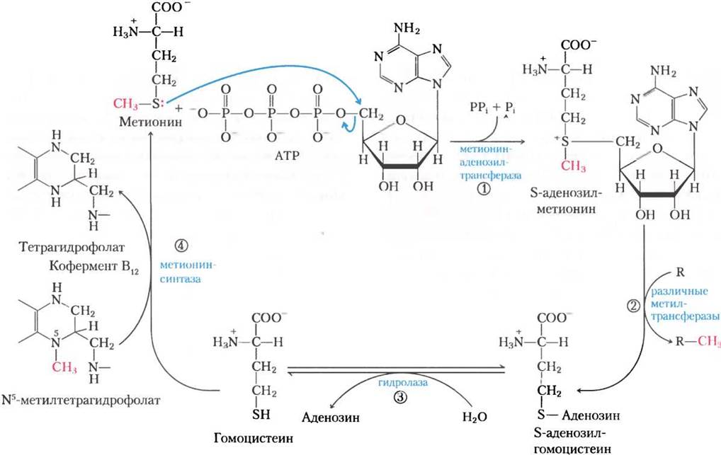
S-Adenosylmethionine is a powerful alkylating agent owing to the destabilizing properties of its sulfonium ion. The methyl group of S-adenosylmethionine is primed for nucleophilic attack, making it approximately 1,000 times more reactive than the methyl group of N5-methyltetrahydrofolate.
The transfer of a methyl group from S-adenosylmethionine to an acceptor yields S-adenosylhomocysteine (Fig. 18-18, step (2)), which is subsequently cleaved into homocysteine and adenosine (step (3)). Methionine is regenerated by the transfer of a methyl group to homocysteine in a reaction catalyzed by methionine synthase (step (4)); methionine is then converted back into S-adenosylmethionine, thus completing the methyl-activation cycle.
One form of methionine synthase, common to bacteria, uses N5-methyltetrahydrofolate as a methyl donor. Another form of the enzyme, present in certain bacteria and mammals, also utilizes N5-methyltetrahydrofolate, but the methyl group is first transferred to a vitamin B12 coenzyme derivative, cobalamin. This forms methylcobalamin, which then serves as the methyl donor in the synthesis of methionine. This reaction and the conversion of L-methylmalonyl-CoA to succinyl-CoA (see Box 17-2, Fig. 17-2a) are the only known B12-dependent reactions in mammals.
Vitamin B12 and folate are closely linked in these metabolic pathways. Vitamin B12 deficiency (pernicious anemia) is a rare condition occurring only in individuals with impaired intestinal absorption of the vitamin (see Box 17-2) or in strict vegetarians (as vitamin B12 is absent from plant foods). The disease develops slowly because the body requires very small amounts of vitamin B12, and hepatic reserves are sufficient to last for three to five years. The symptoms include not only anemia but also various neurological disorders.
The anemia can be traced to the reaction catalyzed by methionine synthase. As noted above, the methyl group of methylcobalamin is derived from N5-methyltetrahydrofolate, and in mammalian Cells, this is the sole reaction that utilizes N5-methyltetrahydrofolate. The Conversion of the N5,N10-methylene form to the N5-methyl form of tetrahydrofolate is irreversible (Fig. 18-17). Consequently, if coenzyme B12 is unavailable for the synthesis of methylcobalamin, folate becomes trapped in the N5-methyl form. The anemia resulting from vitamin B12 malabsorption is known as megaloblastic anemia, and it is accompanied by
a decrease in mature erythrocytes and the appearance of immature precursor cells, or megaloblasts, in the Bone Marrow. In the Blood, normal red Blood Cells are progressively replaced by fewer, abnormally large erythrocytes called macrocytes. This impaired red blood Cell development is a direct consequence of the depletion of N5,N10-methylenetetrahydrofolate, which is required for the synthesis of thymidine NUCLEOTIDES essential for DNA Replication (see Chapter 22). Folate deficiency, which exhausts all stores of tetrahydrofolate coenzymes, also leads to anemia via approximately the same mechanism. The symptoms of vitamin B12 deficiency can be alleviated by administering either the vitamin itself or folate.
However, treating pernicious anemia with Folic acid alone is dangerous because the neurological symptoms of vitamin B12 deficiency will continue to progress. These symptoms are unrelated to a defect in methionine synthase; rather, the abnormal enzyme methylmalonyl-CoA mutase causes an accumulation of unusual fatty acids containing an odd number of carbon atoms in neuronal membranes. Therefore, in cases of anemia associated with folate deficiency where the exact metabolic cause has not been definitively established, both folate and vitamin B12 are frequently prescribed. Early Diagnosis of vitamin B12 deficiency is critically important, as some of the associated neurological damage may be irreversible.
Folate deficiency also results in a shortage of N5-methyltetrahydrofolate required for the methionine synthase reaction. This leads to elevated blood levels of homocysteine, which can trigger myocardial infarction, Hypertension, and stroke. It is estimated that high homocysteine levels account for 10% of all myocardial infarctions. This condition can be corrected by administering folic acid. ■
Another cofactor in amino acid catabolism, tetrahydrobiopterin, resembles the pterin moiety of tetrahydrofolate, but it is not involved in one-carbon transfer reactions; instead, it participates in oxidation reactions. We will examine its function when discussing phenylalanine degradation (see Fig. 18-24).
Six Amino Acids Are Degraded to Pyruvate
The carbon skeletons of six amino acids are converted, entirely or in part, into pyruvate. Pyruvate can subsequently form either acetyl-CoA, which is ultimately oxidized in the citric acid cycle, or oxaloacetate, which enters gluconeogenesis. These six amino acids are Alanine, tryptophan, Cysteine, serine, glycine, and threonine (Fig. 18-19). Alanine is converted directly to pyruvate in a transamination reaction with α-ketoglutarate, whereas the side chain of tryptophan is released as alanine and thus ultimately yields pyruvate. Cysteine is converted to pyruvate in a two-step process involving the initial removal of the sulfur atom followed by transamination. Serine is converted to pyruvate by serine dehydratase. In this pyridoxal phosphate-dependent reaction, both the β-hydroxyl and α-amino groups of serine are removed (Fig. 18-20a).
Glycine is degraded via three pathways, only one of which leads to pyruvate. Glycine is converted to serine through an enzymatic hydroxymethyl addition reaction (Figs. 18-19 and 18-20b). This reaction, catalyzed by serine hydroxymethyltransferase, requires the coenzymes tetrahydrofolate and pyridoxal phosphate. Serine is then converted to pyruvate as described above. In the second pathway, which is more common in animals, glycine undergoes oxidative Cleavage to CO2, NH+4, and a methylene group (-CH2-) (Figs. 18-19 and 18-20c). This readily reversible reaction, catalyzed by the glycine cleavage system (glycine synthase), also requires tetrahydrofolate to accept the methylene group. In this oxidative degradation pathway, two carbon atoms do not enter the citric acid cycle: one is lost as CO2, and the other is converted into the methylene group of N5,N10-methylenetetrahydrofolate (Fig. 18-17), which serves as a one-carbon donor in various biosynthetic pathways. The operation of this second glycine degradation pathway is likely critical for mammals. Individuals with severe defects in glycine cleavage system activity suffer from a condition known as nonketotic hyperglycinemia, characterized by elevated serum glycine levels that lead to severe mental retardation and early childhood death. High concentrations of glycine—an inhibitory neurotransmitter—provide a plausible explanation for the neurological consequences of this disorder. Numerous Genetic Defects in Amino acid metabolism have been identified in humans (Table 18-2), and we will encounter several more in this chapter. ■
Figure 18-19. Catabolic pathways of alanine, glycine, serine, cysteine, tryptophan, and threonine. The Fate of the indole group of tryptophan is shown in Figure 18-21. Details of most reactions involving Serine and Glycine are presented in Figure 18-20. The threonine degradation pathway depicted here represents only the final third of total threonine catabolism (alternative routes are shown in Fig. 18-27). Some cysteine degradation pathways lead to pyruvate; the sulfur atom of cysteine can enter various metabolic routes, one of which is illustrated in Figure 22-15. Carbon atoms in this and subsequent figures are color-coded to trace their fate through metabolic intermediates.
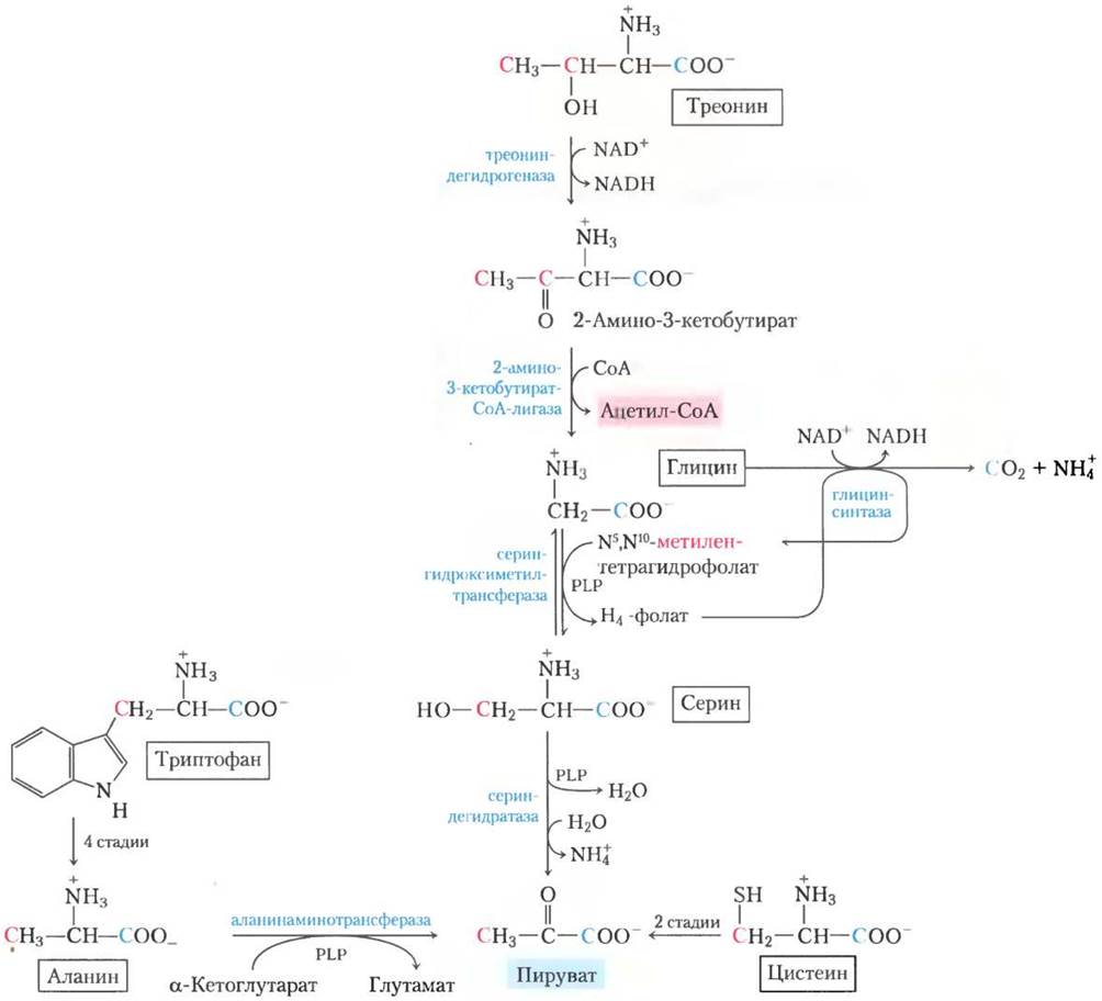
Figure 18-20. Reaction mechanisms. Roles of the cofactors pyridoxal phosphate and tetrahydrofolate in glycine and serine metabolism. The initial stage of each reaction (not shown) involves The formation of a covalent imine linkage between enzyme-bound PLP and The amino acid substrate: serine in (a), and glycine in (b) and (c). (a) PLP-catalyzed elimination of Water in the serine dehydratase reaction (step 1) leads to the subsequent formation of pyruvate. (b) In the hydroxymethyltransferase reaction, a PLP-stabilized carbanion (the product of step 1) acts as a key intermediate in transferring a methylene group (as -CH2-OH) from N5,N10-methylenetetrahydrofolate to yield serine. (c) The glycine cleavage system (glycine synthase) is a multienzyme complex comprising P, H, T, and L subunits. Ultimately, this reversible reaction releases CO2 and NH+4, while the second carbon atom of glycine is attached to tetrahydrofolate to yield N5,N10-methylenetetrahydrofolate. Pyridoxal phosphate activates the α-carbon of amino acids at critical stages of all these reactions, whereas tetrahydrofolate carries one-carbon units in two of them (see Figs. 18-6 and 18-17).

Table 18-2. Selected Genetic Diseases Caused by Defects in Amino Acid Catabolism
Disease |
Approximate incidence (per 100,000 newborns) |
Defective process |
Defective enzyme |
Symptoms and effects |
Albinism |
<3 |
Melanin synthesis from tyrosine |
Tyrosine 3-monooxygenase (tyrosinase) |
|
Alkaptonuria |
<0.4 |
Tyrosine degradation |
Homogentisate 1,2-dioxygenase |
Black urine; late-onset Arthritis |
Argininemia |
<0.5 |
Arginase |
Mental retardation |
|
Argininosuccinic aciduria |
<1.5 |
Urea cycle |
Argininosuccinate lyase |
Vomiting; convulsions |
Carbamoyl phosphate synthetase I deficiency |
<0.5 |
Urea cycle |
Carbamoyl phosphate synthetase I |
Lethargy; convulsions; early death |
Homocystinuria |
<0.5 |
Methionine degradation |
Cystathionine β-synthase |
Defective bone development; mental retardation |
Maple syrup urine disease (ketoaciduria) |
<0.4 |
Degradation of Isoleucine, Leucine, and Valine |
Branched-chain α-keto acid dehydrogenase complex |
Vomiting; convulsions; mental retardation; early death |
Methylmalonic acidemia |
<0.5 |
Conversion of propionyl-CoA to succinyl-CoA |
Methylmalonyl-CoA mutase |
Vomiting; convulsions; mental retardation; early death |
Phenylketonuria |
<8 |
Conversion of phenylalanine to tyrosine |
Phenylalanine hydroxylase |
Vomiting in neonates; mental retardation |
In the third and final pathway of glycine degradation, the achiral glycine molecule serves as a substrate for D-Amino Acid Oxidase. Glycine is thus converted to glyoxylate, an alternative substrate for hepatic Lactate dehydrogenase (p. 91). Glyoxylate is then oxidized in an NAD+-dependent reaction to oxaloacetate:
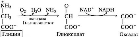
The primary function of D-amino acid oxidase, which is present in high concentrations in the Kidneys, is the detoxification of ingested D-Amino Acid Derivatives originating from bacterial cell walls and dietary sources (cooking at high temperatures causes spontaneous racemization of a small fraction of L-amino acids in proteins). Oxalate, whether ingested or enzymatically generated in the kidneys, has significant health implications. Calcium oxalate crystals account for up to 75% of all Kidney stones. ■
There are two primary pathways for The breakdown of threonine. One pathway leads to the formation of pyruvate via glycine (Fig. 18-19). This conversion to glycine occurs in two steps, beginning with the threonine dehydrogenase-catalyzed conversion of threonine to 2-amino-3-ketobutyrate. This pathway accounts for only 10% to 30% of total threonine catabolism in humans, though it may be more prominent in other mammals. In humans, the major pathway leads to the formation of succinyl-CoA, as described below.
In vitro, serine hydroxymethyltransferase can catalyze the single-step conversion of threonine to glycine and acetaldehyde, but this pathway is virtually inactive in mammals.
Seven Amino Acids Are Degraded to Acetyl-CoA
Portions of the carbon skeletons of seven amino acids—tryptophan, lysine, phenylalanine, tyrosine, leucine, isoleucine, and threonine—yield acetyl-CoA and/or acetoacetyl-CoA, with the latter being subsequently converted to acetyl-CoA (Fig. 18-21). Some of the final steps in the degradation of leucine, lysine, and tryptophan resemble the Oxidation of Fatty acids. Threonine (not shown in Fig. 18-21) is converted to acetyl-CoA via the minor pathway illustrated in Figure 18-19.
Figure 18-21. Catabolic pathways of tryptophan, lysine, phenylalanine, tyrosine, leucine, and isoleucine. Specific carbon atoms of these amino acids (highlighted in red) are converted to acetyl-CoA. Tryptophan, phenylalanine, and isoleucine also contain carbon atoms (shown in blue) that become incorporated into pyruvate or other citric acid cycle intermediates. The phenylalanine degradation pathway is shown in greater detail in Figure 18-23. The fate of nitrogen atoms is omitted from this scheme; in most cases, they are transferred to α-ketoglutarate to yield glutamate.

The catabolic pathways of two of these seven amino acids deserve special attention. Tryptophan is degraded via the most complex pathway of all amino acid catabolism in animal Tissues; a portion of the tryptophan molecule (four of its carbon atoms) is converted to acetyl-CoA via acetoacetyl-CoA. Several intermediates of tryptophan catabolism serve as precursors in the Synthesis of Other Biomolecules (Fig. 18-22), including nicotinate, a precursor of NAD and NADP in animals; serotonin, a vertebrate neurotransmitter; and indoleacetate, a plant growth factor. Some of these biosynthetic pathways are described in greater detail in Chapter 22 (see Figs. 22-28, 22-29).
Fig. 18-22. Tryptophan as a precursor of several biologically important molecules. Nicotinate (niacin), indoleacetate, and serotonin are synthesized from the aromatic ring of tryptophan. The atoms that form the nicotinate ring are highlighted in color.

Genetic defects in the Enzymes involved in phenylalanine degradation pathways lead to several inherited human disorders (Fig. 18-23); these will be discussed shortly. Phenylalanine and its oxidation product tyrosine (both containing nine carbon atoms) are degraded into two fragments that can enter The Tricarboxylic Acid Cycle: four of the nine carbon atoms form acetoacetate, which is converted to acetoacetyl-CoA and then to acetyl-CoA, while the second four-carbon fragment yields fumarate. Thus, eight of the nine carbon atoms of these two amino acids enter the tricarboxylic acid cycle, and the ninth is lost as CO2. Following a hydroxylation reaction, phenylalanine is converted to tyrosine, which is also a precursor of the neurotransmitter dopamine and the Hormones norepinephrine and epinephrine secreted by The adrenal medulla (see Fig. 22-29). In addition, tyrosine serves as the precursor for melanin, the dark pigment of skin and hair.
Fig. 18-23. Catabolic pathways of Phenylalanine and Tyrosine. In healthy humans, these amino acids are converted to acetoacetyl-CoA and fumarate. Genetic defects in various enzymes (shown in yellow) cause inherited disorders.

Phenylalanine catabolism can be impaired in some individuals
Many amino acids act as Neurotransmitters, neurotransmitter precursors, or neurotransmitter antagonists; consequently, genetic defects in amino acid metabolism can lead to neurological disorders and mental retardation. Most of these conditions are characterized by the accumulation of a specific intermediate. For example, defects in the Gene encoding phenylalanine hydroxylase, the first enzyme in phenylalanine catabolism (Fig. 18-23), cause phenylketonuria, the most common cause of elevated phenylalanine levels (hyperphenylalaninemia).
Phenylalanine hydroxylase (phenylalanine 4-monooxygenase) belongs to a major class of enzymes known as mixed-function oxidases (see Box 21-1). They catalyze the incorporation of one atom of O2 into a substrate via hydroxylation, while the other oxygen atom is reduced to H2O. Phenylalanine hydroxylase uses tetrahydrobiopterin as a cofactor, which transfers electrons from NADH to O2 and is oxidized to dihydrobiopterin in the process (Fig. 18-24). It is subsequently reduced back by the enzyme dihydrobiopterin reductase in an NADH-dependent reaction.
Fig. 18-24. The Role of tetrahydrobiopterin in the reaction catalyzed by phenylalanine hydroxylase. The hydrogen atom highlighted in pink is transferred directly from C-4 to C-3. This feature, discovered at the U.S. National Institutes of Health (NIH), was named the NIH shift.

In phenylketonuria, a secondary metabolic pathway of phenylalanine, which is normally used to a minimal extent, becomes activated. In this pathway, phenylalanine undergoes transamination with pyruvate to yield phenylpyruvate (Fig. 18-25).
Fig. 18-25. Alternative pathways of phenylalanine catabolism in phenylketonuria. In phenylketonuria, phenylpyruvate accumulates in tissues, blood, and urine. The urine may also contain phenylacetate and phenyllactate.

Phenylalanine and phenylpyruvate accumulate in the blood and tissues and are excreted in the urine—hence the name "phenylketonuria." Rather than being excreted, much of the phenylpyruvate is either decarboxylated to phenylacetate or reduced to phenyllactate. Phenylacetate imparts a characteristic odor to the urine, which is commonly used to screen for phenylketonuria in newborns. The accumulation of phenylalanine or its metabolites early in life impairs normal Brain development, resulting in severe mental retardation. This may be caused by an excess of phenylalanine competing with Other Amino Acids for the transport protein that carries amino acids across the blood-brain barrier, ultimately leading to a deficiency of essential metabolites.
Phenylketonuria was one of the first inherited human Metabolic Disorders to be discovered. If diagnosed in early infancy, The Development of mental retardation can be prevented by a strictly controlled diet. The diet must supply only The amount of phenylalanine and tyrosine strictly required for Protein Synthesis. The intake of protein-rich foods must be restricted. Natural proteins, such as milk casein, must first be hydrolyzed, and the bulk of the phenylalanine removed to provide an adequate diet, at least during childhood. The artificial sweetener aspartame is a dipeptide composed of aspartate and phenylalanine methyl ester (see Fig. 1-23, b); therefore, foods sweetened with aspartame are hazardous to individuals who must maintain a low-phenylalanine diet.
Phenylketonuria can also be caused by a defect in the enzyme that catalyzes the regeneration of tetrahydrobiopterin (Fig. 18-24). Treatment in this case is more complex than simply restricting dietary phenylalanine and tyrosine. Tetrahydrobiopterin is also required for the synthesis of L-3,4-dihydroxyphenylalanine (L-dopa) and 5-hydroxytryptophan, which are precursors of the neurotransmitters norepinephrine and serotonin. In this variant of phenylketonuria, these precursors must be included in the diet. Dietary supplementation with tetrahydrobiopterin itself is ineffective because it is unstable and does not cross the blood-brain barrier, and thus cannot reach the enzyme.
Neonatal screening for Genetic Disorders can be highly effective, particularly in the case of phenylketonuria. The tests employed (which go far beyond simply smelling the urine!) are relatively inexpensive, and the detection and early treatment of phenylketonuria in infancy (which occurs in eight to ten out of every 100,000 newborns) save millions of dollars each year that would otherwise be required for the management of the advanced disease. More importantly, early detection through these simple tests spares families immense emotional trauma—a benefit that cannot be measured in monetary terms.
Another inherited defect in phenylalanine catabolism is alkaptonuria, which is caused by a deficiency of the enzyme homogentisate dioxygenase (Fig. 18-23). This condition is considered less severe than phenylketonuria and has less tragic consequences; however, due to The excretion of large amounts of homogentisate, which becomes oxidized, the patient's urine turns black. Individuals with alkaptonuria are also prone to developing certain forms of arthritis. Alkaptonuria holds significant historical interest: in the early 1900s, Archibald Garrod discovered that the condition was inherited and deduced that it resulted from the absence of a single enzyme. Garrod was the first to link an inherited disease to an enzyme defect, marking a major breakthrough in the development of science that inevitably led to our modern understanding of genes and the biochemical pathways described in Part III. ■
Five amino acids are converted to α-ketoglutarate
The carbon skeletons of five amino acids (Proline, glutamate, glutamine, Arginine, and histidine) enter the tricarboxylic acid cycle as α-ketoglutarate (Fig. 18-26). Proline, glutamate, and glutamine each contain five carbon atoms. The cyclic Structure of proline is opened upon oxidation of the carbon atom most distant from the carboxyl group, yielding a Schiff base; subsequent Hydrolysis of this intermediate produces linear glutamate-γ-semialdehyde. This intermediate is further oxidized at the same carbon atom to yield glutamate. Glutamine is converted to glutamate through the action of glutaminase or any of several enzymatic reactions in which glutamine donates its amide nitrogen to an acceptor. Transamination or deamination of glutamate then yields α-ketoglutarate.
Arginine and histidine possess a five-carbon backbone to which a sixth carbon is attached via a nitrogen atom. Consequently, the catabolic conversion of these amino acids into glutamate is more complex than the pathway from proline to glutamine (Fig. 18-26). Arginine is converted to the five-carbon skeleton of Ornithine within The urea cycle (Fig. 18-10), after which ornithine undergoes transamination to form glutamate-γ-semialdehyde. The conversion of histidine to five-carbon glutamate is a multistep process in which the extra carbon atom is removed in a reaction requiring tetrahydrofolate as a cofactor.
Fig. 18-26. Catabolic pathways of arginine, histidine, glutamate, glutamine, and proline. These amino acids are converted to α-ketoglutarate. The numbered steps of the histidine pathway are catalyzed by (1) histidine ammonia-lyase, (2) Urocanase, (3) imidazolonepropionate hydrolase, and (4) glutamate formiminotransferase.

Four amino acids are degraded to succinyl-CoA
The carbon skeletons of methionine, isoleucine, threonine, and valine are degraded via a pathway that leads to succinyl-CoA (Fig. 18-27), an intermediate of the citric acid cycle. Methionine donates its methyl group to one of several possible acceptors via S-adenosylmethionine, and three of its four remaining carbon atoms enter propionate as propionyl-CoA, a precursor of succinyl-CoA. Isoleucine undergoes transamination followed by The oxidative decarboxylation of the resulting α-keto acid.
Fig. 18-27. Catabolic pathways of methionine, isoleucine, threonine, and valine. The degradation of these amino acids converges on succinyl-CoA; in addition, two carbon atoms of isoleucine yield acetyl-CoA (see Fig. 18-21). The threonine degradation pathway shown here occurs in humans, whereas the pathway found in other organisms is shown in Fig. 18-19. The conversion of methionine to homocysteine is detailed in Fig. 18-18; homocysteine to α-ketobutyrate in Fig. 22-14; and propionyl-CoA to succinyl-CoA in Fig. 17-11.
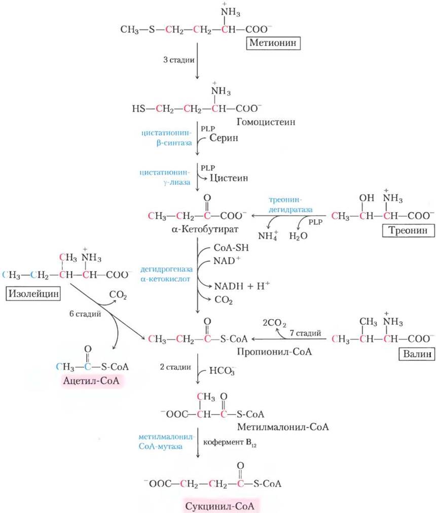
The remaining five-carbon skeleton is further oxidized to acetyl-CoA and propionyl-CoA. Valine undergoes transamination and decarboxylation, followed by a series of oxidative reactions that convert the remaining four carbon atoms into propionyl-CoA. Several steps in the degradation pathways of valine and isoleucine resemble the stages of fatty acid degradation (see Fig. 17-8a). In human tissues, threonine is also converted to propionyl-CoA in a two-step process. This is the primary pathway of threonine degradation in humans (for an alternative pathway, see Fig. 18-19). The Mechanism of the first reaction is analogous to that catalyzed by serine dehydratase; in fact, serine dehydratase and Threonine dehydratase may well be the same enzyme.
Propionyl-CoA, formed from these Three amino acids, is converted to succinyl-CoA via the pathway described in Chapter 17: carboxylation to methylmalonyl-CoA, epimerization of methylmalonyl-CoA, and conversion to succinyl-CoA by the B12-dependent enzyme methylmalonyl-CoA mutase (see Fig. 17-11). In a rare genetic disease known as methylmalonic acidemia, methylmalonyl-CoA mutase is deficient, which has severe metabolic consequences (Table 18-2, Box 18-2).
Box 18-2. MEDICINE. Scientists Solve a Mysterious Murder
Sometimes truth is stranger than fiction and reads more like a movie plot. Take, for example, the case of Patricia Stallings. She was sentenced to life imprisonment for the murder of her newborn son, but later exonerated through the forensic expertise of three persistent scientists.
The story began in the summer of 1989, when Stallings brought her three-month-old son Ryan to the pediatric intensive care unit at Cardinal Glennon Children's Hospital in St. Louis. The infant experienced respiratory distress, uncontrollable vomiting, and gastrointestinal illness. Based on these symptoms, the attending toxicologist diagnosed Ethylene glycol poisoning (a component of antifreeze). This Conclusion was subsequently supported by test results from a commercial laboratory.
When the child recovered, he was placed in a foster home, and Stallings and her husband David were permitted to visit him. However, the baby fell ill and died shortly after a brief visit with Stallings; she was subsequently charged with first-degree murder and arrested without bail. The test results from both the commercial and hospital laboratories seemed indisputable: both labs detected large amounts of ethylene glycol in the boy's blood and trace amounts of the substance in the bottle of milk Stallings had used to feed her son during the visit.
Yet, astonishingly, without realizing it, Stallings had set up a brilliant experiment. While incarcerated, she became pregnant and gave birth to another son, David Stallings Jr., in February 1990. He was immediately placed in a foster home, but two weeks later he began exhibiting symptoms similar to Ryan's. David was promptly diagnosed with a rare metabolic disorder, methylmalonic acidemia (MMA). This recessive genetic amino acid metabolism disorder occurs in approximately one out of every 48,000 newborns, and its symptoms are virtually identical to those caused by ethylene glycol poisoning.
Stallings had no opportunity to poison her second son, but Missouri prosecutors were reluctant to embrace new research technologies and insisted on prosecuting her regardless. The court refused to admit the MMA diagnosis of the second child as evidence, and in January 1991, Patricia Stallings was convicted of first-degree murder and sentenced to life in prison.
Fortunately for Stallings, William Sly, chairman of the Department of Biochemistry and Molecular Biology at Saint Louis University, and James Shoemaker, director of the university's metabolic screening laboratory, became interested in the case after seeing it on a television news program. Shoemaker re-analyzed Ryan's blood and found no ethylene glycol. The scientists contacted Piero Rinaldo, an expert in metabolic diseases at the Yale University School of Medicine, who had a laboratory equipped to diagnose MMA from blood samples.
Upon analyzing Ryan's blood, Rinaldo found high concentrations of methylmalonic acid, a breakdown product of the branched-chain amino acids isoleucine and valine; methylmalonic acid accumulates in MMA patients due to a defect in the enzyme required to convert it to the next step in the metabolic pathway. Notably, he observed that the child's blood and urine contained massive amounts of ketones—another metabolic hallmark of the disease. Like Shoemaker, he found no ethylene glycol in the child's body fluid samples. The formula bottle could not be tested because it had mysteriously disappeared. Rinaldo's analysis convinced him that Ryan had died of MMA. But how could two laboratories have reported finding ethylene glycol in his blood? Could both have made a mistake?
Rinaldo reviewed the laboratory records and found what he described as "horrifying." The first laboratory reported that Ryan Stallings's blood sample contained ethylene glycol, even though the blood chromatogram did not match that of a control sample containing ethylene glycol. "This is not just a matter of questionable interpretation of test results. The quality of their analysis is simply unacceptable," Rinaldo stated. And what about the second laboratory? Like Rinaldo, this lab detected abnormal substances in Ryan's blood and simply "assumed it was ethylene glycol." "Although nothing unusual was found in the bottle samples," Rinaldo indignantly noted, "the lab nevertheless claimed ethylene glycol was present there as well."
Rinaldo presented his findings to prosecutor George McElroy, who announced at a press conference the following day: "I no longer have confidence in these laboratories' test results." After reviewing the conclusion that Ryan Stallings had died of MMA, McElroy dropped all charges against Patricia Stallings on September 20, 1991.
Adapted from Michelle Hoffman (1991). Science 253, 931. © American Association for the Advancement of Science, 1991
Branched-Chain Amino Acids Are Not Degraded in the Liver
While the catabolism of Most amino acids occurs in the liver, the three branched-chain amino acids with side chains (leucine, isoleucine, and valine) are oxidized for energy primarily in Muscle, adipose tissue, kidneys, and the brain. These extrahepatic tissues contain an aminotransferase that is absent in the liver. This enzyme converts all three branched-chain amino acids into their corresponding α-keto acids (Fig. 18-28). Next, the branched-chain α-keto acid dehydrogenase complex catalyzes the Oxidative Decarboxylation of all three α-keto acids, releasing the carboxyl group as CO2 in each case and forming an acyl-CoA derivative. This reaction is formally analogous to two other oxidative decarboxylation processes encountered in Chapter 16: the Oxidation of Pyruvate to acetyl-CoA by the pyruvate dehydrogenase complex (see Fig. 16-6) and The oxidation of α-ketoglutarate to succinyl-CoA by the α-ketoglutarate dehydrogenase complex (p. 194). All three enzyme complexes are structurally similar and employ essentially the same reaction mechanism. They involve five cofactors (thiamine pyrophosphate, FAD, NAD, lipoate, and coenzyme A), and the three proteins of each complex catalyze homologous reactions. This is an instance where enzymatic blueprints originally "designed" to catalyze one reaction were "borrowed" via Gene Duplication and subsequently adapted to carry out similar reactions in other pathways.
Fig. 18-28. Catabolic Pathways of the three branched-chain amino acids valine, isoleucine, and leucine. In the three pathways operating in extrahepatic tissues, the first two enzymes are shared, as shown here. The branched-chain α-keto acid dehydrogenase complex is analogous to the pyruvate and α-ketoglutarate dehydrogenase complexes and utilizes the same five cofactors (some not shown here). A defect in this enzyme causes maple syrup urine disease in humans.

Experiments with rats have shown that the branched-chain α-keto acid dehydrogenase complex is regulated by covalent modification in response to dietary levels of branched-chain amino acids. When dietary levels of branched-chain amino acids are low or deficient, the enzyme complex is phosphorylated by a protein kinase and thereby inactivated. The addition of excess branched-chain amino acids to the diet triggers dephosphorylation and subsequent activation of the enzyme. Note that the pyruvate dehydrogenase complex is regulated in a similar manner via phosphorylation and dephosphorylation (p. 208).
A relatively rare genetic disorder exists in which the three branched-chain α-keto acids (along with their amino acid precursors, particularly leucine) accumulate in the blood and appear in excess in the urine. This disorder is named maple syrup urine disease due to the characteristic odor imparted to the urine by the α-keto acids produced by the defective α-keto acid dehydrogenase complex. If left untreated, the disease leads to neurological impairment, mental retardation, and death in early childhood. Treatment involves strict dietary management, minimizing the intake of valine, isoleucine, and leucine to the level strictly required for normal growth. ■
Asparagine and Aspartate Are Degraded to Oxaloacetate
The carbon skeletons of asparagine and aspartate ultimately enter the citric acid cycle as oxaloacetate. The enzyme asparaginase catalyzes the hydrolysis of asparagine to aspartate, which then undergoes transamination with α-ketoglutarate to yield glutamate and oxaloacetate (Fig. 18-29).
Fig. 18-29. Catabolic pathway of asparagine and aspartate. Both amino acids are converted to oxaloacetate.

Thus, we have examined how the 20 standard amino acids, having lost their amino groups, are degraded via dehydrogenation, decarboxylation, and other reactions into carbon skeletons of six central metabolites that can enter the citric acid cycle. Once degraded to acetyl-CoA, these components are fully oxidized to carbon dioxide and water, yielding ATP via Oxidative Phosphorylation.
As with Introduction/36.html">CARBOHYDRATES and Lipids, the breakdown of amino acids in the citric acid cycle ultimately generates reducing equivalents (NADH and FADH2). Our overview of catabolic pathways concludes in the next chapter with a Discussion of cellular Respiration, in which these reducing equivalents fuel the oxidative and energy-yielding processes in aerobic organisms.
■ Depending on the ultimate degradation product, some Amino acids can be converted into ketone bodies, some into glucose, and others into both. Thus, amino acid catabolism is fully integrated into Intermediary Metabolism and can be critical for survival under conditions where amino acids serve as a major source of metabolic energy.
■ The carbon skeletons of amino acids enter the citric acid cycle as five intermediates: acetyl-CoA, α-ketoglutarate, succinyl-CoA, fumarate, and oxaloacetate. Several are also converted into pyruvate, which can then be transformed into either acetyl-CoA or oxaloacetate.
■ The amino acids converted into pyruvate are alanine, cysteine, glycine, serine, threonine, and tryptophan. Leucine, lysine, phenylalanine, and tryptophan are degraded to acetyl-CoA via acetoacetyl-CoA. Isoleucine, leucine, threonine, and tryptophan can be converted directly into acetyl-CoA.
■ Arginine, glutamate, glutamine, histidine, and proline yield α-ketoglutarate; isoleucine, methionine, threonine, and valine form succinyl-CoA; all four atoms of phenylalanine and tyrosine contribute to the formation of fumarate; and asparagine and aspartate are converted into oxaloacetate.
Summary of Section 18.3 Pathways of Amino Acid Carbon Skeleton Degradation
■ Following the removal of amino groups, the carbon skeletons of amino acids undergo oxidation, and the resulting intermediates can then enter the citric acid cycle, being oxidized to CO2 and H2O. These pathways require several cofactors: tetrahydrofolate and S-adenosylmethionine in one-carbon transfer reactions, and tetrahydrobiopterin in the oxidation of phenylalanine by phenylalanine hydroxylase.
■ Branched-chain amino acids (isoleucine, leucine, and valine), unlike most other amino acids, are catabolized exclusively in extrahepatic tissues.
■ A number of severe human diseases can be caused by genetic defects in the enzymes of amino acid catabolism.
Last update: 06/08/2026
Editorial and Educational Adaptation: This material has been compiled based on the primary/original source text. The project team performed an editorial review, corrected technical inaccuracies, structured sections, and adapted the content for an educational format.
What was processed:
- elimination of formatting defects (OCR errors, structural breaks, corrupted characters);
- editorial organization of content;
- standardization of terminology in accordance with academic sources;
- verification of factual statements against the original source text.
All mentions of the author, publication year, and origin of the primary text have been preserved in accordance with the source.