Biological Chemistry - Berezov T. T., Korovkin B. F. 1998
Nervous Tissue
Metabolic Features of Nervous Tissue
Amino Acid and Protein Metabolism
The total amino acid content in human Brain tissue is 8 times higher than their concentration in the Blood. The Amino Acid Composition of the brain exhibits a distinct Specificity. For instance, the concentration of free glutamic acid in the brain is higher than in any other mammalian organ (10 µmol/g). Glutamic acid, together with its amide glutamine and the tripeptide Glutathione, accounts for more than 50% of the $α$-amino nitrogen in the brain. The brain contains A number of free Amino Acids that are found in only negligible amounts in other mammalian tissues. These include $γ$-aminobutyric acid, N-acetylaspartic acid, and cystathionine (see Chapter 1).
It is known that Amino acid METABOLISM in brain tissue proceeds in various directions. First and foremost, the free amino acid pool serves as a source of "raw Materials" for the synthesis of Proteins and biologically active amines. One of the Functions of dicarboxylic amino acids in the brain is the binding of ammonia released during nerve Cell excitation.
The influx of amino acids into the brain tissue and their efflux, as well as the utilization of blood glucose for the synthesis of neuronal and glial amino acids, vary across different Regions of the brain. These differences are largely determined by the blood-brain barrier, which must be considered specifically for each substance or Class of substances. The blood-brain barrier should not be viewed as a uniform structural formation acting as a simple transport barrier; variations in the relative rates of substance uptake into different brain regions may be due to the CHARACTERISTICS OF THE vascular endothelium, the basement membrane, or the arrangement of adjacent glial cell processes. Under in vitro conditions (in the absence of the barrier), many amino acids accumulate in brain Cells via Active Transport, involving several independent Na+-dependent transport systems.
It has been established that proteins in the brain are in a state of active turnover, as evidenced by the rapid incorporation of radioactive amino acids into protein molecules. However, the rates of protein Synthesis and degradation vary across different regions of the brain. The Proteins of the Gray matter in the cerebral hemispheres and the proteins of the Cerebellum exhibit a particularly high rate of turnover. In areas of the brain rich in conduction pathways, namely axons (White matter), The rate of Protein Synthesis AND degradation is lower.
Various functional states of the Central Nervous system are accompanied by Changes in the Rate of protein turnover. For example, when animals are exposed to excitatory agents (such as pharmacological drugs and electric current), the rate of Protein metabolism in the brain increases. Conversely, under The Influence of anesthesia, the rates of protein degradation and synthesis decrease.
Excitation of The Nervous System is accompanied by an increase in ammonia content within the Nervous Tissue. This phenomenon is observed during The stimulation of both peripheral nerves and the brain itself. It is believed that Ammonia Production during excitation primarily results from the deamination of AMP.
Ammonia is a highly toxic substance, particularly to the nervous system. Glutamic acid plays a crucial role in eliminating ammonia. It is capable of binding ammonia to form glutamine, a substance harmless to nervous tissue. This amidation reaction proceeds with the participation of the enzyme Glutamine Synthetase and requires the expenditure of ATP energy (see Chapter 12). The direct source of glutamic acid in brain tissue is the reductive amination pathway of $α$-ketoglutaric acid;

The formation of glutamic acid from $α$-ketoglutaric acid and ammonia serves as an important mechanism for neutralizing ammonia in brain tissue, where The pathway of ammonia elimination via urea synthesis does not play a significant role.
Additionally, glutamic acid in nervous tissue can undergo decarboxylation to form GABA:
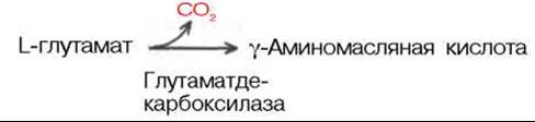
GABA is found in the highest concentrations within the gray matter of the brain. It is present in significantly smaller amounts in the Spinal Cord and peripheral nerves.
Last update: 06/08/2026
Editorial and Educational Adaptation: This material has been compiled based on the primary/original source text. The project team performed an editorial review, corrected technical inaccuracies, structured sections, and adapted the content for an educational format.
What was processed:
- elimination of formatting defects (OCR errors, structural breaks, corrupted characters);
- editorial organization of content;
- standardization of terminology in accordance with academic sources;
- verification of factual statements against the original source text.
All mentions of the author, publication year, and origin of the primary text have been preserved in accordance with the source.