Review of Medical Physiology - William F. Ganong 2002
Endocrine System, Metabolism, and Reproduction
Thyroid Gland
Synthesis and Secretion of Thyroid Hormones
Chemistry
The principal Hormones secreted by The Thyroid Gland are triiodothyronine (T3) and thyroxine (T4). Triiodothyronine is also formed in peripheral Tissues by the deiodination of T4 (see below). Both hormones are iodinated Amino Acids (Fig. 18-4). Small amounts of reverse triiodothyronine (3,3',5'-triiodothyronine, rT3) and Other Compounds have been detected in the thyroid venous Blood. Triiodothyronine is more active than T4, whereas rT3 is inactive. The naturally occurring forms of T4 and its derivatives with an asymmetric carbon atom are L-isomers. At the same time, D-thyroxine accounts for only a small fraction of the L-form's activity.
Thyroglobulin
T3 and T4 hormones are synthesized within the colloid through the iodination and Condensation of Tyrosine molecules peptide-linked to thyroglobulin. This glycoprotein consists of two subunits and has a Molecular Weight of 660,000. CARBOHYDRATES account for about 10% of its mass. It also contains 123 tyrosine residues, though typically only four to eight of these are incorporated into THYROID HORMONES. The synthesis of thyroglobulin occurs in the follicular epithelial Cells of the thyroid gland, while its secretion into the colloid takes place via exocytosis in granules that also contain thyroperoxidase (see below). The hormones remain bound to thyroglobulin until secretion. Following secretion, the colloid is engulfed by thyroid cells, and the peptide linkages are hydrolyzed, releasing free T3 and T4 into the capillaries. Thus, the follicular cells of the thyroid perform three Functions: they accumulate and transport iodide; they synthesize thyroglobulin and secrete it into the colloid; and they cleave the thyroid hormones from thyroglobulin and secrete them into the blood.
Class="center">
Fig. 18-3. Thyroid Cell. Left: normal. Right: after marked TSH stimulation. The arrows on the left indicate the secretion of thyroglobulin into the colloid. The right side illustrates the endocytosis of the colloid and the fusion of the colloid-containing vacuole with a lysosome. The cells are adjacent to capillaries with fenestrations in the endothelial wall.
Thyroglobulin enters the blood along with the colloid. The normal plasma concentration of thyroglobulin is about 6 ng/mL, and this level rises in hyperthyroidism and certain types of thyroid Cancer. However, the function of circulating thyroglobulin, if any, remains unknown.

Fig. 18-4. Thyroid hormones. The numbers in the rings of the T4 formula indicate the positions of iodine atoms in the molecule.
Iodine METABOLISM
Iodine serves as the raw material for the synthesis of thyroid hormones. Ingested iodine is converted into iodide and absorbed. The fraction of absorbed I is shown in Fig. 18-5. The minimum daily iodine intake required to maintain normal thyroid function is 150 µg in adults (see Table 17-7), although in developed countries the average dietary intake is 500 µg/day. The normal plasma level of I is about 0.3 µg/dL, distributed in a volume of 25 L (35% of body weight). The primary Organs that accumulate I are the thyroid gland, which utilizes it to synthesize thyroid hormones, and the Kidneys, which excrete it in the urine. Under normal rates of thyroid hormone synthesis and secretion, 120 µg/day enters the thyroid gland. The thyroid secretes 80 µg/day as iodine incorporated into T3 and T4. Forty milligrams of I per day diffuses into the ECF. Secreted T3 and T4 are metabolized in The Liver and other tissues, releasing 60 µg of I daily into the ECF. Some thyroid hormone derivatives are excreted in the Bile, and a small portion of their iodine is reabsorbed (enterohepatic Circulation), but the net loss of I via feces is about 20 µg/day. Consequently, the total influx of I into the ECF is 500 + 40 + 60 = 600 µg/day; 20% of this I enters the thyroid gland, while 80% is excreted in the urine.
Iodide Concentration
The thyroid gland concentrates iodide through its active Transport from the circulation into the colloid. This transport mechanism is often referred to as the iodide-trapping mechanism, or the iodide pump. The pump is an example of secondary Active Transport (see Chapter 1); Na+ and I are cotransported into thyroid cells, while intracellular Na+ is accumulated in the interstitium by the Na+-K+-ATPase. The Na+-I cotransporter has been cloned; it possesses 12 transmembrane domains with both the amino and carboxyl terminals located intracellularly.

Fig. 18-5. Iodine metabolism.
The thyroid cell is approximately 50 mV more negative relative to the interstitial region of the colloid; in other words, it has a resting Membrane Potential of -50 mV. Iodide accumulates within The Cell against this electrical gradient and then diffuses down its gradient into the colloid. Iodine uptake can be studied by administering radioactive iodine in tracer doses—amounts too small to significantly alter the body's iodide levels. Within the gland, iodide is rapidly oxidized and bound to tyrosine. Despite this, The ratio of free thyroid iodide to plasma serum iodide (the T/S ratio) remains greater than 1 under normal conditions. If binding to tyrosine residues is blocked by antithyroid drugs such as propylthiouracil (see below), iodide accumulates within the thyroid gland, and the T/S ratio increases markedly. Perchlorate and many other anions reduce iodide transport through competitive inhibition.
It is worth noting that the thyroid gland's response to iodide is unique; iodide is essential for normal thyroid function (as described in detail below), yet both its deficiency and excess inhibit the organ's activity.
The Salivary Glands, intestinal mucosa, Placenta, ciliary body of the eye, choroid plexus, and Mammary Glands also transport iodide against a concentration gradient, though their uptake is unaffected by TSH. Mammary glands also bind iodine; diiodotyrosine is formed in mammary tissue, whereas T3 and T4 are not. The physiological significance of all these extrathyroidal iodide-concentrating mechanisms remains fully unresolved.
Synthesis of Thyroid Hormones
Within the thyroid gland, iodide is oxidized to iodine and, within seconds, becomes bound to three positions of the tyrosine molecules coupled to thyroglobulin (Fig. 18-6). The enzyme responsible for iodine oxidation and binding is thyroperoxidase, operating alongside hydrogen peroxide, which acts as an electron acceptor. Following this, the resulting monoiodotyrosine (MIT) is iodinated at position 5 to form diiodotyrosine (DIT). Two molecules of DIT undergo oxidative condensation to yield T4, accompanied by the Cleavage of the Alanine side chain from the molecule forming the outer ring. There are two prevailing theories regarding how the coupling reaction occurs. According to one, coupling takes place between two DIT molecules attached to thyroglobulin (intramolecular coupling); According to the other, the DIT molecule that forms the outer ring first detaches from thyroglobulin (intermolecular coupling). In either case, thyroperoxidase is involved in both iodination and coupling. T3 is presumably formed through the condensation of MIT and DIT. The condensation of DIT and MIT likely also produces small quantities of rT3. In a normal human thyroid gland, the average distribution of iodinated compounds is 23% MIT, 33% DIT, 35% T4, and 7% T3, with only trace amounts of rT3 and other compounds.
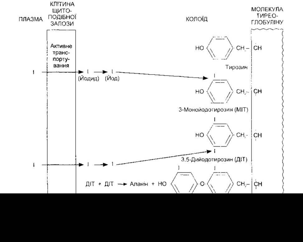
Fig. 18-6. Schematic diagram of thyroid hormone Biosynthesis. Tyrosine iodination occurs at the apical surface of thyroid cells, while the molecules remain peptide-linked to thyroglobulin.
Secretion
The human thyroid gland secretes approximately 80 µg (103 nmol) of T4, 4 µg (7 nmol) of T3, and 2 µg (3.5 nmol) of rT3 per day (Fig. 18-7); however, MIT and DIT are not secreted. Thyroid cells engulf the colloid via endocytosis (see Chapter 1). This continuous uptake at the colloid border forms resorption lacunae, which are visible in active glands (see Fig. 18-2). Within the cells, the colloid globules fuse with Lysosomes (see Fig. 18-3). The peptide bonds linking the iodinated residues to thyroglobulin are cleaved by proteases within the lysosomes, releasing T4, T3, DIT, and MIT into the Cytoplasm. The iodinated tyrosine residues are deiodinated by microsomal iodotyrosine deiodinase. This enzyme does not act upon iodinated thyronines, allowing T4 and T3 to escape into the blood. The iodine released from the deiodination of MIT and DIT is recycled by the gland, providing twice as much iodide for hormone synthesis as supplied by the iodide pump. In patients and subjects with an inherited deficiency of iodotyrosine deiodinase, MIT and DIT appear in the urine alongside symptoms of iodine deficiency (see below).

Fig. 18-7. Secretion and interconversion of thyroid hormones in normal adults. Values are given in micrograms per day. Note that the majority of T3 and rT3 is derived from T4 via peripheral tissue deiodination, with only small amounts secreted by the thyroid gland.
Last update: 10/08/2026
Editorial and Educational Adaptation: This material has been compiled based on the primary/original source text. The project team performed an editorial review, corrected technical inaccuracies, structured sections, and adapted the content for an educational format.
What was processed:
- elimination of formatting defects (OCR errors, structural breaks, corrupted characters);
- editorial organization of content;
- standardization of terminology in accordance with academic sources;
- verification of factual statements against the original source text.
All mentions of the author, publication year, and origin of the primary text have been preserved in accordance with the source.