Principles of Biochemistry Volume 3 - A. Lehninger 1985
Selected Aspects of Human Biochemistry
Digestion, nutrient transport, and metabolic interconnection
Adipose tissue is characterized by an active metabolism
Adipose tissue, consisting of fat Cells, or adipocytes (Fig. 24-16), is amorphous and distributed throughout the body: it is found beneath the Skin, surrounding deeply situated Blood Vessels, and within the Abdominal cavity. The total amount of adipose tissue in an average-weight young adult male is about 20 kg, which is roughly equal to total Muscle mass. Approximately 65% of the weight of adipose tissue is accounted for by stored triacylglycerols. Although adipose tissue appears inert at first glance, it is actually endowed with very high metabolic activity. Thus, adipose tissue rapidly responds to metabolic and hormonal stimuli and participates in the active crosstalk that exists between the Liver, skeletal Muscles, and The Heart.
Class="center">
Fig. 24-16. Scanning electron micrograph of adipose tissue cells (adipocytes). In adipose tissue, adipocytes are surrounded by a supporting network composed of blood capillaries and Collagen fibers. The adipocytes are filled with fat droplets that are actively involved in METABOLISM.
Just as in other Cell types, Glycolysis actively proceeds in fat cells (adipocytes), Pyruvate and Fatty acids are oxidized in The Citric Acid Cycle, and Oxidative Phosphorylation takes place. When CARBOHYDRATES are abundantly supplied to the Organism, glucose in adipose tissue is converted into fatty acids via The intermediate formation of pyruvate and acetyl-CoA; the fatty acids are utilized to form triacylglycerols, which accumulate as large lipid globules (Fig. 24-16). In this process of glucose-to-fat conversion, the reducing equivalent is NADPH, which is generated in the Pentose Phosphate Pathway as well as by the malic enzyme (malate dehydrogenase; Section 21.5,d).
Adipocytes also actively accumulate triacylglycerols derived from the gastrointestinal tract in the form of chylomicrons (Sections 12.8 and 24.1,c), especially after a fatty meal. Chylomicrons reaching adipose tissue are acted upon by lipoprotein lipase, which is localized in the cells of blood capillaries. This enzyme cleaves one or more fatty acids from the triacylglycerols within the chylomicrons. The fatty acids released by lipoprotein lipase are subsequently taken up by adipose tissue cells, where they are enzymatically converted into triacylglycerols and thereby stored as reserves. However, the fatty acids released by lipoprotein lipase in the capillaries of skeletal muscles and the heart serve as fuel for these Organs and are oxidized within them. As they lose triacylglycerols through the action of lipoprotein lipase, blood chylomicrons decrease in size, yet they retain their Phospholipids, Cholesterol esters, and Proteins. These residual structures—chylomicron remnants—are cleared from the bloodstream and taken up by the liver. The triacylglycerols stored within adipose tissue cells are not subjected to the action of the lipoprotein lipase localized in blood capillary cells, but are instead hydrolyzed by intracellular lipases: the resulting free fatty acids can then enter the blood, where they bind to serum albumin.
Each serum albumin molecule is capable of binding two long-chain fatty acid molecules very tightly and another one or two molecules less tightly. Because serum albumin is present in Blood Plasma at a very high concentration, it serves as the primary carrier of fatty acids in the blood. In this albumin-bound form, fatty acids are delivered to skeletal muscles and the heart, where they are predominantly utilized.
The rate of fatty acid release from adipocytes increases dramatically under the Influence of the hormone epinephrine (Chapter 25), which binds to cell-surface receptors and promotes The conversion of the inactive form of adipocyte lipase into the active form via phosphorylation. The binding of Insulin to The surface of fat cells counteracts The Effect of epinephrine and decreases adipocyte lipase activity.
Cases of genetic lipoprotein lipase deficiency are known to occur. In patients with this deficiency, chylomicrons persist in the bloodstream for extended periods after a fatty meal. Triacylglycerols that cannot be properly utilized due to the lack of lipoprotein lipase are deposited as yellow, lipid-engorged nodules beneath the skin. Genetic Defects in Other types of plasma Lipoproteins disrupt their normal metabolism. There is evidence suggesting that atherosclerosis and myocardial infarction occur more frequently in individuals with elevated blood levels of triacylglycerols.
Humans, much like many animals—especially those that undergo hibernation—possess a specialized type of adipose tissue known as brown fat (Fig. 24-17). The presence of this tissue is particularly characteristic of newborns, in whom it is located in the neck and the upper Regions of the chest and back. The color of brown fat is due to the presence of A large number of Mitochondria rich in Cytochromes (Section 17.17). Brown fat is specialized for heat production rather than ATP generation during the Oxidation of Fatty acids. The inner mitochondrial membranes in brown adipose tissue contain specific pores through which H+ ions are transferred, and their H+-ion-translocating capacity is regulated. Through these pores, H+ ions pumped out of the mitochondria during electron transport (Section 17.15,e) can re-enter the respiring mitochondria; the net result is an unregulated or "futile" cycle of H+ ions that dissipates energy as heat instead of forming ATP (Section 17.17). When the organism has no need for heat, these H+ pores close, and the mitochondria of brown adipose tissue resume ATP synthesis.
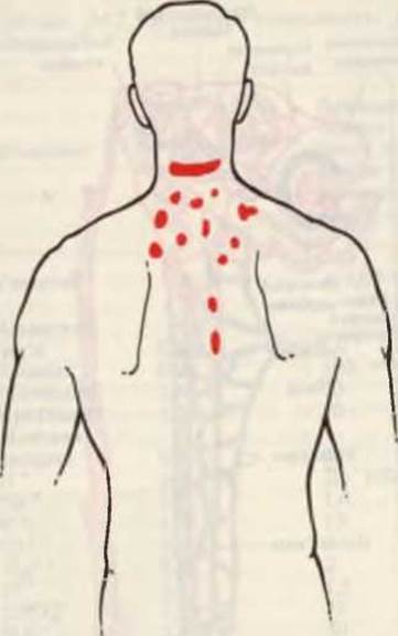
Fig. 24-17. Localization of brown adipose tissue (highlighted in red) in the neck and back region of an adult human.
Last update: 06/08/2026
Editorial and Educational Adaptation: This material has been compiled based on the primary/original source text. The project team performed an editorial review, corrected technical inaccuracies, structured sections, and adapted the content for an educational format.
What was processed:
- elimination of formatting defects (OCR errors, structural breaks, corrupted characters);
- editorial organization of content;
- standardization of terminology in accordance with academic sources;
- verification of factual statements against the original source text.
All mentions of the author, publication year, and origin of the primary text have been preserved in accordance with the source.