BIOCHEMISTRY: A TEXTBOOK FOR MEDICAL UNIVERSITIES - E. S. Severin - 2004
CHAPTER 7. CARBOHYDRATE METABOLISM
IX. Glucose Synthesis in the Liver (Gluconeogenesis)
Certain Tissues, such as the Brain, require a continuous supply of glucose. When dietary carbohydrate intake is insufficient, Blood glucose levels are maintained within the normal range for some time through The breakdown of hepatic Glycogen. However, glycogen reserves in the Liver are limited. They decrease significantly after 6 — 10 hours of fasting and are almost completely depleted following a 24-hour fast. Under these conditions, the liver initiates de novo glucose synthesis — Gluconeogenesis. Gluconeogenesis is The process of synthesizing glucose from non-carbohydrate precursors. Its primary function is to maintain blood glucose levels during prolonged fasting and intense physical exertion. The process occurs predominantly in The Liver and, to a lesser extent, in the renal cortex and the intestinal mucosa. These tissues can supply 80 — 100 g of glucose per day. During fasting, the brain accounts for the majority of the body's glucose demand. This is because brain Cells, unlike other tissues, cannot meet their Energy Requirements through the Oxidation of Fatty acids (see Chapter 8).
In addition to the brain, glucose is essential for tissues and cells where the aerobic pathway of breakdown is impossible or restricted, such as erythrocytes (which lack Cell/35.html">Mitochondria), retinal cells, and adrenal medullary cells, among others.
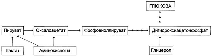
The primary precursors of gluconeogenesis are lactate, Amino Acids, and glycerol. The incorporation of these substrates into gluconeogenesis depends on the physiological state of the Organism.
✵ Lactate is the end product of anaerobic Glycolysis. It is produced in erythrocytes and working Muscles under various physiological conditions. Consequently, lactate is continuously utilized in gluconeogenesis.
✵ Glycerol is released during the Hydrolysis of fats in adipose tissue during fasting or prolonged physical activity.
✵ Amino acids are generated through the breakdown of Muscle Proteins and are channeled into gluconeogenesis during extended fasting or prolonged muscular work.
Figure 7-44 illustrates the points of entry for the primary precursors into gluconeogenesis.
Class="center">Fig. 7-44. Incorporation of substrates into gluconeogenesis.
A. Reactions of Gluconeogenesis
Most reactions of gluconeogenesis proceed via the reversible steps of glycolysis (Fig. 7-45, reactions 9, 8, 7, 6, 5, 4, 2) and are catalyzed by the same Enzymes. However, Three Reactions of glycolysis are thermodynamically irreversible. At these stages, the reactions of gluconeogenesis proceed through alternative pathways.
Fig. 7-45. Glycolysis and gluconeogenesis. Enzymes of the reversible reactions of glycolysis and gluconeogenesis: 2 — phosphoglucoisomerase; 4 — aldolase; 5 — Triosephosphate isomerase; 6 — glyceraldehyde-3-phosphate dehydrogenase; 7 — phosphoglycerate kinase; 8 — phosphoglycerate mutase; 9 — enolase. Enzymes of the irreversible reactions of gluconeogenesis: 11 — Pyruvate carboxylase; 12 — phosphoenolpyruvate carboxykinase; 13 — fructose-1,6-bisphosphatase; 14 — glucose-6-phosphatase. I-III — Substrate Cycles.
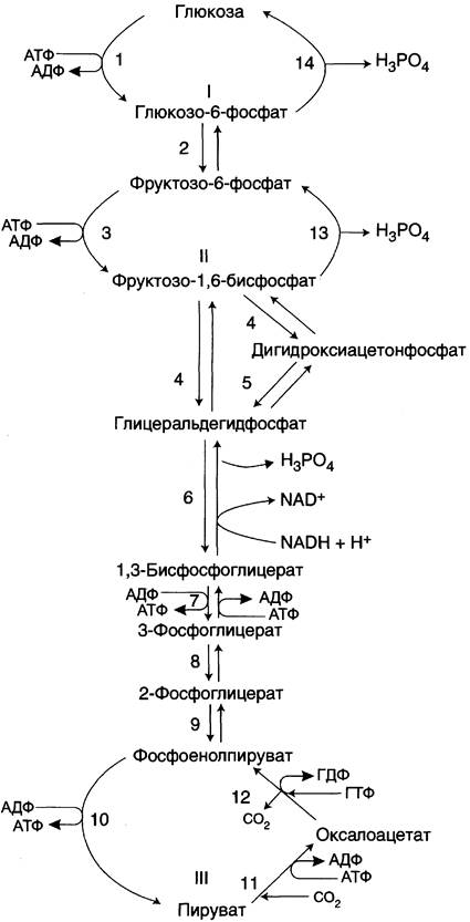
It should be noted that glycolysis takes place in the Cytosol, whereas certain reactions of gluconeogenesis occur within the mitochondria.
Let us examine in greater detail those reactions of gluconeogenesis that differ from the reactions of glycolysis and involve alternative enzymes. We will first consider the process of glucose synthesis from pyruvate.
1. Formation of phosphoenolpyruvate from pyruvate — the first irreversible step of gluconeogenesis
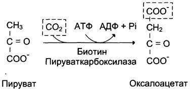
The formation of phosphoenolpyruvate from pyruvate occurs via two reactions (Fig. 7-45, reactions 11, 12), the first of which takes place in the mitochondria. Pyruvate derived from lactate or Certain amino acids is transported into the mitochondrial matrix, where it is carboxylated to form oxaloacetate (Fig. 7-46). Pyruvate carboxylase, The enzyme catalyzing this reaction, is a mitochondrial enzyme that requires biotin as a coenzyme. The reaction proceeds with the consumption of ATP.
Fig. 7-46. Formation of oxaloacetate from pyruvate.
Subsequent transformations of oxaloacetate take place in the cytosol. Therefore, a transport system must exist at this stage to shuttle oxaloacetate across the mitochondrial membrane, which is otherwise impermeable to it. Within the mitochondrial matrix, oxaloacetate is reduced to malate (Fig. 7-47) with the participation of NADH (the reverse reaction of The Citric Acid Cycle). The resulting malate then crosses the mitochondrial membrane via specific carrier proteins. Additionally, oxaloacetate can be transported from the mitochondria to the cytosol in the form of aspartate via the malate-aspartate shuttle mechanism discussed previously (Fig. 7-39).
Fig. 7-47. Conversion of oxaloacetate to malate.
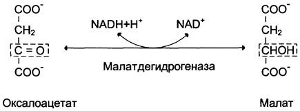
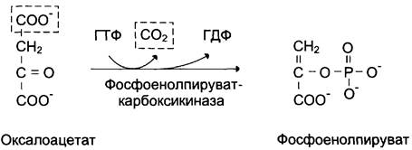
In the cytosol, malate is converted back into oxaloacetate via an oxidation reaction involving the coenzyme NAD+. Both reactions—the reduction of oxaloacetate and The oxidation of malate—are catalyzed by malate dehydrogenase; however, the former utilizes the mitochondrial enzyme, whereas the latter employs the cytosolic one. The oxaloacetate produced from malate in the cytosol is subsequently converted into phosphoenolpyruvate in a reaction catalyzed by phosphoenolpyruvate carboxykinase, a GTP-dependent enzyme (Fig. 7-48). The enzyme is named after the reverse reaction.
Fig. 7-48. Conversion of oxaloacetate into phosphoenolpyruvate.
The pathway of all reactions occurring in the first irreversible stage of gluconeogenesis is illustrated in Fig. 7-49.
Fig. 7-49. Formation of oxaloacetate, transport into the cytosol, and conversion into phosphoenolpyruvate. 1 — transport of pyruvate from the cytosol into the mitochondria; 2 — conversion of pyruvate into oxaloacetate (OA); 3 — conversion of OA into malate or aspartate; 4 — transport of aspartate and malate from the mitochondria into the cytosol; 5 — conversion of aspartate and malate into OA; 6 — conversion of OA into phosphoenolpyruvate.
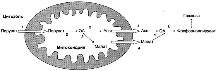
It should be noted that this bypass pathway of gluconeogenesis requires the consumption of two high-energy phosphate molecules (ATP and GTP) per molecule of the starting substance, pyruvate. When calculated for the synthesis of a single glucose molecule from two pyruvate molecules, the expenditure is 2 mol of ATP and 2 mol of GTP, or a total of 4 mol of ATP (for simplicity, the energy cost for the synthesis of ATP and GTP is assumed to be equal). Following the formation of phosphoenolpyruvate, all subsequent reactions also
proceed in the cytosol up to the Formation of fructose-1,6-bisphosphate and are catalyzed by glycolytic enzymes.
2. Hydrolysis of Fructose-1,6-Bisphosphate and Glucose-6-Phosphate
The removal of the phosphate group from fructose-1,6-bisphosphate and glucose-6-phosphate represents another irreversible step in gluconeogenesis. In glycolysis, these reactions are catalyzed by specific Kinases utilizing ATP energy. In contrast, during gluconeogenesis, they proceed without the involvement of ATP and ADP, being accelerated not by kinases, but by Phosphatases—enzymes belonging to the hydrolase class. The enzymes fructose-1,6-bisphosphatase and glucose-6-phosphatase catalyze the removal of the phosphate group from fructose-1,6-bisphosphate and glucose-6-phosphate, respectively. Afterward, free glucose is released from The Cell into the bloodstream. The overall scheme of all gluconeogenesis reactions is presented in Fig. 7-45.
Thus, the liver contains four enzymes that participate exclusively in gluconeogenesis, catalyzing the bypass Reactions of the irreversible stages of glycolysis. These are pyruvate carboxylase, phosphoenolpyruvate carboxykinase, fructose-1,6-bisphosphatase, and glucose-6-phosphatase.
3. Energetic Balance of Gluconeogenesis from Pyruvate
This process consumes 6 mol of ATP for the synthesis of 1 mol of glucose from 2 mol of pyruvate. Four moles of ATP are expended during the synthesis of phosphoenolpyruvate from oxaloacetate, and an additional 2 mol of ATP are utilized at the stages of 1,3-bisphosphoglycerate formation from 3-phosphoglycerate.
The net equation for gluconeogenesis from pyruvate is expressed as follows:
2 Pyruvate + 4 ATP + 2 GTP + 2 (NADH + H+) + 4 H2O —> Glucose + 4 ADP + 2 GDP + 6 H3PO4 + 2 NAD+.
B. SYNTHESIS OF GLUCOSE from Lactate
Lactate produced during anaerobic glycolysis is not a metabolic dead end. The utilization of lactate involves its conversion into pyruvate in the liver. Lactate serves as a source of pyruvate not only during starvation but also under normal physiological conditions. Its conversion into pyruvate and the subsequent utilization of the latter constitute a key mechanism for lactate clearance.
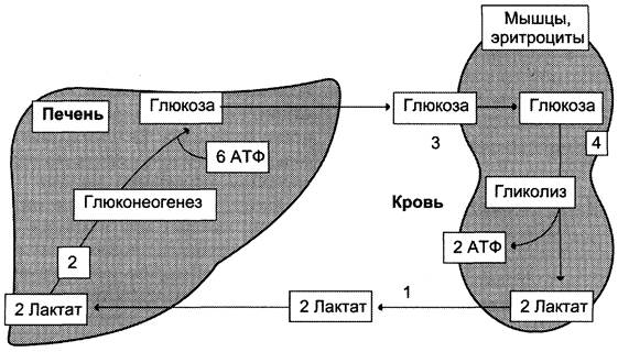
Lactate formed in vigorously working muscles or in cells with a predominantly anaerobic pathway of Glucose Catabolism enters the bloodstream and is subsequently delivered to the liver. Because the NADH/NAD+ ratio in the liver is lower than in contracting muscle, the Lactate dehydrogenase reaction proceeds in the reverse direction—that is, toward the formation of pyruvate from lactate. Subsequently, pyruvate enters The gluconeogenesis pathway, and the newly synthesized glucose is released into the blood to be taken up by skeletal muscles. This
sequence of events is referred to as the “glucose-lactate cycle” or the “Cori cycle” (Fig. 7-50). The Cori cycle fulfills two critical Functions: 1 — it ensures the clearance and utilization of lactate; 2 — it prevents the accumulation of lactate and the resultant dangerous drop in pH (lactic acidosis). A portion of the pyruvate derived from lactate is oxidized in the liver to CO2 and H2O. The energy released from this oxidation can be harnessed to drive the ATP-requiring reactions of gluconeogenesis.
Fig. 7-50. The Cori cycle (glucose-lactate cycle). 1 — transport of lactate from contracting muscle via the bloodstream to the liver; 2 — hepatic synthesis of glucose from lactate; 3 — transport of glucose from the liver via the bloodstream to active muscle; 4 — utilization of glucose as an energy substrate by contracting muscle and generation of lactate.
Lactic Acidosis. The term “acidosis” denotes an increase in the acidity of the Body Fluids (a drop in pH) beyond the normal physiological range. Acidosis results either from increased proton production or decreased proton excretion (and in some cases, both). Metabolic acidosis develops when there is an elevation in the concentration of acidic intermediary metabolites due to enhanced synthesis or reduced rates of degradation and elimination. When the body's acid-base balance is disturbed, buffer compensation mechanisms are rapidly engaged (within 10–15 minutes). Pulmonary compensation stabilizes the HCO3-/H2CO3 ratio, which normally stands at 1:20 but declines during acidosis. Pulmonary compensation is achieved by increasing the volume of venti-
lation and, consequently, accelerating the elimination of CO2 from the body. However, the primary role in compensating for acidosis belongs to renal mechanisms involving the ammonia buffer system (see Section 9). One of the potential causes of metabolic acidosis is the accumulation of lactic acid. Under normal conditions, hepatic lactate is either reconverted into glucose via gluconeogenesis or oxidized. Alongside the liver, the Kidneys and Heart muscle serve as major consumers of lactate, where it can be oxidized to CO2 and H2O and utilized as an energy source, particularly during physical exertion.
Blood lactate levels reflect a dynamic equilibrium between its production and utilization rates. Transient, compensated lactic acidosis is quite common even in healthy individuals during intense muscular work. In untrained subjects, exercise-induced lactic acidosis develops fairly rapidly as a consequence of a relative oxygen deficit in the muscles, with compensation achieved through hyperventilation.
In uncompensated lactic acidosis, blood lactate levels rise up to 5 mmol/L (normal values are up to 2 mmol/L), while blood pH may drop to 7.25 or lower (normal range is 7.36–7.44).
Elevated blood lactate levels can result from impaired pyruvate METABOLISM (Fig. 7-51).
Fig. 7-51. Impairments in pyruvate metabolism in lactic acidosis. 1 — impaired utilization of pyruvate in gluconeogenesis; 2 — impaired pyruvate oxidation.
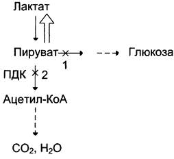
Thus, in Hypoxia caused by impaired tissue oxygenation or blood supply, The activity of the pyruvate dehydrogenase complex decreases, leading to suppressed Oxidative Decarboxylation of pyruvate. Under these conditions, the equilibrium of the pyruvate <-> lactate reaction shifts toward lactate production. Furthermore, hypoxia reduces ATP synthesis, which subsequently slows down gluconeogenesis—another pathway for lactate utilization. Elevated lactate concentrations and decreased intracellular pH negatively affect the activity of all enzymes, including pyruvate carboxylase, which catalyzes the initial reaction of gluconeogenesis.
Lactic acidosis is also promoted by impaired gluconeogenesis resulting from hepatic failure of various etiologies. Additionally, vitamin B1 deficiency can lead to lactic acidosis, as the derivative of this vitamin (Thiamine diphosphate) acts as a coenzyme within the PDC during The oxidative decarboxylation of pyruvate (see Section 6). Thiamine deficiency may occur, for instance, in malnourished individuals with alcohol dependence.
Thus, the causes of lactic acid accumulation and The Development of lactic acidosis may include:
✵ activation of anaerobic glycolysis due to tissue hypoxia of various origins;
✵ liver disorders (toxic dystrophies, cirrhosis, etc.);
✵ impaired lactate utilization due to hereditary defects in gluconeogenesis enzymes or glucose-6-phosphatase deficiency;
✵ impaired PDC function resulting from enzymatic defects or hypovitaminosis;
✵ administration of certain medications, such as biguanides (gluconeogenesis blockers used in the Treatment of Diabetes Mellitus).
B. Synthesis of glucose from amino acids
Under fasting conditions, a portion of muscle proteins is degraded into amino acids, which subsequently enter Catabolic pathways. Amino acids that are converted into pyruvate or Tricarboxylic Acid Cycle intermediates during catabolism can be considered potential precursors of glucose and glycogen and are termed glucogenic. For instance, oxaloacetate derived from aspartic acid is an intermediate in both The Tricarboxylic Acid Cycle and gluconeogenesis.
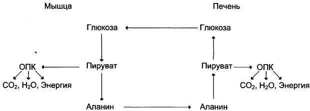
Of all the amino acids delivered to the liver, approximately 30% are accounted for by Alanine. This is because the breakdown of muscle proteins yields amino acids, many of which are converted directly into pyruvate or first into oxaloacetate and then into pyruvate. The latter is transformed into alanine by acquiring an amino group from Other Amino Acids. Alanine is transported via the bloodstream from muscles to the liver, where it is reconverted into pyruvate, which is partially oxidized and partially channeled into gluconeogenesis. Consequently, the following sequence of events takes place (glucose-alanine cycle): glucose in muscles —> pyruvate in muscles —> alanine in muscles —> alanine in the liver —> glucose in the liver —> glucose in muscles (Fig. 7-52). The entire cycle does not result in a net increase of glucose in the muscles, but it solves Structure/149.html">The problem of transporting amino nitrogen from muscles to the liver and prevents lactic acidosis.
Fig. 7-52. The glucose-alanine cycle.
G. Synthesis of glucose from glycerol
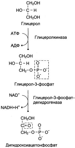
Glycerol is produced during the hydrolysis of triacylglycerols, primarily within adipose tissue. It can be utilized only by tissues that contain the enzyme glycerol kinase, such as the liver and kidneys. This ATP-dependent enzyme catalyzes The conversion of glycerol to α-glycerophosphate (glycerol-3-phosphate). When glycerol-3-phosphate enters gluconeogenesis, it undergoes dehydrogenation by an NAD-dependent dehydrogenase to form dihydroxyacetone phosphate (Fig. 7-53), which is subsequently converted into glucose.
Fig. 7-53. Conversion of glycerol to dihydroxyacetone phosphate.
Last update: 06/08/2026
Editorial and Educational Adaptation: This material has been compiled based on the primary/original source text. The project team performed an editorial review, corrected technical inaccuracies, structured sections, and adapted the content for an educational format.
What was processed:
- elimination of formatting defects (OCR errors, structural breaks, corrupted characters);
- editorial organization of content;
- standardization of terminology in accordance with academic sources;
- verification of factual statements against the original source text.
All mentions of the author, publication year, and origin of the primary text have been preserved in accordance with the source.