Harper's Illustrated Biochemistry, Volume 1 - Murray R. 1993
Bioenergetics and Carbohydrate and Lipid Metabolism
Glycogen Metabolism
Mechanisms for the Control of Glycogenolysis and Glycogenesis
The Key Enzymes controlling Glycogen METABOLISM—Glycogen phosphorylase and glycogen synthase—are regulated by a complex series of reactions involving both allosteric mechanisms (see p. 104) and covalent modification via phosphorylation and dephosphorylation of the enzyme (see p. 108).
Activation and inactivation of phosphorylase (Fig. 19.5)
In the Liver, phosphorylase exists in both active and inactive forms. In active phosphorylase (phosphorylase a), the hydroxyl group of one of the Serine residues is phosphorylated. Upon the action of a specific phosphatase (protein phosphatase-1), the enzyme is converted into inactive phosphorylase b As a result of the hydrolytic Cleavage of phosphate from the serine residue. Reactivation occurs via rephosphorylation by ATP under the action of a specific enzyme, phosphorylase kinase.
Muscle phosphorylase is immunologically and genetically distinct from the corresponding liver enzyme. It can exist in two forms: phosphorylase a, a phosphorylated enzyme that is active both in the presence and absence of AMP (its allosteric modulator), and phosphorylase b, which is dephosphorylated and active only in the presence of AMP. Phosphorylase a is the normal physiologically active form of the enzyme. It is a dimer, with each monomer containing one molecule of Pyridoxal phosphate.
cAMP-mediated activation
In muscle, Phosphorylase is activated by adrenaline (Fig. 19.5). However, this effect is not direct but mediated through cAMP (3',5'-cyclic adenylic acid; cyclic AMP) (Fig. 19.6 and Chapter 44). cAMP is an intracellular intermediate that acts as a second messenger in the action of A number of Hormones. It is formed from ATP through the action of the enzyme adenylate cyclase, located on the inner surface of The Cell membrane. Adenylate cyclase is activated (indirectly) by the hormones adrenaline and noradrenaline—ligands of ß-adrenergic receptors localized in the cell membrane; in the liver, it is activated by Glucagon acting via a specific glucagon receptor. cAMP is degraded by the action of phosphodiesterase; it is this enzyme that normally maintains cAMP concentration at a low level. Evidence indicates that Insulin increases phosphodiesterase activity in the liver, leading to a decrease in cAMP concentration.
Class="center">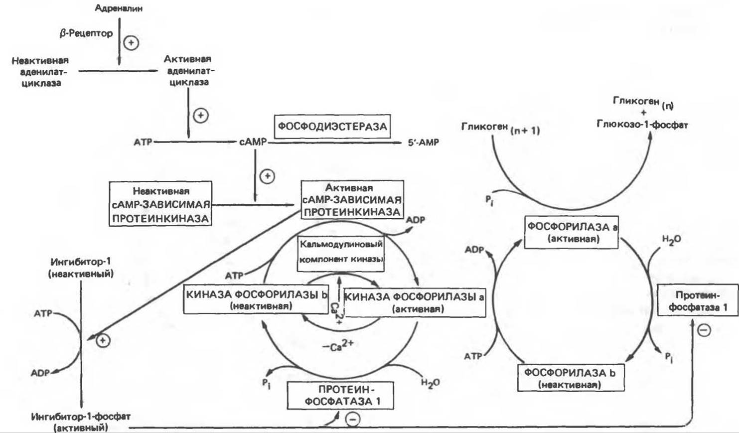
Fig. 19.5. Regulation of phosphorylase activity in muscle (n is the number of glucose residues). The reaction sequence forms a cascade, allowing the hormonal signal to be amplified at each stage.
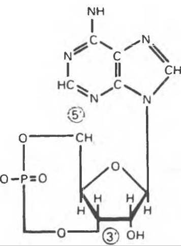
Fig. 19.6. 3',5'-Adenylic acid (cyclic AMP, cAMP).
An increase in cAMP concentration activates an enzyme with very broad Specificity: cAMP-dependent protein kinase. This kinase catalyzes the ATP-dependent phosphorylation of inactive phosphorylase kinase to form active phosphorylase kinase, which in turn phosphorylates and activates phosphorylase b, converting it into phosphorylase a (Fig. 19.5).
Inactive cAMP-dependent protein kinase consists of two pairs of subunits; each pair includes a regulatory subunit (R), capable of binding two cAMP molecules, and a catalytic subunit (C), whose Structure includes the Active Site. The binding of cAMP to the R2C2 complex causes its dissociation, resulting in the release of active C monomers (see Chapter 44):

Activation by Ca2+ ions and synchronization with Muscle contraction
Immediately after the onset of muscle contraction, Glycogenolysis increases several hundred-fold. The process involves the rapid activation of phosphorylase due to the activation of phosphorylase kinase by Ca2+ ions—the same signal that initiates contraction. Muscle phosphorylase kinase consists of four types of subunits: a, ß, y, and δ. Its structure is (a, ß, y, δ)4. The a and ß subunits contain serine residues phosphorylated by cAMP-dependent protein kinase. The ß subunit binds four Ca2+ ions and is identical to the Ca2+-binding protein calmodulin. The binding of Ca2+ ions activates the catalytic center of the y subunit, although the molecule remains in the dephosphorylated b-conformation. At the same time, full activity of the phosphorylated a-form is achieved only in the presence of Ca2+ ions. Notably, calmodulin shares structural similarities with the muscle Ca2+-binding protein troponin C (TnC). A second molecule of calmodulin or TnC can interact with phosphorylase kinase, causing additional enzyme activation. Thus, the activation of muscle contraction and glycogenolysis are mediated by the same Ca2+-binding protein. Calmodulin is a protein involved in many aspects of calcium action within the cell (see Chapter 44).
Glycogenolysis in the liver
It has been established that during The stimulation of glycogenolysis by catecholamines in the liver, a1 receptors act as the primary mediators. This process involves a cAMP-independent mobilization of Ca2+ ions and their transfer from the Mitochondria to the Cytosol, where they stimulate the Ca2+/calmodulin-sensitive phosphorylase kinase. Unlike liver phosphorylase, Skeletal Muscle phosphorylase is not activated by glucagon. Notably, cardiac muscle phosphorylase is activated by this hormone. Another important difference is the inhibition of liver protein phosphatase-1 by the active form of phosphorylase.
Inactivation of phosphorylase
Phosphorylase a and phosphorylase kinase a are dephosphorylated and inactivated by protein phosphatase-1. An inhibitor of protein phosphatase-1 is a protein known as inhibitor-1; the latter becomes active only after phosphorylation by cAMP-dependent protein kinase. Thus, cAMP controls both the activation and inactivation of phosphorylase (Fig. 19.5).
Activation and inactivation of glycogen synthase (Fig. 19.7)
Like phosphorylase, glycogen synthase can exist in either a phosphorylated or unphosphorylated state. However, unlike phosphorylase, the dephosphorylated form (glycogen synthase a) is active, which can be inactivated to form glycogen synthase b through the phosphorylation of seven serine residues by at least five different protein Kinases. All seven phosphorylation sites are located on each of the four identical subunits. Two of these protein kinases are Ca2+/calmodulin-dependent. One is phosphorylase kinase, and the other is cAMP-dependent protein kinase; it is this protein kinase that mediates cAMP-dependent hormonal effects that simultaneously inhibit glycogen synthesis and activate glycogenolysis. The remaining kinases are known as glycogen synthase kinases-3, -4, and -5.
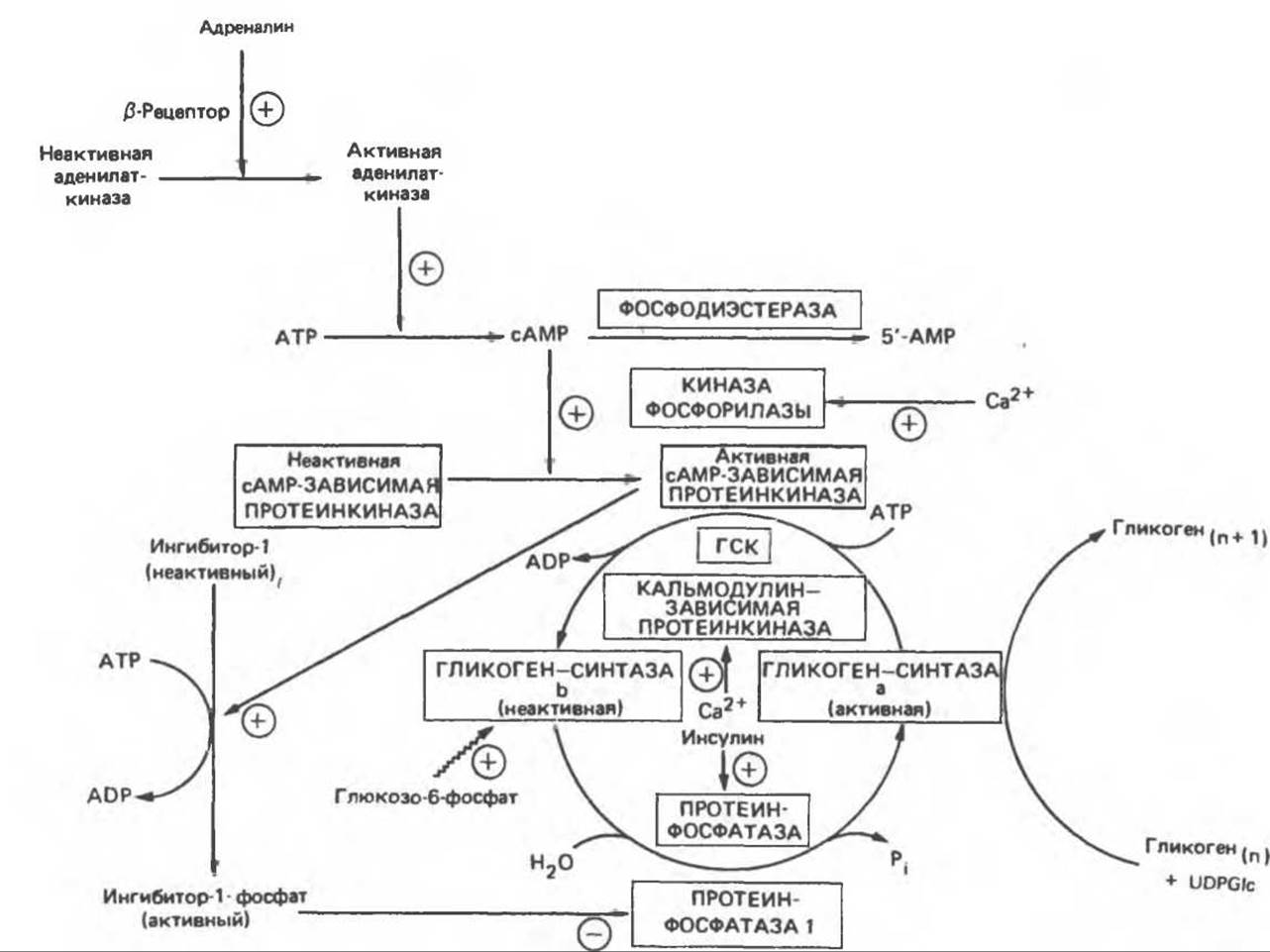
Fig. 19.7. Regulation of Glycogen synthase activity in muscle (n is the number of glucose residues). The reaction sequence forms a cascade that allows signal Amplification at each stage; nanomolar quantities of a hormone can cause significant changes in glycogen concentration. GSK: glycogen synthase kinases-3, -4, and -5. The wavy arrow indicates allosteric activation.
Glucose-6-phosphate is an allosteric activator of glycogen synthase b, causing a decrease in the Km for UDP-glucose and thereby enabling glycogen synthesis by the phosphorylated form of the enzyme. Glycogen exerts an inhibitory effect on its own synthesis; insulin stimulates glycogen synthesis in muscle by promoting the dephosphorylation and activation of glycogen synthase b. Normally, the dephosphorylation of glycogen synthase b is carried out by protein phosphatase-1, which is controlled by cAMP-dependent protein kinase (Fig. 19.7).
Other Aspects of The regulation of glycogen metabolism will be discussed on p. 219.
Last update: 06/08/2026
Editorial and Educational Adaptation: This material has been compiled based on the primary/original source text. The project team performed an editorial review, corrected technical inaccuracies, structured sections, and adapted the content for an educational format.
What was processed:
- elimination of formatting defects (OCR errors, structural breaks, corrupted characters);
- editorial organization of content;
- standardization of terminology in accordance with academic sources;
- verification of factual statements against the original source text.
All mentions of the author, publication year, and origin of the primary text have been preserved in accordance with the source.